Review
. 2023 Apr 3;13(4):a041156.
doi: 10.1101/cshperspect.a041156.

Extracellular Matrix Regulation of Vascular Morphogenesis, Maturation, and Stabilization

Affiliations
Review

Extracellular Matrix Regulation of Vascular Morphogenesis, Maturation, and Stabilization

George E Davis et al. Cold Spring Harb Perspect Med. .

Abstract

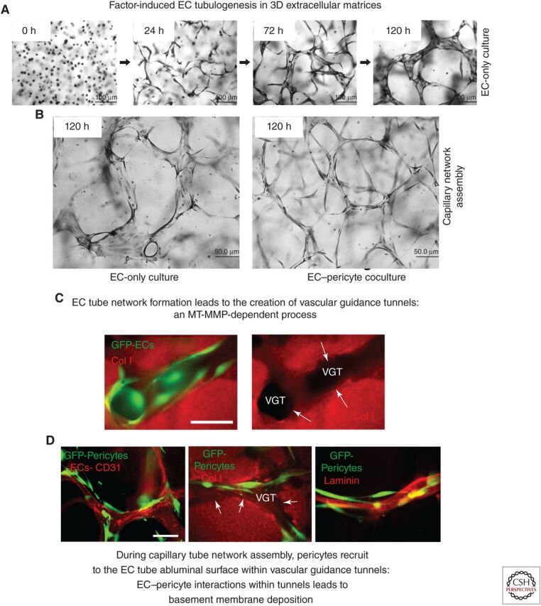
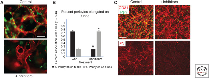
The extracellular matrix represents a critical regulator of tissue vascularization during embryonic development and postnatal life. In this perspective, we present key information and concepts that focus on how the extracellular matrix controls capillary assembly, maturation, and stabilization, and, in addition, contributes to tissue stability and health. In particular, we present and discuss mechanistic details underlying (1) the role of the extracellular matrix in controlling different steps of vascular morphogenesis, (2) the ability of endothelial cells (ECs) and pericytes to coassemble into elongated and narrow capillary EC-lined tubes with associated pericytes and basement membrane matrices, and (3) the identification of specific growth factor combinations ("factors") and peptides as well as coordinated "factor" and extracellular matrix receptor signaling pathways that are required to form stabilized capillary networks.


Figures

Figure 1.
Figure 1.
Extracellular matrix and growth factor–induced signaling stimulates endothelial tubulogenesis, and endothelial–pericyte tube coassembly to generate capillary networks with associated basement membrane matrices. (A) Using a serum-free defined model system with added SCF, IL-3, SDF-1α, FGF-2, and insulin (“factors”) and 3D collagen matrices, endothelial cells (ECs) were seeded alone and were allowed to assemble into networks of EC-lined tubes for the indicated times. Cultures were fixed and stained with toluidine blue. Scale bar, 100 µm. (B) EC-only versus EC and pericyte cocultures were seeded in the presence of the “factors” in 3D collagen matrices and were fixed and stained after 120 h. Scale bar, 50 µm. (C) GFP-labeled EC-only cultures were seeded in collagen matrices and, after 72 h, were fixed and stained with anticollagen type I antibodies (red). (VGTs) Vascular guidance tunnels. White arrows indicate the borders of these tunnel spaces. Scale bar, 50 µm. (D) ECs and GFP-labeled pericytes were cocultured in 3D collagen matrices for 120 h, and, after fixation, were immunostained with antibodies to CD31 to label ECs, collagen type I to delineate the VGT spaces, and laminin to label the vascular basement membrane matrix. White arrows indicate the borders of the tunnel spaces. Scale bar, 50 µm.
Figure 2.
Figure 2.
“Factor”-dependent molecule and signaling requirements for endothelial cell (EC) tubulogenesis, EC–pericyte tube coassembly, and basement membrane deposition, all necessary steps to create capillary networks. (AC) EC–pericyte cocultures were established in 3D collagen matrices, and, after 120 h, cultures were fixed, photographed under light microscopy (A), or immunostained with antibodies directed to CD31 to visualize ECs (B), or to collagen type IV to visualize basement membrane deposition. Scale bars, 100 µm (A); 50 µm (B,C). (D) Schematic diagram showing key molecule and signaling requirements for ECs to respond to a five-“factor” combination of SCF, IL-3, SDF-1α, FGF-2, and insulin, which stimulates EC tubulogenesis in 3D matrices in conjunction with integrin- and MT1-MMP-dependent signaling events. (Col I) Collagen type I, (FN) fibronectin, (FG) fibrinogen/fibrin. The indicated key molecules and signaling pathways are required for EC lumen and tube formation and include key kinase cascades, small GTPases, and their effectors, and the production of EC-derived factors (PDGF-BB, PDGF-DD, ET-1, TGF-β1, and HB-EGF) that drive pericyte recruitment, pericyte proliferation, and pericyte-induced basement membrane assembly.
Figure 3.
Figure 3.
Distinct vascular morphogenic (“fire”) versus vascular stabilization (“ice”) signals delivered to endothelial cells (ECs) by exposed interstitial matrices, such as collagen type I or vascular basement membranes (BMs), respectively. Schematic diagram discussing how EC exposure to different types of extracellular matrix (ECM) environments can either lead to initiation/stimulation of vascular morphogenesis (“fire”) or can suppress morphogenesis and lead to stabilization of the vasculature (“ice”). Exposure of ECs to collagen matrices increases Src and Rho activities, which leads to disassembly of VE-cadherin junctions and increases actin stress fibers. These signals cause increased sprouting behavior and EC cord assembly. In contrast, exposure to laminins, such as laminin-511, leads to EC wall stabilization with enhanced VE–cadherin junctions due to increased Rac, Rap, EPAC, and PKA activities. Overall, this leads to suppression of EC activation and morphogenesis. EC adhesive interactions with the vascular BM enhances apicobasal polarization, which further enhances EC junctional stability, and the vascular BM matrix may also facilitate this stabilization by sequestering or inactivating proinflammatory or growth factor/peptide/small molecule mediators that contribute to vascular destabilization.
Figure 4.
Figure 4.
Endothelial cell (EC)–pericyte interactions control vascular basement membrane (BM) matrix deposition, which changes integrin requirements necessary for vascular wall maturation and stabilization compared to those controlling vascular morphogenesis. (A) EC-only cultures versus EC–pericyte cocultures were established in 3D collagen matrices in the presence of the “factors” and, after 120 h, were fixed and imaged by transmission electron microscopy. Collagen fibrils are visualized on the basal aspects of the tubes and the luminal (L) spaces were devoid of collagen fibrils. The black arrows indicate the vascular BM. Scale bars, 0.5 µm (left panel); 1 µm (right panel). (B) Key morphologic features comparing EC-only cultures to EC–pericyte cocultures in 3D collagen matrices. Blocking antibody experiments revealed complete dependence on the α2β1 integrin in EC-only cultures, while EC–pericyte cocultures demonstrated involvement of the BM-binding integrins, α6β1, α3β1, α1β1, and α5β1, while showing much less involvement of α2β1. EC-only cultures were more susceptible to MMP-1-dependent tube regression (leading to collagen matrix degradation and tube collapse) compared to EC–pericyte cocultures when serine proteinases such as plasminogen (converted to plasmin by ECs) or plasma kallikrein were added due to EC production of TIMP-2 and pericyte production of TIMP-3.
Figure 5.
Figure 5.
Blockade of pericyte recruitment during capillary assembly induces less elongated and markedly widened endothelial cell (EC) tubes with strongly reduced deposition of basement membrane matrices. A mixture of chemical inhibitors directed to pericyte receptors, which recognize EC-derived factors that stimulate pericyte recruitment and proliferation, were added to EC–pericyte cocultures for 120 h. The combined chemical inhibitors included imatinib (which blocks PDGFRβ), CI 1020 (which blocks endothelin receptor A), SB431542 (to block the TGF-β receptor, Alk5), and gefitinib (which blocks EGF receptors), and their addition was compared to control cultures. (A) Untreated versus inhibitor-treated cultures were stained with anti-CD31 antibodies to label ECs, revealing marked shortening and widening of EC tubes following addition of the inhibitors. Pericytes were labeled with GFP. Scale bar, 50 µm. (B) Quantitation of pericyte recruitment following inhibitor treatment reveals marked blockade of pericyte recruitment and elongation along the EC-lined tubes compared to control conditions. Asterisks indicate significance at p < 0.05 (n = 6). (C) Control versus inhibitor-treated cocultures were fixed at 120 h and were immunostained with anti-CD31 and anti-fibronectin (FN) to stain the vascular basement membrane matrix. Scale bar, 50 µm.
Figure 6.
Figure 6.
Hypothesis: Healthy capillaries and their associated basement membrane matrices are disease suppressors in that they promote the stability and quiescence of the vasculature and its associated tissue parenchyma. Endothelial cell (EC)–pericyte cocultures were established in 3D collagen matrices and, after 120 h, were fixed and stained with antibodies to CD31 to label ECs or collagen-type IV to label basement membranes. Pericytes were labeled with GFP and are shown to recruit to the abluminal surface of EC-lined tubes where the collagen-type IV–rich basement membrane assembles in between the two cell types. We hypothesize that healthy assembled capillaries and their basement membranes stabilize the capillary networks and their adjacent tissue parenchymal by decreasing the availability or activity of proinflammatory mediators or other activating growth factors, peptides, and other small molecules. In this manner, the stabilized capillaries and tissues protect against key mechanisms of disease pathogenesis (listed in the left lower box), and this protection further decreases the development of key diseases that are directly linked to capillary dysfunction and loss (right lower box).

Similar articles

Cited by

References

    1. Adams RH, Alitalo K. 2007. Molecular regulation of angiogenesis and lymphangiogenesis. Nat Rev 8: 464–478. 10.1038/nrm2183 - DOI - PubMed
    1. Alavi A, Hood JD, Frausto R, Stupack DG, Cheresh DA. 2003. Role of Raf in vascular protection from distinct apoptotic stimuli. Science 301: 94–96. 10.1126/science.1082015 - DOI - PubMed
    1. Aplin AC, Fogel E, Zorzi P, Nicosia RF. 2008. The aortic ring model of angiogenesis. Methods Enzymol 443: 119–136. 10.1016/S0076-6879(08)02007-7 - DOI - PubMed
    1. Apte RS, Chen DS, Ferrara N. 2019. VEGF in signaling and disease: beyond discovery and development. Cell 176: 1248–1264. 10.1016/j.cell.2019.01.021 - DOI - PMC - PubMed
    1. Armulik A, Genové G, Betsholtz C. 2011. Pericytes: developmental, physiological, and pathological perspectives, problems, and promises. Dev Cell 21: 193–215. 10.1016/j.devcel.2011.07.001 - DOI - PubMed

Publication types

MeSH terms

Substances

LinkOut - more resources