Review
. 2022 Jul 11:practneurol-2021-003324.
doi: 10.1136/practneurol-2021-003324. Online ahead of print.

Classical infratentorial superficial siderosis of the central nervous system: pathophysiology, clinical features and management

Affiliations
Review

Classical infratentorial superficial siderosis of the central nervous system: pathophysiology, clinical features and management

Natallia Kharytaniuk et al. Pract Neurol. .

Abstract

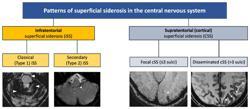
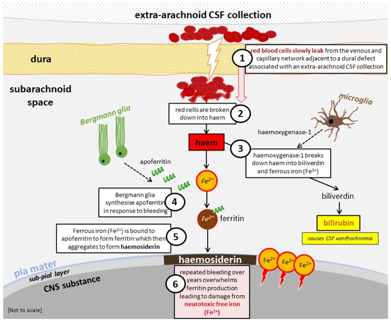
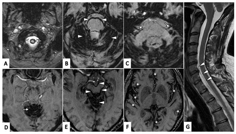
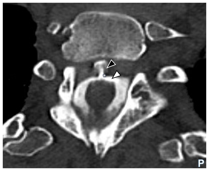
The term superficial siderosis (SS) is derived from the Greek word 'sideros', meaning iron. It includes two subtypes, distinguished by their anatomical distribution, causes and clinical features: 'classical' infratentorial SS (iSS, which sometimes also affects supratentorial regions) and cortical SS (cSS, which affects only supratentorial regions). This paper considers iSS, a potentially disabling disorder usually associated with very slow persistent or intermittent subarachnoid bleeding from a dural defect, and characterised by progressive hearing and vestibular impairment, ataxia, myelopathy and cognitive dysfunction. The causal dural defect-most often spinal but sometimes in the posterior fossa-typically follows trauma or neurosurgery occurring decades before diagnosis. Increasing recognition of iSS with paramagnetic-sensitive MRI is leading to an unmet clinical need. Given the diagnostic challenges and complex neurological impairments in iSS, we have developed a multidisciplinary approach involving key teams. We discuss pathophysiology, diagnosis and management of iSS, including a proposed clinical care pathway.

Keywords: MRI; NEUROSURGERY; SUPERFICIAL SIDEROSIS.


Conflict of interest statement

Competing interests: None declared.

Figures

Figure 1
Figure 1
Classification and types of superficial siderosis of the central nervous system (CNS). Shown in the left hand panels, type 1 (classical) infratentorial superficial siderosis (iSS) refers to a symmetrical pattern of infratentorial siderosis affecting 2/3 areas of the cerebellum, brainstem or craniocervical junction, often associated with the clinical syndrome of hearing loss, ataxia and imbalance, and myelopathy while type 2 iSS is distinguished by limited and often asymmetrical infratentorial haemosiderin (white arrow). In cortical superficial siderosis (cSS; shown in the right hand panels), haemosiderin is limited to supratentorial structures and can be focal (asterisk) or disseminated (multiple asterisks).
Figure 2
Figure 2
Diagram of pathophysiological processes implicated in infratentorial superficial siderosis (classical, Type 1); adapted, with permission from Chan et al, 2021 . Legend: CNS central nervous system; CSF cerebrospinal fluid.
Figure 3
Figure 3. Typical (classical) radiological appearances of haemosiderin deposits in infratentorial superficial siderosis (axial susceptibility weighted MR images, SW MRI, A-F)
A. Craniocervical junction (arrow); B. Haemosiderin involving cerebellar folia with a rim of haemosiderin surrounding the pons (all marked with arrow heads); C. Haemosiderin involving the vestibulo-cochlear nerves (CN VIII) bilaterally (arrows); D. Haemosiderin involving superior vermis (arrow); E. Haemosiderin involving superior vermis with a rim of haemosiderin surrounding the midbrain(all marked with arrowheads); F. Haemosiderin involving supratentorial regions (including the Sylvian fissures, temporal and frontal lobes and cingulate gyri, all marked with asterisks); G. Saggital T2-weighted MRI demonstrating ventral cerebrospinal fluid spinal collection (arrowheads).
Figure 4
Figure 4
Axial CT myelogram image showing contrast (white arrowhead) in the cerebrospinal fluid (CSF) and clearly visible extravasation of contrast (black arrowhead) at the site of dural defect (asterisk) in the thoracic region. Legend: CT computerised tomography; P posterior.
Figure 5
Figure 5
(A-E). A, B. Intra-operative myelogram showing contrast injected through the ventral dural defect (white arrow) which had shown accumulation of dye in the ventral epidural space (white arrowheads) (B) prior to dural derfet repair; C. intra-operative images showing the 2mm ventral dural defect (black arrow); the arachnoid had prolapsed into the defect thus creating a CSF fistula; in the image, spinal cord has been retracted, and dorsal dura reflected to expose the ventral dural defect; D. following defect repair with 6-0 suture (black arrowhead) and Anastoclips® and (E) reinforced with Durepair™ dural patch and Evicel® tissue glue. Legend: CSF cerebrospinal fluid.

References

    1. Wilson D, Chatterjee F, Farmer SF, et al. Infratentorial superficial siderosis: Classification, diagnostic criteria, and rational investigation pathway. Ann Neurol. 2017;81(3):333–43. doi: 10.1002/ana.24850. [published Online First: 2016/12/27] - DOI - PubMed
    1. Charidimou A, Linn J, Vernooij MW, et al. Cortical superficial siderosis: detection and clinical significance in cerebral amyloid angiopathy and related conditions. Brain. 2015;138(Pt 8):2126–39. doi: 10.1093/brain/awv162. [published Online First: 2015/06/28] - DOI - PubMed
    1. ORPHANET. Disease: superficial siderosis. 2022. [accessed 2022/05/23]. [Available from https://www.orpha.net/consor/cgi-bin/OC_Exp.php?lng=EN&Expert=247245]
    1. Fearnley JM, Stevens JM, Rudge P. Superficial siderosis of the central nervous system. Brain. 1995;118(Pt 4):1051–66. [published Online First: 1995/08/01] - PubMed
    1. Hamill RC. Report of a case of melanosis of the brain, cord, and meninges. J Nerv Ment Dis. 1908;35:594.