Review
. 2022 Nov:91:101093.
doi: 10.1016/j.preteyeres.2022.101093. Epub 2022 Jul 8.

Cell fate decisions, transcription factors and signaling during early retinal development

Affiliations
Review

Cell fate decisions, transcription factors and signaling during early retinal development

Raven Diacou et al. Prog Retin Eye Res. 2022 Nov.

Abstract

The development of the vertebrate eyes is a complex process starting from anterior-posterior and dorso-ventral patterning of the anterior neural tube, resulting in the formation of the eye field. Symmetrical separation of the eye field at the anterior neural plate is followed by two symmetrical evaginations to generate a pair of optic vesicles. Next, reciprocal invagination of the optic vesicles with surface ectoderm-derived lens placodes generates double-layered optic cups. The inner and outer layers of the optic cups develop into the neural retina and retinal pigment epithelium (RPE), respectively. In vitro produced retinal tissues, called retinal organoids, are formed from human pluripotent stem cells, mimicking major steps of retinal differentiation in vivo. This review article summarizes recent progress in our understanding of early eye development, focusing on the formation the eye field, optic vesicles, and early optic cups. Recent single-cell transcriptomic studies are integrated with classical in vivo genetic and functional studies to uncover a range of cellular mechanisms underlying early eye development. The functions of signal transduction pathways and lineage-specific DNA-binding transcription factors are dissected to explain cell-specific regulatory mechanisms underlying cell fate determination during early eye development. The functions of homeodomain (HD) transcription factors Otx2, Pax6, Lhx2, Six3 and Six6, which are required for early eye development, are discussed in detail. Comprehensive understanding of the mechanisms of early eye development provides insight into the molecular and cellular basis of developmental ocular anomalies, such as optic cup coloboma. Lastly, modeling human development and inherited retinal diseases using stem cell-derived retinal organoids generates opportunities to discover novel therapies for retinal diseases.

Keywords: Cell determination; Ciliary marginal zone; Differentiation; Ectoderm; Homeodomain; Lhx2; Neuroectoderm; Optic cup; Otx2; Pax6; Retinal pigmented epithelium; Retinal progenitor cells; Six3; Six6.


Figures

Fig. 1.
Fig. 1.
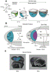
The earliest stages of neuroectoderm formation and eye field formation. A) Cell fate maps of the mouse E7.0-E8.5 gastrula (Peng et al., 2016). B) Anterior neural plate (blue) patterning in zebrafish prior and during the formation of the eye field (purple) (see Giger and Houart, 2018). Two opposite gradients of Sfrps (anterior-high/posterior-low) and Wnts (anterior-low/posterior-high) control regionalization of the anterior neural plate (see Cavodeassi et al., 2013). C) 3D-sketches of the E8.0 and E8.5 mouse embryos showing folding of the neural tube (prospective diencephalon, blue), location and bilateral split of the single eye field (purple). At E8.5, the observable optic vesicle is marked (purple). Abbreviations: Embryonic mesoderm, EMB-MES; endoderm, ENDO; extraembryonic mesoderm, EXM; prospective forebrain, FB; prospective midbrain and hindbrain, M/HB; neuroectoderm, NE; primitive streak, PS; spinal cord, SC; surface ectoderm, SE. Crossed arrows indicate three embryonic axes.
Fig. 2.
Fig. 2.
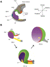
3D-formation of the optic cup (mouse E9.0-E12.5). A) Diagrammatic visualization of the mouse E9.0 embryo showing formation of the optic vesicle. B) 3D-formation of the optic cup. Note that ventral retinal blood vessels leaving the fissure are not shown for simplicity. Abbreviations: ciliary margin zone, CMZ; dorsal/ventral optic stalk, dOS/vOS; neuroretina, NE; optic fissure, OF; optic nerve, ON; optic stalk, OS; prospective lens ectoderm, PLE; prospective neuroretina, PNR; periocular mesenchyme, POM; prospective RPE, PRPE; surface ectoderm, SE.
Fig. 3.
Fig. 3.
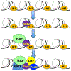
Chromatin opening by pioneering transcription factors. Pioneering transcription factors such as SOX2 move through the chromatin and scan potential target sites on nucleosomal DNA (Dodonova et al., 2020). When sites are located, chromatin opening requires recruitment of chromatin remodelers, eviction and/or remodeling of one or a few nucleosomes and may include displacement of linker histone H1. The initial opening allows other DNA-binding transcription factors such as Pax6 to join the pioneering transcription factors and recruit additional transcription factors, such as transcription factors such as signal regulated transcription factors (SRTFs) and other enzymes and complexes to facilitate transcription.
Fig. 4.
Fig. 4.
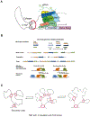
Summary models of tissue-specific transcriptional control. A) Regulation of transcription by promoter-enhancer looping mediated by CTCF and cohesin. The physical proximity of the distal enhancer is brought together by protein-protein interactions involving enhancer-bound transcriptional factors (TFs), their associated chromatin remodelers (BAF) and the Mediator (Med) complex with the promoter-bound TFs. Transcriptional activity is illustrated by RNA polymerase II at the core promoter (see Carter and Zhao, 2021). Nucleosomes (not shown for simplicity) are decorated by specific combinations of histone PTMs, such as H3K4me1 and H3K27ac (enhancer) and H3K4me3 and H3K27ac (promoter) (Ernst et al., 2011; Rada-Iglesias et al., 2011). Nascent bidirectional formation of two eRNAs is also shown. B) Cis-regulatory grammar (positioning) of model enhancers. Three different motifs (arrows) are shown with variable affinity (color gradients) to their cognate transcription factors (TFs). Motif number, orientation and order are shown by eight representative examples. Spacing between individual sites is based on 10.5 bp per DNA turn. If two centers of adjacent 6–10 bp long binding sites are separated by 10–11 bp, both TF1 and TF2 are on the same side of DNA and probability of their cooperative action due to permissive steric requirements is high (cooperative binding). In contrast, other alignments are functionally suboptimal (non-cooperative binding) (see Long et al., 2016 for additional details). C) 3D-organization of a representative topologically associated domains (TADs) showing formation and dynamics of multiple sub-TAD loops. During cellular differentiation, different enhancers and employed and topologically associated domains organization changes (see Atlasi and Stunnenberg, 2017; Hansen et al., 2018).
Fig. 5.
Fig. 5.
Molecular structures and DNA-binding of Otx2, Lhx2, Six3 and Six6. A) Otx2 (mouse: 289 amino acids, 31.6 kDa), phosphorylation sites: Y18, S28, and Y31; ubiquitylation K59; N-terminal HD (amino acids: 38–98). DNA-binding logo from ChIP-seq data (Samuel et al., 2014). B) Lhx2 (mouse: 406 amino acids; 44.4 kDa), internal HD (amino acids: 266–325), N-terminal LIM1 domain 1 (LD1, amino acids: 53–105) and LIM2 domain 2 (LD2, amino acids 115–168). C) Six3 (mouse 333 amino acids, 35.6 kDa), HD (amino acids: 207–266), Six domain (SD), mutation F88E (Q domain) disrupts interaction with Tle4 and Tle5 (Zhu et al., 2002). N-terminal portion of the HD (Hu et al., 2008) does not interact with DNA as it lacks basic Arg residues. The DNA-binding logo (weblogo.berkeley.edu/logo.cgi) was generated using known sites (Liu et al., 2006, 2010). D) Six6 (mouse 246 amino acids, 27.7 kDa). Phosphorylation sites: T212, S221, S225, S227 and S228. DNA-binding logo was obtained from ISMARA (https://www.ismara.unibas.ch/ISMARA/scratch/NHBE_SC2/ismara_report/pages/SIX6.html). Structural modeling: We first obtained crystal structure of OTX2 from PDB id 2dms and structurally aligned it with Aristaless HD which is in complex with DNA (PDB id:3lnq). The final model obtained shows OTX2 bound to linear DNA with important residues K74, R90, and Q97 highlighted. We next generated a homology model for human SIX3 sequence, using the M4T modeling server (Fernandez-Fuentes et al., 2007). We then performed structural alignment of human SIX3 homology model and the crystal structure of HoxA9 and Pbx1 HDs bound to DNA (PDB id: 1PUF). The HD domain of SIX3 is shown in yellow and residues R282, S283, and L284 are highlighted. Finally, we generated structural alignment of the homology model of SIX6 and the protein component of 1B72 structure using the same strategy as described for SIX3. The resulting model shows SIX6 bound to linear DNA with N-terminal residues R140, H141, and L142 highlighted. The HD domain of SIX6 is shown in yellow.
Fig. 6.
Fig. 6.
Molecular structure and interactions of Pax6 with DNA and chromatin. A) Mouse/human Pax6, 422 amino acids, 46 kDa; Pax6 (5a), 436 amino acids, 48 kDa. Phosphorylation: S413 by ERK1/2 and p38 (Mikkola et al., 1999); Y281, Y304 and Y373 by HIPK2 (Kim et al., 2006); sumoylation: various lysine residues such as K53 and K89 of Pax6 (5a) (Yu et al., 2020; Yan et al., 2010). Our studies show functional interactions between Pax6 and Pax6 (5a) regulation of crystallin promoters (Chauhan et al., 2004). B) Model of Pax6 interaction with the nucleosome. PAX6 PAI domain (blue), RED domain (red), and HD domain (yellow) bound to the DNA that is wrapped around the nucleosome. Using the crystal structure of PAX6 (6PAX) (Xu et al., 1999a), we first identified nucleotide binding motifs along the DNA. We next performed nucleotide motif alignment of 6PAX (PAI-linker region) and the corresponding motif of 6S01. The PAX6 HD domain with its binding motif was placed along the nucleotide motif of 6S01. The final model obtained shows PAX6 bound to DNA that is wrapped around the nucleosome with important residues G18, R26, N50, T63 (PAI domain), R128 (RED domain), and R242 (HD domain) highlighted. The individual histone subunits are shown as H3 (light blue), H4 (dark green), H2A (olive), and H2B (light red). Our data show that there are sequence similarities between Pax6 consensus sequence and other transcription factors raising the possibility that some Pax6 sites evolved from pre-existing cis-sites recognized by stress regulatory transcription factors (Cvekl et al., 2017). C) In vivo DNA-binding motifs of Pax6 proteins based on our ChIP-seq studies (Sun et al., 2015). D) Analysis of Pax6 intrinsically disordered regions (IDRs) (iupred2a.elte.hu) (Mészáros et al., 2018) showing highly disordered C-terminal transcriptional activation domain, S/T/P-rich intrinsically disordered domain, IDD (Epstein et al., 1994a).
Fig. 7.
Fig. 7.
Schematic representations of representative model interactions between signaling molecules, DNA-binding transcription factors and patterning processes at the eye field and optic vesicle stages. A) Wnt signaling (canonical and non-canonical) at the anterior neural plate (anterior-posterior, A/P) including the zebrafish eye field (see Esteve and Bovolenta, 2006; Giger and Houart, 2018). B) Complex cross-talks between BMP and Shh signaling in the mouse eye field and its dorso-ventral (D/V) patterning (Zhao et al., 2010). C) and D) Polarization of the distal (proximal) chick optic vesicle by Pax6/Fst/Tgfb2 Turing network (Grocott et al., 2020), respectively.
Fig. 8.
Fig. 8.
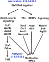
Model of Six3 and Six6 joint functions in the optic cup formation. Pathways downstream of Six3 and Six6 involved in the mouse optic cup patterning (E14.5-E15.5). Six3 and Six6 are jointly required for maintenance of multipotent RPCs through suppressing Wnt/β-catenin signaling and activating retinogenic factors (Diacou et al., 2018). Transcription factors, TFs; signal regulated transcription factors, SRTFs.

References

    1. Abouzeid H, Boisset G, Favez T, Youssef M, Marzouk I, Shakankiry N, Bayoumi N, Descombes P, Agosti C, Munier FL, Schorderet DF, 2011. Mutations in the SPARC-related modular calcium-binding protein 1 gene, SMOC1, cause waardenburg anophthalmia syndrome. Am. J. Hum. Genet 88, 92–98. - PMC - PubMed
    1. Acampora D, Mazan S, Lallemand Y, Avantaggiato V, Maury M, Simeone A, Brûlet P, 1995. Forebrain and midbrain regions are deleted in Otx2−/− mutants due to a defective anterior neuroectoderm specification during gastrulation. Development 121, 3279–3290. - PubMed
    1. Adler R, Belecky-Adams TL, 2002. The role of bone morphogenetic proteins in the differentiation of the ventral optic cup. Development 129, 3161–3171. - PubMed
    1. Adler R, Canto-Soler MV, 2007. Molecular mechanisms of optic vesicle development: complexities, ambiguities and controversies. Dev. Biol 305, 1–13. - PMC - PubMed
    1. Agoston Z, Schulte D, 2009. Meis2 competes with the Groucho co-repressor Tle4 for binding to Otx2 and specifies tectal fate without induction of a secondary midbrainhindbrain boundary organizer. Development 136, 3311–3322. - PubMed

Publication types

Substances