Review
. 2023 Jan;44(1):8-18.
doi: 10.1038/s41401-022-00934-2. Epub 2022 Jul 11.

Protein O-GlcNAcylation in cardiovascular diseases

Affiliations
Review

Protein O-GlcNAcylation in cardiovascular diseases

Hui-Fang Wang et al. Acta Pharmacol Sin. 2023 Jan.

Abstract

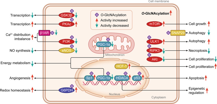
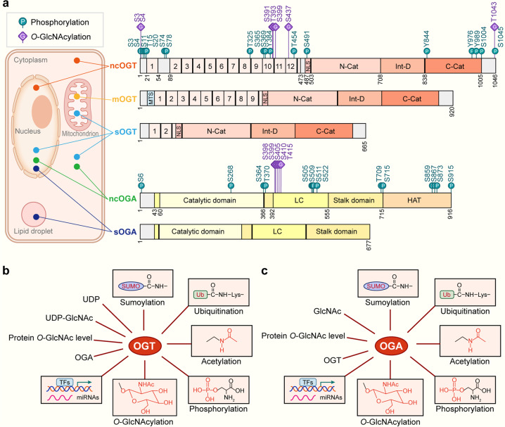
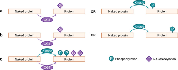
O-GlcNAcylation is a post-translational modification of protein in response to genetic variations or environmental factors, which is controlled by two highly conserved enzymes, i.e. O-GlcNAc transferase (OGT) and protein O-GlcNAcase (OGA). Protein O-GlcNAcylation mainly occurs in the cytoplasm, nucleus, and mitochondrion, and it is ubiquitously implicated in the development of cardiovascular disease (CVD). Alterations of O-GlcNAcylation could cause massive metabolic imbalance and affect cardiovascular function, but the role of O-GlcNAcylation in CVD remains controversial. That is, acutely increased O-GlcNAcylation is an adaptive heart response, which temporarily protects cardiac function. While it is harmful to cardiomyocytes if O-GlcNAcylation levels remain high in chronic conditions or in the long run. The underlying mechanisms include regulation of transcription, energy metabolism, and other signal transduction reactions induced by O-GlcNAcylation. In this review, we will focus on the interactions between protein O-GlcNAcylation and CVD, and discuss the potential molecular mechanisms that may be able to pave a new avenue for the treatment of cardiovascular events.

Keywords: O-GlcNAcylation; cardiovascular disease; glycomics; glycosylation.


Conflict of interest statement

The authors declare no competing interests.

Figures

Fig. 1
Fig. 1. The types, cellular distribution, and functions of proteins that could be O-GlcNAcylated in cells.
O-GlcNAcylated proteins, mainly located in the cytoplasm, nucleus, mitochondrion, and cell membrane, include the protein kinases (in red color), transcription factors and coactivators (in green color), metabolic proteins (in blue color), membrane receptors (in pink color), and other signaling molecules (in yellow color). They are responsible for essential activities with regard to epigenetic regulation, gene transcription, energy metabolism, cell cycle behavior, vascular function, Ca2+ distribution, NO production and redox homeostasis. Most of these proteins can be O-GlcNAcylated (in purple squares with the letter ‘G’) and their activities and functions can be changed directly. Differently, mTOR and SNAP29 are directly triggered by the higher intracellular O-GlcNAcylation levels. Proteins with enhanced activity after O-GlcNAcylation modification are indicated by red arrows, and proteins with decreased activity after O-GlcNAcylation modification are shown with green arrows. GSK-3β Glycogen synthase kinase-3β, PKAc PKA catalytic subunit, β1AR β1-adrenoceptor, PI3K Phosphoinositide 3-kinase, eNOS Endothelial nitric oxide synthase, G6PDH Glucose 6-phosphatedehydrogenase, PGC-1α Peroxisome proliferator-activated receptor-γ coactivator-1α, Sp1 Specificity protein 1, HDAC4 histone deacetylase 4, mTOR Mechanistic/mammalian target of rapamycin, SNAP29 Synaptosomal-associated protein 29, ULK1 Unc-51-like autophagy activating kinase 1, RIPK3 Receptor-interacting protein kinase 3, HCF-1 Host cell factor-1.
Fig. 2
Fig. 2. The molecular structure, localization and regulation of OGT and OGA.
a Structure and localization of OGT and OGA isoforms. All the identified phosphorylation sites are shown as green circles with the letter ‘P’, and O-GlcNAcylation sites are shown as purple squares with the letter ‘G’. The N-terminal region of three OGT isoforms is consisted of TPRs and an NLS. The catalytic region is composed of an N-cat domain, an Int-D, and a C-cat domain. Additionally, mOGT has a typical MTS. These two OGA splicing variants are constituted by a catalytic domain in the N-terminal region and two stalk domains (including N-terminal and C-terminal) and two LC regions, but they differ in a pseudo-HAT domain. The localization of ncOGT and ncOGA mainly concentrates in the cytoplasm and nucleus. mOGT gathers in the mitochondrion, and sOGT is present in the cytoplasm, nucleus, and mitochondrion. sOGA is centered in the nucleus and lipid droplet. b Regulation of OGT. OGT is modulated at several aspects containing HBP substrate availability such as UDP and UDP-GlcNAc. The intracellular protein O-GlcNAc level and the counterpart magnitude of OGA can regulate OGT in a feedback style. OGT can be regulated through TFs and miRNAs at the transcriptional level. Post-translational modifications e.g., O-GlcNAcylation, phosphorylation, acetylation, ubiquitination, and sumoylation can also regulate OGT. c Regulation of OGA. OGA is regulated by various factors containing feedback inhibition by intracellular GlcNAc level. The intracellular protein O-GlcNAc level and the counterpart magnitude of OGT can regulate OGA. OGA can be regulated through TFs and miRNAs at the transcriptional level. Post-translational modifications by O-GlcNAcylation, phosphorylation, acetylation, ubiquitination, and sumoylation can also regulate OGA. OGT O-GlcNAc transferase, OGA Protein O-GlcNAcase, UDP-GlcNAc Uridine diphosphate N-acetylglucosamine, TPRs Tetratricopeptide repeats, NLS Nuclear localization signal, N-cat N-terminal catalytic domain, Int-D Intervening sequence, C-cat C-terminal catalytic domain, MTS Mitochondrial targeting sequence, LC Low complexity, pseudo-HAT Pseudohistone acetyl transferase, TFs Transcription factors, miRNAs MicroRNAs, ncOGT Nucleocytoplasmic OGT, mOGT Mitochondrial OGT, sOGT Short OGT, ncOGA Nucleocytoplasmic OGA, sOGA Short isoform OGA. The databases https://glygen.org/, https://www.uniprot.org/, and https://research.bioinformatics.udel.edu/iptmnet/ are used to search for known modification sites.
Fig. 3
Fig. 3. The crosstalk between protein phosphorylation and O-GlcNAcylation.
a Protein substrate can be either O-GlcNAcylated or phosphorylated at the same amino acid residue in a substitutable and competitive way. b Protein substrate is either phosphorylated at given sites or O-GlcNAcylated at other different sites, which is both reciprocal and alternative. c Protein substrate is simultaneously under O-GlcNAcylation and phosphorylation at different locations.

Similar articles

Cited by

References

    1. Roth GA, Mensah GA, Johnson CO, Addolorato G, Ammirati E, Baddour LM, et al. Global Burden of Cardiovascular Diseases and Risk Factors, 1990–2019: Update From the GBD 2019 Study. J Am Coll Cardiol. 2020;76:2982–3021. doi: 10.1016/j.jacc.2020.11.010. - DOI - PMC - PubMed
    1. Torres CR, Hart GW. Topography and polypeptide distribution of terminal N-acetylglucosamine residues on the surfaces of intact lymphocytes. Evidence for O-linked GlcNAc. J Biol Chem. 1984;259:3308–17. doi: 10.1016/S0021-9258(17)43295-9. - DOI - PubMed
    1. Butkinaree C, Park K, Hart GW. O-linked β-N-acetylglucosamine (O-GlcNAc): Extensive crosstalk with phosphorylation to regulate signaling and transcription in response to nutrients and stress. Biochim Biophys Acta. 2010;1800:96–106. doi: 10.1016/j.bbagen.2009.07.018. - DOI - PMC - PubMed
    1. Hanover JA, Krause MW, Love DC. The hexosamine signaling pathway: O-GlcNAc cycling in feast or famine. Biochim Biophys Acta. 2010;1800:80–95. doi: 10.1016/j.bbagen.2009.07.017. - DOI - PMC - PubMed
    1. Tran DH, Wang ZV. Glucose metabolism in cardiac hypertrophy and heart fail- ure. J Am Heart Assoc. 2019;8:e012673. doi: 10.1161/JAHA.119.012673. - DOI - PMC - PubMed

MeSH terms

Substances