. 2022 Jul 10;22(1):228.
doi: 10.1186/s12935-022-02637-4.

LINC00893 inhibits the progression of prostate cancer through miR-3173-5p/SOCS3/JAK2/STAT3 pathway

Affiliations

LINC00893 inhibits the progression of prostate cancer through miR-3173-5p/SOCS3/JAK2/STAT3 pathway

Chuigong Yu et al. Cancer Cell Int. .

Abstract

Background: Prostate cancer (PCa) is one of the most common malignant tumors in the male urinary system. In recent years, the morbidity and mortality of PCa have been increasing due to the limited effects of existing treatment strategies. Long non-coding RNA (lncRNA) LINC00893 was reported to inhibit the proliferation and metastasis of papillary thyroid cancer cells, but its role in PCa has not been reported. This study aims to investigate the role and underlying mechanism of LINC00893 in regulating the progression of PCa cells.

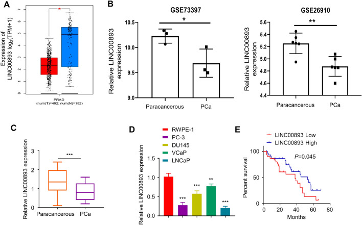
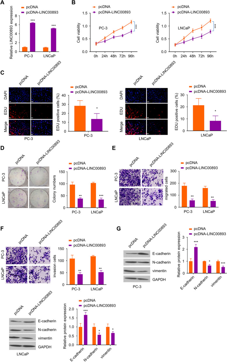
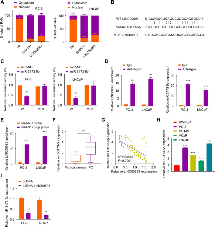
Methods: We first compared LINC00893 expression levels between PCa tissues and normal prostate tissues through TCGA database. The relative LINC00893 expression levels were further validated in 66 pairs of PCa tissues and para-cancerous normal tissues, as well as in PCa cell lines. Gain-of-function experiment was performed by transfecting PCa cell with LINC00893 expression vector, and CCK (Cell count kit)-8, 5-Ethynyl-2'-deoxyuridine (EdU) incorporation, colony information and transwell assays were conducted to assess the functional phenotypes. Dual-luciferase reporter, RNA-binding protein immunoprecipitation (RIP) and RNA pull-down assays were performed to evaluate the molecular interactions.

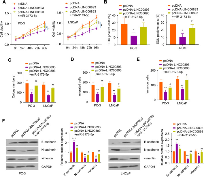
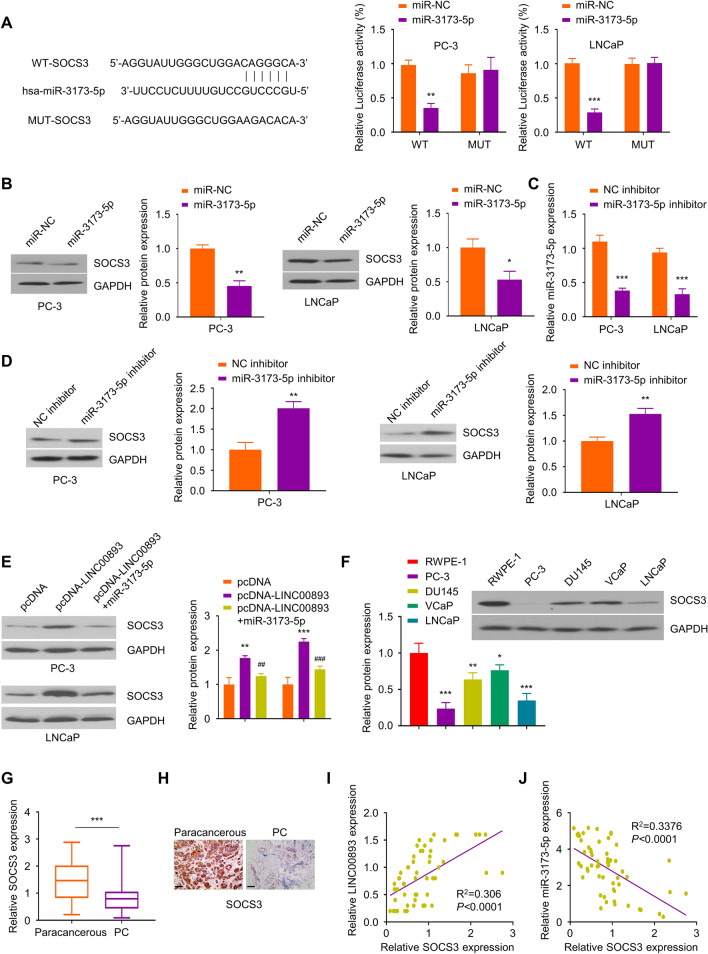
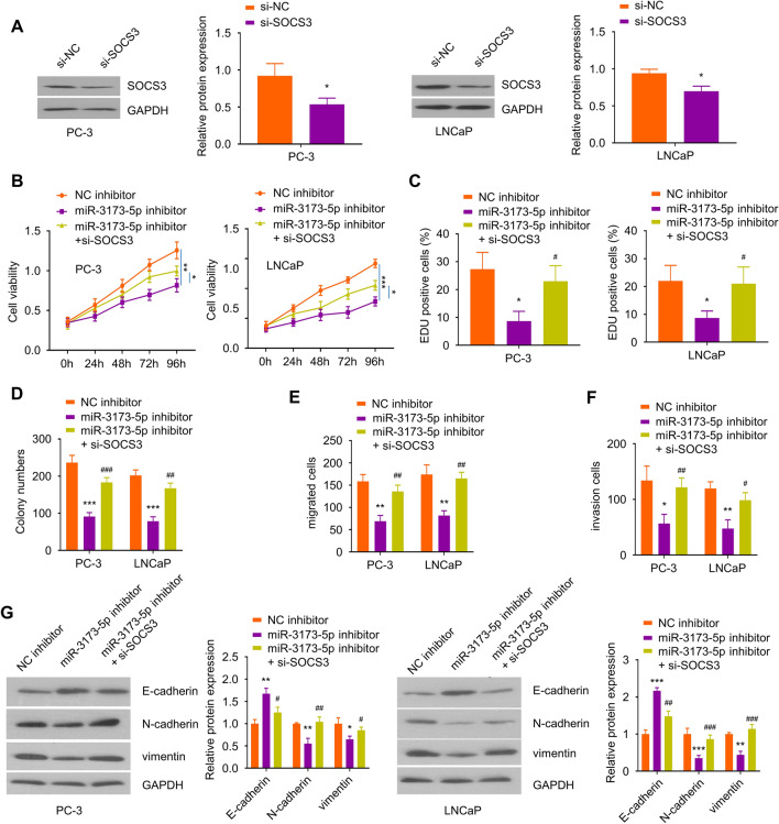
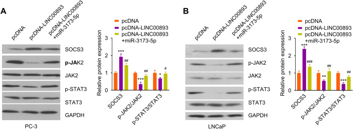
Results: LINC00893 was downregulated in PCa tissues and cell lines, and patients with low expression of LINC00893 were associated with a poorer overall survival rate. LINC00893 overexpression hindered the proliferation, epithelial-mesenchymal transition (EMT) as well as the migratory ability of PCa cells, and suppressed the tumorigenesis of PCa cells in nude mice. We further demonstrated that LINC00893 acted as a sponge for miR-3173-5p and inhibited its activity, which in turn regulated the suppressor of cytokine signaling 3 (SOCS3)/Janus Kinase 2 (JAK2)/signal transducer and activator of transcription 3 (STAT3) signaling axis.

Conclusions: Our study demonstrated that LINC00893 suppresses the progression of PCa cells through targeting miR-3173-5p/SOCS3/JAK2/STAT3 axis. Our data uncovers a novel tumor-suppressor role of LINC00893 in PCa, which may serve as a potential strategy for targeted therapy in PCa.

Keywords: JAK2/STAT3 signaling pathway; LINC00893; Prostate cancer; SOCS3; miR-3173-5p.


Conflict of interest statement

The authors declare no conflict of interests.

Figures

Fig. 1
Fig. 1
LINC00893 is downregulated in PCa tissues and cell lines. A, B LINC00893 expression levels in PCa tissues and paired para-cancerous tissues were compared in TCGA database (A), and in GSE73397 & GSE26910 datasets from GEO database (B). C The relative LINC00893 expression level was examined in 66 pairs of PCa tissues and para-cancerous tissues by RT-qPCR. D LINC00893 expression levels were examined in PCa cell lines (PC-3, DU145, VCaP and LNCaP) and human normal prostate epithelial cell line (RWPE-1) by RT-qPCR. E 66 PCa patients were divided into LINC00893low (n = 33) and LINC00893high (n = 33) group according to the median value of LINC00893 expression. Kaplan–Meier survival analysis was used to evaluate the overall survival rate of patients in the two groups. Three independent assays were performed with three technical replicates in C and D. The error bars are defined as s.d. *, P < 0.05, **, P < 0.01, and ***, P < 0.001
Fig. 2
Fig. 2
LINC00893 functions as a tumor suppressor factor to inhibit the proliferation, migration, invasion and EMT of PCa cells. PC-3 and LNCaP cells were transfected with pcDNA3.1-LINC00893 plasmids or pcDNA3.1-vector for 48 h. A LINC00893 expression level was examined by RT- qPCR in cells transfected with pcDNA3.1-LINC00893 or vector. B CCK-8 proliferation assay in cells transfected with pcDNA3.1-LINC00893 or vector was perfromed at indicated time points. C EdU incorporation assay in cells transfected with pcDNA3.1-LINC00893 or vector. Scale bar: 50 μM. D Colony formation assay in cells transfected with pcDNA3.1-LINC00893 or vector. E, F Transwell migration assay (E) and invasion assay (F) in cells transfected with pcDNA3.1-LINC00893 or vector. G The protein levels of E-cadherin and mesenchymal markers N-cadherin and vimentin were assessed by Western blot in cells transfected with pcDNA3.1-LINC00893 or vector. Three independent assays were performed with three technical replicates. The error bars are defined as s.d. *, P < 0.05, **, P < 0.01, and ***, P < 0.001
Fig. 3
Fig. 3
LINC00893 binds to miR-3173-5p and inhibits its expression. A Relative LINC00893 levels were quantified by RT-qPCR in the cytoplasmic and nuclear fractions of PC-3 and LNCaP cells. U6 and GAPDH were used as marker for the nuclear and cytoplasmic fraction respectively. B The binding sites between LINC00893 and miR-3173-5p were predicted from Starbase database. C Dual luciferase reporter assay was performed using pmirGLO-WT-LINC00893 and pmirGLO-MUT-LINC00893 reporter in the presence of miR-3173-5p mimic or miR-NC. Renilla luciferase control plasmids were co-transfected into cells, and the relative firefly luciferase activity in the reporter was normalized to renilla luciferase activity in the control plasmid. D RIP assay using non-specific IgG or anti-Ago2 antibody. The relative enrichment of LINC00893 and miR-3173-5p was normalized to the input samples. E RNA pull-down assay using biotin-labeled miR-3173-5p probe or miR-NC control probe. The relative enrichment of LINC00893 was normalized to the input samples. F miR-3173-5p expression levels in 66 pairs of PCa tissues and para-cancerous tissues were assessed by RT-qPCR. G The correlation between LINC00893 and miR-3173-5p expression level was evaluated by Spearman correlation coefficient analysis. H The relative miR-3173-5p expression levels in PCa cell lines (PC-3, DU145, VCaP and LNCaP) and human normal prostate epithelial cell line RWPE-1 were detected by RT-qPCR. I miR-3173-5p expression levels were examined by RT-qPCR in cells transfected with pcDNA3.1-LINC00893 or vector. Three independent assays were performed with three technical replicates. The error bars are defined as s.d. *, P < 0.05, **, P < 0.01, and ***, P < 0.001
Fig. 4
Fig. 4
miR-3173-5p mediates the effects of LINC00893 on PCa cell phenotype. PC-3 and LNCaP cells were transfected with pcDNA vector, pcDNA-LINC00893 or pcDNA-LINC00893 + miR-3173-5p mimic for 48 h. A, B The proliferation ability of PCa cells under different conditions was detected by CCK-8 (A) and EdU incorporation assay (B). C Colony formation assay in PC-3 and LNCaP cells under the above experimental conditions. Transwell migration assay (D) and invasion assay (E) in PC-3 and LNCaP cells under the above experimental conditions. F EMT related proteins E-cadherin, N-cadherin and vimentin were evaluated by Western blot in PC-3 and LNCaP cells under the above experimental conditions. Three independent assays were performed with three technical replicates. The error bars are defined as s.d. *, P < 0.05, **, P < 0.01, and ***, P < 0.001
Fig. 5
Fig. 5
MiR-3173-5p binds to the 3'UTR of SOCS3 mRNA and inhibits its expression. A The binding sites of miR-3173-5p on SOCS3 mRNA 3'UTR region were predicted through Starbase database. Dual luciferase reporter assay was performed using pmirGLO-WT-SOCS3 and pmirGLO-MUT-SOCS3 reporter in the presence of miR-3173-5p mimic or miR-NC. Renilla luciferase control plasmids were co-transfected into cells, and the relative firefly luciferase activity in the reporter was normalized to renilla luciferase activity in the control plasmid. B SOCS3 protein levels were examined by Western blot after the transfection of miR-3173-5p mimic in PC-3 and LNCaP cells. C miR-3173-5p expression level was examined by RT-qPCR in PC-3 and LNCaP cells after the transfection with miR-3173-5p inhibitor or NC inhibitor. D SOCS3 protein levels were assessed by Western blot after the transfection with miR-3173-5p inhibitor or NC inhibitor. E Cells were transfected with pcDNA vector, pcDNA-LINC00893 plasmid or pcDNA-LINC00893 + miR-3173-5p mimic for 48 h, and SOCS3 protein levels were examined by Western blot. F SOCS3 protein levels in PCa cell lines and human normal prostate epithelial cell line RWPE-1 were evaluated by Western blot. G Relative SOCS3 mRNA levels in 66 pairs of PCa tissues and para-cancerous tissues were analyzed by RT-qPCR. H. SOCS3 protein levels in PCa tissues and matched para-cancerous tissues were evaluated by IHC. Scale bar: 100 μM. I, J The correlations between LINC00893 and SOCS3 mRNA, and between MiR-3173-5p and SOCS3 mRNA were analyzed by Spearman correlation coefficient analysis. Three independent assays were performed with three technical replicates. The error bars are defined as s.d. *, P < 0.05, **, P < 0.01, and ***, P < 0.001
Fig. 6
Fig. 6
SOCS3 silencing attenuated the effects of miR-3173-5p inhibitor in PCa cells. A The knockdown efficiency of SOCS3 siRNA in PC-3 and LNCaP cells was examined by Western blot. For B-G, PC-3 and LNCaP cells were transfected with NC inhibitor, miR-3173-5p inhibitor or miR-3173-5p inhibtor + si-SOCS3. B, C The proliferation ability of cells in the above experimental groups was evaluated by CCK-8 (B) and EdU incorporation assay (C). D Colony formation assay in PC-3 and LNCaP cells of the above experimental groups. Transwell migration assay (E) and invasion assay (F) in PC-3 and LNCaP cells of the above experimental groups. G The protein levels of EMT markers E-cadherin, N-cadherin and vimentin in different treatment groups were detected by Western blot. Three independent assays were performed with three technical replicates. The error bars are defined as s.d. *, P < 0.05, **, P < 0.01, and ***, P < 0.001
Fig. 7
Fig. 7
LINC00893/miR-3173-5p axis regulates SOCS3/JAK2/STAT3 signaling pathway. PC-3 and LNCaP cells were transfected with pcDNA, pcDNA-LINC00893 or pcDNA-LINC00893 + miR-3173-5p mimic. The protein levels of p-JAK2, p-STAT3, JAK2, STAT3 and SOCS3 in PC-3 (A) and LNCaP (B) cells were assessed by Western blot. Three independent assays were performed with three technical replicates. The error bars are defined as s.d. *, P < 0.05, **, P < 0.01, and ***, P < 0.001
Fig. 8
Fig. 8
Overexpression of LINC00893 suppresses the tumorigenesis of PCa cells in nude mice. PC-3 cells stably expressing LINC00893 (PC-3-LINC00893) or the control cells (PC-3-vector) were inoculated subcutaneously into NOD/SCID mice (n = 6 in each group). A The volume of the xenograft tumor was measured every 5 days. B The weight of the subcutaneous xenograft tumor was measured on day 30. C LINC00893 and miR-3173-5p expression levels in xenograft tumor tissues were assessed by RT-qPCR. D The protein levels of SOCS3, E-cadherin, vimentin and N-cadherin in xenograft tumor tissues was detected by Western blot. E Ki-67 and SOCS3 protein expression levels in xenograft tumor sections were detected by IHC staining: Scale bar: 100 μM. Three independent assays were performed with three technical replicates. The error bars are defined as s.d. *, P < 0.05, **, P < 0.01, and ***, P < 0.001

Similar articles

Cited by

References

    1. Schatten H. Brief overview of prostate cancer statistics, grading, diagnosis and treatment strategies. Adv Exp Med Biol. 2018;1095:1–14. doi: 10.1007/978-3-319-95693-0_1. - DOI - PubMed
    1. Gomella LG. Prostate Cancer Statistics: Anything You Want Them To Be. Can J Urol. 2017;24(1):8603–8604. - PubMed
    1. Bostwick DG, Burke HB, Djakiew D, Euling S, Ho SM, Landolph J, Morrison H, Sonawane B, Shifflett T, Waters DJ, et al. Human prostate cancer risk factors. Cancer. 2004;101(10 Suppl):2371–2490. doi: 10.1002/cncr.20408. - DOI - PubMed
    1. Pienta KJ, Esper PS. Risk factors for prostate cancer. Ann Intern Med. 1993;118(10):793–803. doi: 10.7326/0003-4819-118-10-199305150-00007. - DOI - PubMed
    1. Kolonel LN, Altshuler D, Henderson BE. The multiethnic cohort study: exploring genes, lifestyle and cancer risk. Nat Rev Cancer. 2004;4(7):519–527. doi: 10.1038/nrc1389. - DOI - PubMed

LinkOut - more resources