

Biomechanical Evaluation of Spinal Column after Percutaneous Cement Discoplasty: A Finite Element Analysis
- PMID: 35818350
- PMCID: PMC9363717
- DOI: 10.1111/os.13314
Biomechanical Evaluation of Spinal Column after Percutaneous Cement Discoplasty: A Finite Element Analysis
Abstract
Objective: To compare the biomechanical properties of percutaneous cement discoplasty (PCD) in the spinal column between different implant-endplate friction.
Methods: A validated L3-Scarumfinite element (FE) model was modified for simulation. In the PCD model, the L4/5 level was modified based on model 1 (M1) and model 2 (M2). In M1, the interaction between bone cement and endplate was defined as face-to-face contact with a friction coefficient of 0.3; in M2, the contact was defined as a Tie constraint. 7.5 N m moments of four physiological motions and axial load of 15, 100 and 400 N preload were imposed at the top of L3. The range of motion (ROM) and interface stress analysis of endplates, annulus fibrosus and bone cement of the operated level were calculated for comparisons among the three models.
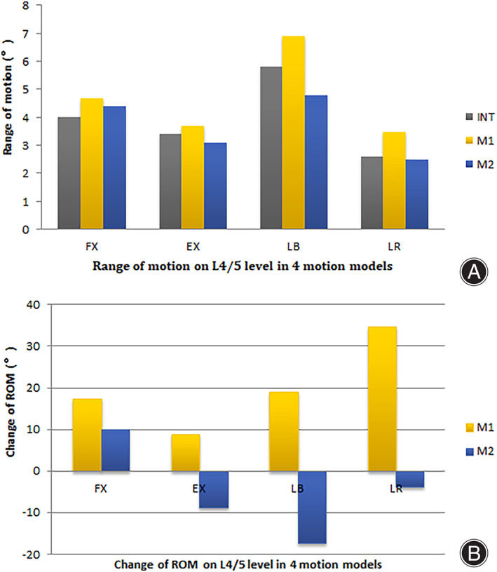
Results: The ROM of M1 and M2 increased when compared with the intact model during flexion (FL) (17.5% vs 10.0%), extension (EX) (8.8% vs -8.8%), left bending (LB) (19.0% vs -17.2%) and left axial rotation (LR) (34.6% vs -3.8%). The stress of annulus fibrosus in M1 and M2 decreased in FL (-48.4% vs -57.5%), EX (-25.7% vs -14.7%), LB (-47.5% vs -52.4%), LR (-61.4% vs -68.7%) and axis loading of 100 N (-41.5% vs -15.3%), and 400 N (-27.9% vs -27.3%). The stress of upper endplate of M1 and M2 increased in FL (24.6% vs 24.7%), LB (82.2% vs 89.5%), LR (119% vs 62.4%) and axis loading of 100 N (64.6% vs 45.5%), and 400 N (58.2% vs 24.3%), but was similar in EX (2.9% vs 0.3%). The stress of lower endplate of M1 and M2 increased in FL (170.9% vs 175.0%), EX (180.8% vs 207.7%), LB (302.6% vs 274.7%), LR (332.4% vs 132.8%) and axis loading of 100 N (350.7% vs 168.6%), and 400 N (165.2% vs 106.7%).
Conclusion: Percutaneous cement discoplasty procedure could make effect on the mobility or stiffness. The fusion of bone cement and endplate might have more biomechanical advantages, including of the decreasing rate of implant subsidence and dislocation, and the increase spine stability.
Keywords: Biomechanical; Bone cement; Finite element analysis; Lumbar spinal stenosis; PMMA; Percutaneous cement discoplasty.
© 2022 The Authors. Orthopaedic Surgery published by Tianjin Hospital and John Wiley & Sons Australia, Ltd.
Conflict of interest statement
The authors declare that they have no conflict of interest.
Figures


References
-
- Varga PP, Jakab G, Bors IB, et al. Experiences with PMMA cement as a stand‐alone intervertebral spacer: percutaneous cement discoplasty in the case of vacuum phenomenon within lumbar intervertebral discs. Orthopade. 2015;44Suppl 1:S1–7. - PubMed
MeSH terms
Substances
Grants and funding
LinkOut - more resources
Full Text Sources
Miscellaneous
