. 2022 Jul 8;11(7):9.
doi: 10.1167/tvst.11.7.9.

Topical Losartan and Corticosteroid Additively Inhibit Corneal Stromal Myofibroblast Generation and Scarring Fibrosis After Alkali Burn Injury

Affiliations

Topical Losartan and Corticosteroid Additively Inhibit Corneal Stromal Myofibroblast Generation and Scarring Fibrosis After Alkali Burn Injury

Lycia Pedral Sampaio et al. Transl Vis Sci Technol. .

Erratum in

Abstract

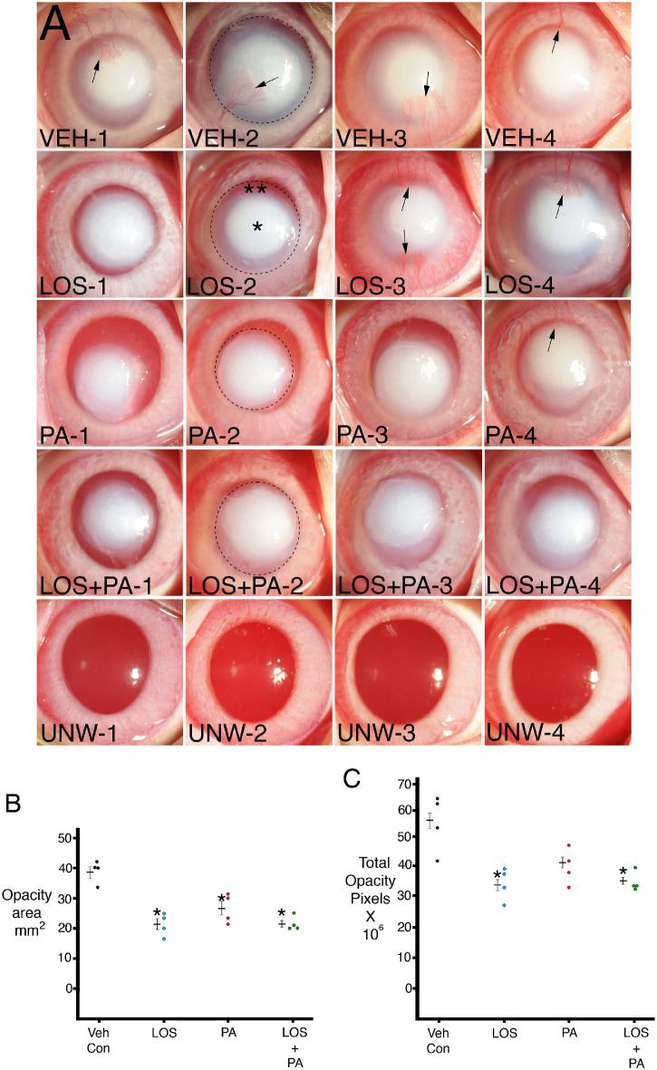
Purpose: To evaluate the efficacy of losartan and prednisolone acetate in inhibiting corneal scarring fibrosis after alkali burn injury in rabbits.

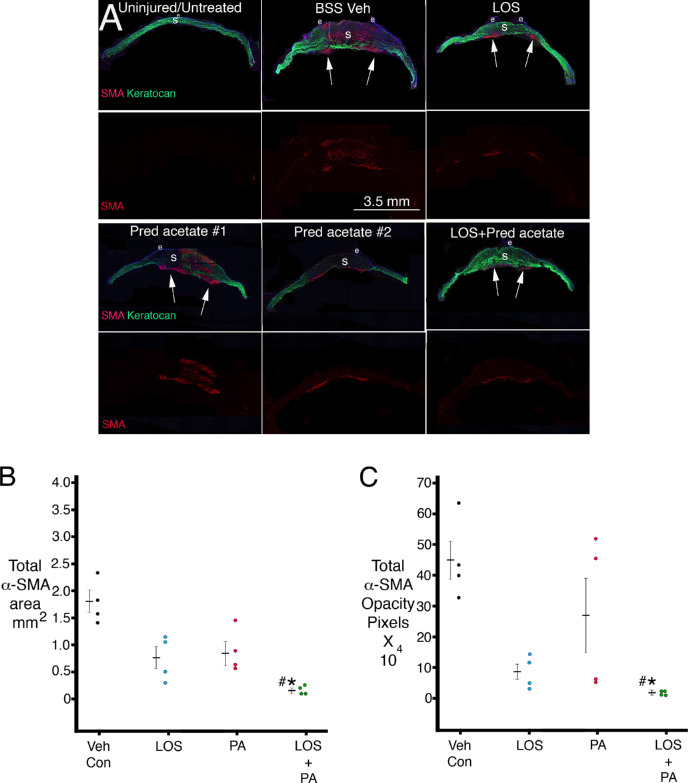
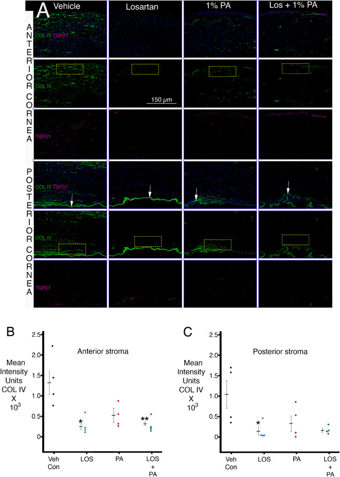
Methods: Sixteen New Zealand White rabbits were included. Alkali injuries were produced using 1N sodium hydroxide on a 5-mm diameter Whatman #1 filter paper for 1 minute. Four corneas in each group were treated six times per day for 1 month with 50 µL of (1) 0.8 mg/mL losartan in balanced salt solution (BSS), (2) 1% prednisolone acetate, (3) combined 0.8 mg/mL losartan and 1% prednisolone acetate, or (4) BSS. Area of opacity and total opacity were analyzed in standardized slit-lamp photos with ImageJ. Corneas in both groups were cryofixed in Optimal cutting temperature (OCT) compound at 1 month after surgery, and immunohistochemistry was performed for alpha-smooth muscle actin (α-SMA) and keratocan or transforming growth factor β1 and collagen type IV with ImageJ quantitation.

Results: Combined topical losartan and prednisolone acetate significantly decreased slit-lamp opacity area and intensity, as well as decreased stromal myofibroblast α-SMA area and intensity of staining per section and confined myofibroblasts to only the posterior stroma with repopulation of the anterior and mid-stroma with keratocan-positive keratocytes after 1 month of treatment. Corneal fibroblasts produced collagen type IV not associated with basement membranes, and this production was decreased by topical losartan.

Conclusions: Combined topical losartan and prednisolone acetate decreased myofibroblast-associated fibrosis after corneal alkali burns that produced full-thickness injury, including corneal endothelial damage. Increased dosages and duration of treatment may further decrease scarring fibrosis.

Translational relevance: Topical losartan and prednisolone acetate decrease myofibroblast-mediated scarring fibrosis after corneal injury.


Conflict of interest statement

Disclosure: L.P. Sampaio, None; G.S.L. Hilgert, None; T.M. Shiju, None; M.R. Santhiago, None; S.E. Wilson, submitted a provisional patent to the US Patent Office regarding topical use of losartan and other angiotensin converting enzyme II receptor inhibitors to decrease corneal scarring fibrosis after injury (P)

Figures

Figure 1.
Figure 1.
(A) Standardized slit-lamp photos of rabbit corneas at 1 month after a 1-minute exposure to 1N NaOH on a 5-mm diameter filter paper and continuous treatment for 1 month with topical vehicle (VEH), 0.2 mg/mL losartan, 1% prednisolone acetate, or 0.2 mg/mL losartan + 1% prednisolone acetate six times per day. Note that opacity in each cornea is made up of central more dense (*) and peripheral less dense (**) zones. Arrows indicate central corneal neovascularization. Dotted circles show examples of ImageJ analysis of total opacity for individual corneas that include the combined more dense central and less dense peripheral zones. UNW-1 to UNW-4 are unwounded and untreated controls for comparison. Magnification 15×. (B) Graph of total opacity area measured with ImageJ in individual corneas. Mean ± standard error of the mean is shown for each group. * indicates the opacity was significantly different from the vehicle BSS control group. Table 2 shows Kruskal–Wallis P values for comparisons between the groups. (C) Graph of total opacity in pixels intensity measured with ImageJ in individual corneas. Mean ± standard error of the mean is shown for each group. * indicates the opacity was significantly different from the vehicle BSS control group. Table 3 shows Kruskal–Wallis P values for comparisons between the groups.
Figure 2.
Figure 2.
(A) Duplex immunohistochemistry for keratocyte-specific marker keratocan (green) and myofibroblast-specific marker α-SMA (red) in uninjured control corneas and after alkali burn injury and 1 month of topical treatment. Fragile peripheral epithelium and persistent epithelial defects were noted in all alkali-burned corneas. In corneas treated with losartan alone or combination losartan + prednisolone acetate, α-SMA–positive myofibroblasts tended to be localized to the posterior cornea, and the anterior cornea was repopulated by keratocan-positive keratocytes. Two examples of corneas treated with prednisolone acetate alone are shown to demonstrate the variability noted in this group. In #1, α-SMA–positive myofibroblasts populated the entire thickness of this cornea, with a persistent epithelial defect. In #2, α-SMA–positive myofibroblasts were present only in the posterior stroma despite the presence of a persistent epithelial defect. All alkali-burned corneas were devoid of corneal endothelium over an 8- to 10-mm diameter area of the posterior cornea. Arrows indicate areas with posterior α-SMA–positive myofibroblasts. LOS is topical losartan. Pred acetate is 1% prednisolone acetate. BSS Veh is balanced salt solution vehicle. e is epithelium. S is stroma. (B) A graph of total α-SMA–positive stromal area determined in central sections of each cornea in each group using ImageJ. * indicates the mean was significantly different from the BSS vehicle control group. # indicates the mean was significantly different from the prednisolone acetate alone group. Table 4 shows Kruskal–Wallis P values for statistical comparisons between the groups. (C) A graph of total α-SMA–positive intensity per corneal section in pixels determined in central sections of each cornea in each group using ImageJ. * indicates the mean was significantly different from the BSS vehicle control group. # indicates the mean was significantly different from the prednisolone acetate–alone group. Note the lower mean and standard error of the mean in the combined losartan + prednisolone acetate group and the higher mean and standard error of the mean in the prednisolone acetate only group. Table 5 shows Kruskal–Wallis P values for statistical comparisons between the groups.
Figure 3.
Figure 3.
Duplex immunohistochemistry for TGF-β1 and collagen type IV in both the anterior stroma and posterior stroma. (A) Representative IHCs for each group are shown. Arrows indicate Descemet's membrane or remnants of Descemet's membrane in each cornea. Representative ImageJ quantitation rectangles (100 × 50 units) for both the anterior stroma (long side of rectangle at anterior stromal surface) and the posterior stroma (long side of rectangle at posterior stromal surface just anterior to Descemet's membrane or its remnants). No differences in TGF-β1 were noted between the groups. (B) ImageJ quantitation of collagen type IV IHC intensity units in the anterior stroma in the groups. * and ** indicate the mean was significantly different from the vehicle group. Table 6 shows Kruskal–Wallis P values for statistical comparisons between the groups. (C) ImageJ quantitation of collagen type IV IHC intensity units in the posterior stroma in the groups. * indicates the mean was significantly different from the vehicle group. Table 6 shows Kruskal–Wallis P values for statistical comparisons between the groups.

References

    1. Witcher J, Srinivasan M, Upadhyay MP.. Corneal blindness: a global perspective. Bull World Health Organ. 2001; 79: 214–221. - PMC - PubMed
    1. Wilson SE. Corneal wound healing. Exp Eye Res. 2020; 197: 109089. - PMC - PubMed
    1. Ljubimov AV, Saghizadeh M.. Progress in corneal wound healing. Prog Retin Eye Res. 2015; 49: 17–45. - PMC - PubMed
    1. Jester JV, Huang J, Petroll WM, Cavanagh HD.. TGFbeta induced myofibroblast differentiation of rabbit keratocytes requires synergistic TGFbeta, PDGF and integrin signaling . Exp Eye Res. 2002; 75: 645–657. - PubMed
    1. de Oliveira RC, Tye G, Sampaio LP, et al.. TGFβ1 and TGFβ2 proteins in corneas with and without stromal fibrosis: delayed regeneration of epithelial barrier function and the epithelial basement membrane in corneas with stromal fibrosis. Exp Eye Res. 2021; 202: 108325. - PMC - PubMed

Publication types

MeSH terms