. 2022 Oct;481(4):621-646.
doi: 10.1007/s00428-022-03375-8. Epub 2022 Jul 11.

EAHP 2020 workshop proceedings, pediatric myeloid neoplasms

Affiliations

EAHP 2020 workshop proceedings, pediatric myeloid neoplasms

R J Leguit et al. Virchows Arch. 2022 Oct.

Abstract

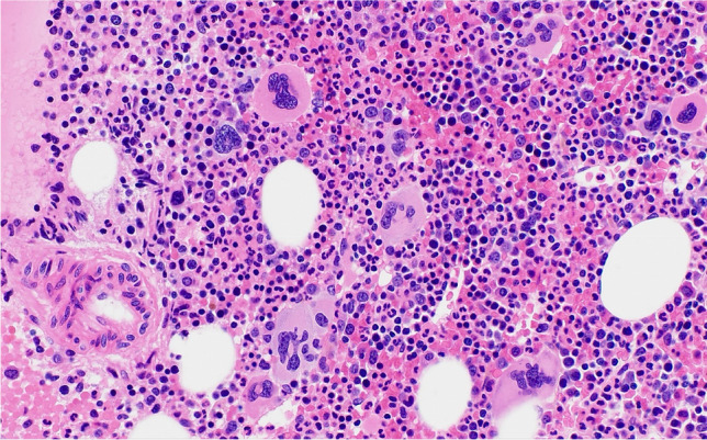
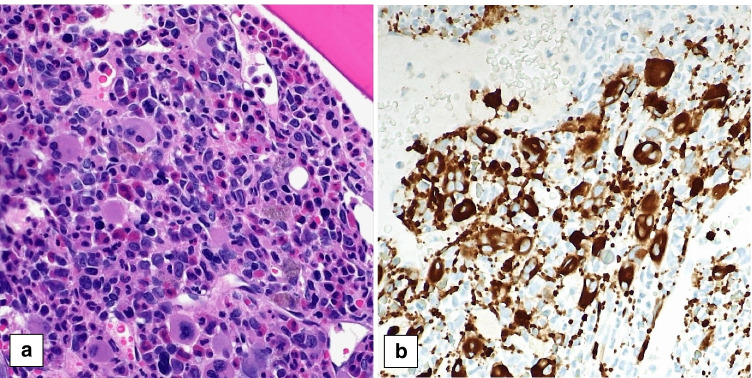
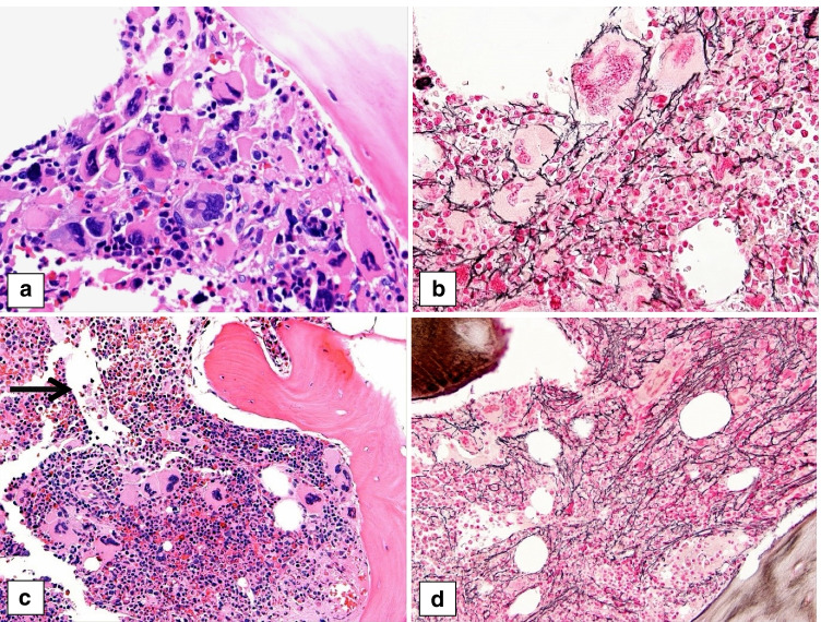
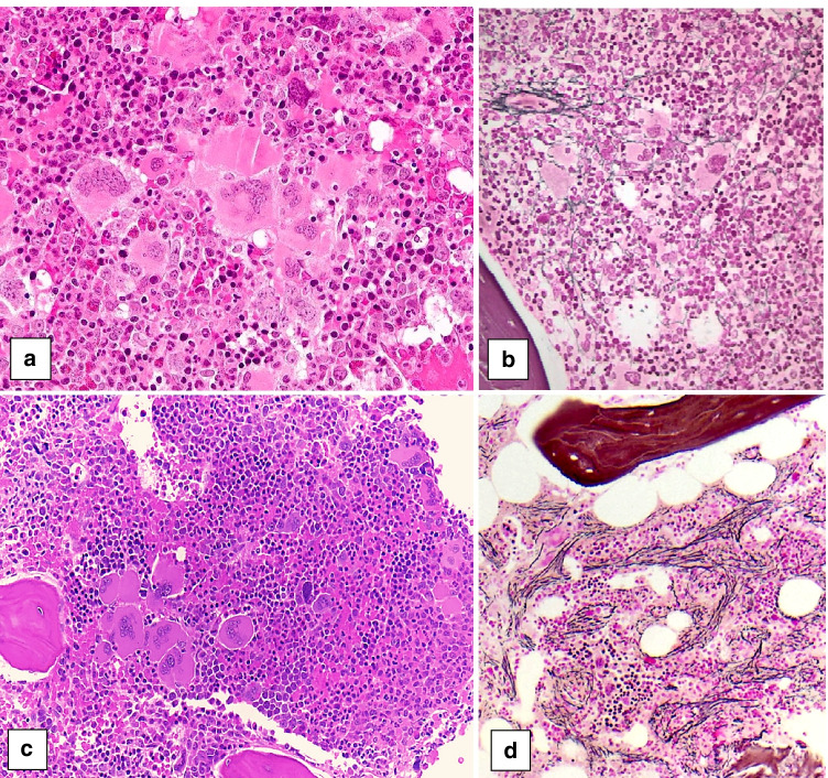
The first section of the bone marrow workshop of the European Association of Haematopathology (EAHP) 2020 Virtual Meeting was dedicated to pediatric myeloid neoplasms. The section covered the whole spectrum of myeloid neoplasms, including myelodysplastic syndromes (MDS), myeloproliferative neoplasms (MPN), myelodysplastic/myeloproliferative neoplasms (MDS/MPN), and acute myeloid leukemia (AML). The workshop cases are hereby presented, preceded by an introduction on these overall rare diseases in this age group. Very rare entities such as primary myelofibrosis, pediatric MDS with fibrosis, and MDS/MPN with JMML-like features and t(4;17)(q12;q21); FIP1L1::RARA fusion, are described in more detail.

Keywords: Acute leukemia; Bone marrow biopsy; EAHP workshop; Juvenile myelomonocytic leukemia; Myelodysplastic syndrome; Myeloproliferative neoplasm; Pediatric.


Conflict of interest statement

Only Dr. Kucine reports a possible conflict of interest: Member, Safety Monitoring Committee-Protagonist Therapeutics.

Figures

Fig. 1
Fig. 1
Pediatric bone marrow biopsies with essential thrombocythemia showing large hyperlobulated megakaryocytes (a case 153, b case 564)
Fig. 2
Fig. 2
Bone marrow biopsy of a 19-year-old young man with panmyelosis and increased, pleomorphic megakaryocytes, consistent with polycythemia vera (case 496)
Fig. 3
Fig. 3
Bone marrow biopsy of a 9-year-old girl with polycythemia vera in accelerated phase (case 700). The HE stain (a) shows increased left shifted granulopoiesis and erythropoiesis, the latter showing megaloblastoid changes, associated with increased megakaryopoiesis characterized by the presence of a mixture of large and small dysplastic forms as highlighted in the CD61 stain (b). Her peripheral blood smear showed 4% blasts and her BM aspirate contained 14% blast by cytology and 13% myeloblasts by flow cytometry
Fig. 4
Fig. 4
Bone marrow biopsies of a 5-year-old boy with primary myelofibrosis (PMF) with a CALR type 1 mutation (case 678), presenting in the prefibrotic stage and showing progression to the fibrotic stage. The first biopsy (a) shows tight clusters of markedly atypical megakaryocytes, including hypolobulated cloud-like and hyperchromatic forms, consistent with PMF. The reticulin stain (b) shows only mild fibrosis, MF1, consistent with the prefibrotic stage. A follow-up biopsy (c) shows hypercellular marrow with increased granulopoiesis, decreased erythropoiesis, tight clusters of atypical, hyperchromatic megakaryocytes and dilated sinusses (arrow). The reticulin stain (d) shows moderate fibrosis, MF2, consistent with progression to fibrotic stage PMF
Fig. 5
Fig. 5
Two cases of pediatric primary myelofibrosis (PMF). a, b Bone marrow biopsy of a 13-year-old girl with a triple negative prefibrotic PMF (case 217) showing hypercellular marrow with tight clusters of atypical megakaryocytes (a HE) and no fibrosis (b reticulin). c, d Bone marrow biopsy a 17-year-old girl with a MPL positive PMF (case 182) showing hypercellular marrow with tight clusters of atypical megakaryocytes (c HE) and moderate fibrosis, MF2 (d reticulin), consistent with PMF, fibrotic stage
Fig. 6
Fig. 6
Myelodysplastic/myeloproliferative neoplasm, with JMML-like features and t(4;17)(q12;q21); FIP1L1::RARA fusion in a 9-month-old girl (case 471). The bone marrow biopsy (a) shows a markedly increased and left-shifted myelopoiesis, decreased erythropoiesis and dysmegakaryopoiesis. The bone marrow aspirate smear (b) demonstrates the expanded population of myelomonocytic cells and atypical promyelocytes with occasional basophilic granules, without Auer rods
Fig. 7
Fig. 7
Two pediatric cases of myelodysplastic syndrome (MDS) with fibrosis showing pronounced dysplasia, espcecially in the megakaryocytes. a, b Case 754 (MDS, unclassifiable, with fibrosis) shows moderate fibrosis, MF2 (a HE, b reticulin stain). c, d Case 322 (MDS with single lineaged dysplasia with fibrosis) shows severe fibrosis, MF3 (c HE, d reticulin stain)
Fig. 8
Fig. 8
Pediatric bone marrow biopsies showing acute myeloid leukemia (AML) with megakaryoblastic differentiation: a, b Case 282, classified as AML-NOS (megakaryoblastic) with KMT2A rearrangement (a HE, b CD61); c, d case 150, classified as AML-MRC due to a complex karyotype, showing partial megakaryoblastic differentiation (c HE, d CD61); e, f case 458, classified as AML-RGA, being an example of an AML (megakaryoblastic) with t(1;22)(p.13.3-q13.1); RBM15::MKL1 (e HE, f CD61)

References

    1. Millot F, Traore P, Guilhot J, Nelken B, Leblanc T, Leverger G, Plantaz D, Bertrand Y, Bordigoni P, Guilhot F. Clinical and biological features at diagnosis in 40 children with chronic myeloid leukemia. Pediatrics. 2005;116:140–143. doi: 10.1542/peds.2004-2473. - DOI - PubMed
    1. Madabhavi I, Patel A, Modi G, Anand A, Panchal H, Parikh S. Pediatric chronic myeloid leukemia: a single-center experience. J Cancer Res Ther. 2020;16:110–115. doi: 10.4103/jcrt.JCRT_833_15. - DOI - PubMed
    1. Millot F, Guilhot J, Baruchel A, Petit A, Bertrand Y, Mazingue F, Lutz P, Verite C, Berthou C, Galambrun C, Sirvent N, Yakouben K, Schmitt C, Gandemer V, Reguerre Y, Couillault G, Mechinaud F, Cayuela JM. Impact of early molecular response in children with chronic myeloid leukemia treated in the French Glivec phase 4 study. Blood. 2014;124:2408–2410. doi: 10.1182/blood-2014-05-578567. - DOI - PubMed
    1. Hijiya N, Schultz KR, Metzler M, Millot F, Suttorp M. Pediatric chronic myeloid leukemia is a unique disease that requires a different approach. Blood. 2016;127:392–399. doi: 10.1182/blood-2015-06-648667. - DOI - PMC - PubMed
    1. Knofler R, Lange BS, Paul F, Tiebel O, Suttorp M. Bleeding signs due to acquired von Willebrand syndrome at diagnosis of chronic myeloid leukaemia in children. Br J Haematol. 2020;188:701–706. doi: 10.1111/bjh.16241. - DOI - PubMed

Publication types

MeSH terms