Review
. 2022 Sep;33(3):335-345.
doi: 10.1007/s12022-022-09726-0. Epub 2022 Jul 11.

Multifocal Fibrosing Thyroiditis: an Under-recognized Mimicker of Papillary Thyroid Carcinoma

Affiliations
Review

Multifocal Fibrosing Thyroiditis: an Under-recognized Mimicker of Papillary Thyroid Carcinoma

Agnese Orsatti et al. Endocr Pathol. 2022 Sep.

Abstract

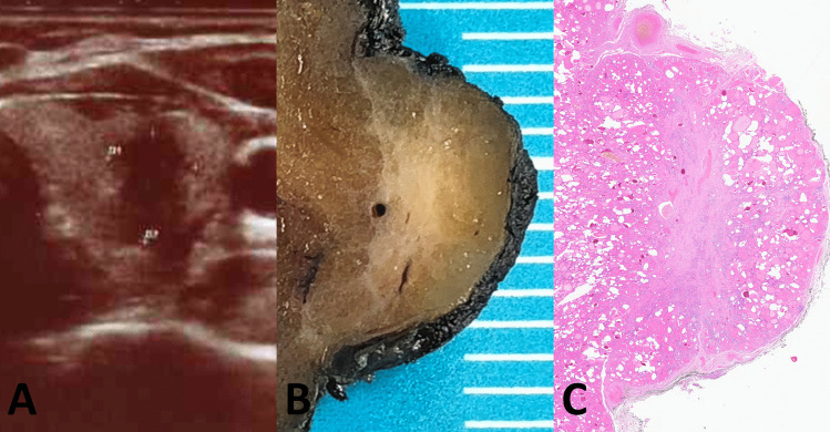
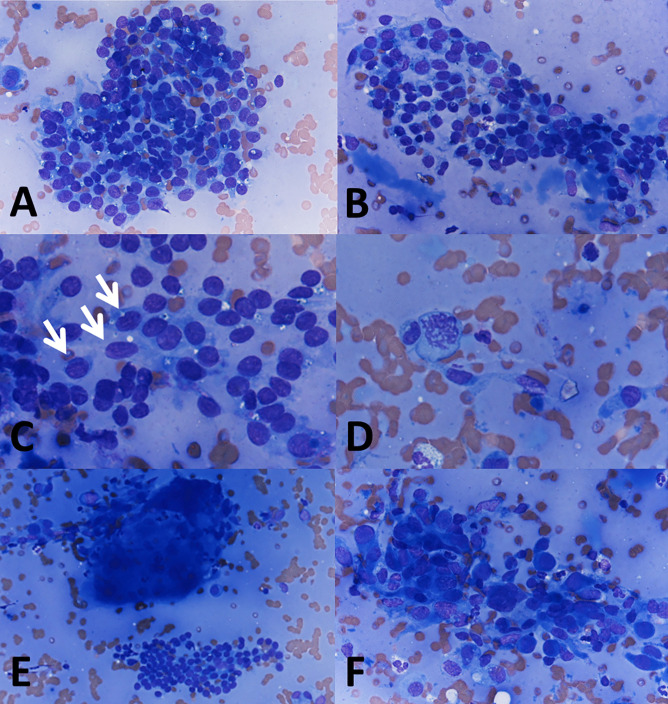
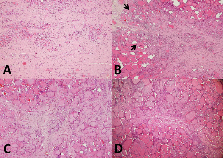
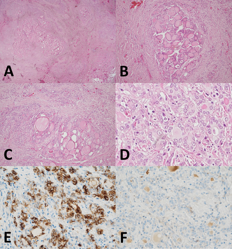
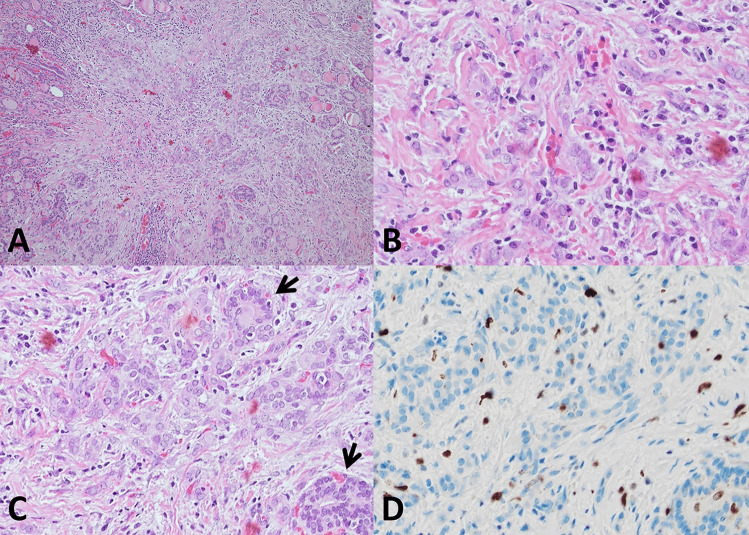
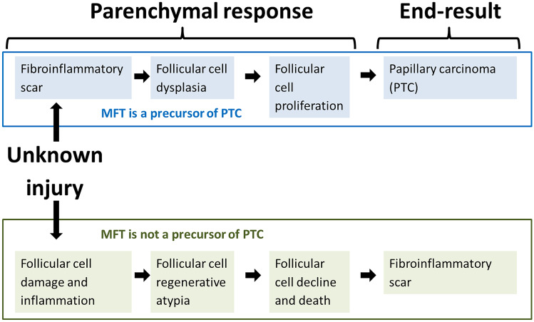
Multifocal fibrosing thyroiditis (MFT) is an enigmatic entity, characterized by multiple fibrotic scar-like lesions with a paucicellular fibrotic center surrounded by a cellular peripheral area with reactive-appearing follicular cell atypia and variable chronic inflammation. Although poorly recognized and likely underreported in surgical pathology, the entity is considered rare with only 65 cases to date-including the current one reported to expand on the preoperative findings of this under-recognized entity. The average age of the patients is 46.8 years (range 15-71 years), 94% are female, with female to male ratio of 15:1. Individual MFT lesions typically have a superficial location. The average number of fibrotic lesions is 15.4 (range 2-51 per MFT case). Their average size is 3.1 mm (range 0.4-15.1). MFT is a disorder of diseased thyroids, typically found postoperatively in glands removed for other reasons, such as chronic lymphocytic/Hashimoto thyroiditis (32.3%), follicular nodular disease (nodular hyperplasia) (30.1%), hyperthyroidism/diffuse hyperplasia (Graves disease) (9.2%). Intriguing is the association with papillary thyroid carcinoma-present in 38.5% of MFT cases, and particularly with sub-centimetric and multifocal papillary thyroid carcinoma, with which MFT can be confused. Cases where MFT is the only thyroid pathology (7.7%) can be preoperatively mistaken for papillary thyroid carcinoma, due to worrisome ultrasound (US) and cytologic features, both of which are here documented for the first time as a component of this article. Wider recognition of MFT and of its cytologic and ultrasound features at preoperative evaluation may reduce unnecessary thyroidectomies.

Keywords: Follicular epithelial dysplasia; Multifocal fibrosing thyroiditis; Papillary thyroid carcinoma; Reactive atypia; Thyroiditis.


Conflict of interest statement

The authors declare no competing interests.

Figures

Fig. 1
Fig. 1
Ultrasound features and macroscopic appearance. Main MFT focus with ultrasound features worrisome for papillary carcinoma that prompted fine needle aspiration (A). Cut surface of the MFT focus on the surgical specimen (B). Low-power microscopic appearance (C)
Fig. 2
Fig. 2
Fine needle aspiration cytology. Scant colloid and small sheets of thyroid cells with anisonucleosis (A, 400 ×). Pseudo-papillary cluster (B, 400 ×). Mildly atypical cells, some with nuclear grooves (C, 600 × ; arrows). Occasional mitosis (D, 600 ×). Multinucleated giant cell (E, 200 ×). Macrophages intermixed with follicular cells showing reactive atypia (F, 400 ×)
Fig. 3
Fig. 3
Histologic features. Zonal distribution of pathologic alterations in the largest 1 cm lesion, with paucicellular fibrotic center (A, 100 ×) and cellular peripheral area with foci of chronic inflammation (B, 40 × ; arrows). Additional fibrosing thyroiditis foci have the same histologic features of the largest focus: 4 mm (C, 40 ×) and 3 mm (D, 40 ×) lesions from the left lobe
Fig. 4
Fig. 4
Histologic features. Small follicles with scant colloid (A, 400 ×), pseudoinfiltrative follicles entrapped in fibrous tissue (B, 400 ×), and vessels with prominent walls (C, 400 ×) at the center of the fibrotic area. Reactive follicular cell atypia (D, 600 ×), elongated follicles lined by crowded follicular cells with overlapping nuclei (E, 600 ×), and multinucleated giant cells with chronic inflammation (F, 200 ×) at the periphery of the central fibrotic core
Fig. 5
Fig. 5
Histologic features. Advanced MFT found incidentally in a thyroid gland removed for a follicular adenoma. The patient’s clinical setting was unremarkable and serum anti-thyroid antibodies were negative. Confluence of fibrotic foci replace large portions of the thyroid parenchyma, separating it into nodules and resulting in a cirrhosis-like pattern (A, 20 × ; B, 100 × ; C, 100 ×). Follicles entrapped in fibrous tissue with enlarged nuclei and chromatin clearing mimicking papillary thyroid carcinoma (D, 600 ×). By immunohistochemistry, atypical follicular cells show partial CD-56 loss (E, 400 ×), HBME-1 is not overexpressed (F, 400 ×)
Fig. 6
Fig. 6
Histologic features. Millimetric MFT focus close to the outer surface of the thyroid gland mimicking at low magnification so-called occult sclerosing papillary thyroid carcinoma (A, 100 ×): MFT was an incidental diagnosis in a thyroid gland removed for other thyroid pathology. Follicles entrapped in the central fibrous MFT core show pseudoinfiltrative growth (B, 400 ×). Elongated follicles with their main axis perpendicular to the fibrotic center lined by crowded follicular cells (C, 200 × ; arrows). Ki67 positivity is largely limited to infiltrating lymphocytes (D, 400 ×)
Fig. 7
Fig. 7
Views on the pathogenesis of Multifocal fibrosing thyroiditis based on divergent interpretation of histologic findings

References

    1. Rosai J, Carcangiu M, DeLellis R. Atlas of tumor pathology, third series, fascicle 5. Washington, DC: Armed Forces Institute of Pathology and American Registry of Pathology. Washington, DC, 1990; 310–311.
    1. Poli F, Trezzi R, Fellegara G, Rosai J. Images in pathology. Multifocal sclerosing thyroiditis. Int J Surg Pathol 17: 144, 2009. - PubMed
    1. Frank R, Baloch ZW, Gentile C, Watt CD, LiVolsi VA. Multifocal fibrosing thyroiditis and its association with papillary thyroid carcinoma using BRAF pyrosequencing. Endocr Pathol. 2014;25:236–240. doi: 10.1007/s12022-013-9289-0. - DOI - PubMed
    1. Fellegara G, Rosai J. Multifocal fibrosing thyroiditis: report of 55 cases of a poorly recognized entity. Am J Surg Pathol. 2015;39:416–424. doi: 10.1097/PAS.0000000000000337. - DOI - PubMed
    1. Fellegara G, Rosai J. Gross appearance of multifocal fibrosing thyroiditis. Am J Surg Pathol. 2015;39:870. doi: 10.1097/PAS.0000000000000431. - DOI - PubMed

LinkOut - more resources