

Analysis of a surface imaging system using a six degree-of-freedom couch
- PMID: 35819973
- PMCID: PMC9359042
- DOI: 10.1002/acm2.13697
Analysis of a surface imaging system using a six degree-of-freedom couch
Abstract
Purpose: To validate surface imaging (SI)-reported offsets using a six degree-of-freedom couch and an anthropomorphic phantom for commissioning and routine quality assurance of an SI system used for stereotactic radiosurgery (SRS).
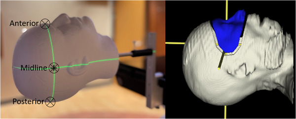
Methods: An anthropomorphic phantom with a radiopaque ball bearing (BB) placed either anterior, midline, or posterior, was tracked with SI with a typical SRS region of interest. Couch motion in all six degrees of freedom was programmed and delivered on a linac. SI system logs were synchronized with linac trajectory logs. Ten random couch positions were selected at couch 0°, 45°, 90°, 270°, 315° with megavolt (MV) images taken to account for couch walkout. The SI residual error (ε), the difference between SI reported offset and MV or trajectory log position, was calculated. Residual errors were measured with and without one SI pod blocked.
Results: The median [range] of magnitude of translational ε was 0.13 [0.07, 0.21], 0.16 [0.11, 0.26], 0.61 [0.50, 0.68], 0.49 [0.42, 0.55], 0.55 [0.38, 0.72] mm for couch rotations of 0°, 45°, 90°, 270°, 315°, respectively, for the midline BB and no pod blocked. The range of all translational ε from all couch angles (with and without pod block) at different BB positions is [0.05, 0.96] mm. The absolute range of difference when changing BB position when no pod is blocked in median translational ε is [0.01, 0.40] mm with the maximum at BB posterior. The absolute range of difference when not changing BB positions with and without pod block in median translational ε is [0.01, 0.37] mm with the maximum at BB posterior and couch 315°.
Conclusion: SI system and linac trajectory log analysis can be used to assess SI system performance with automated couch motion to validate SI accuracy.
Keywords: six degree-of-freedom couch; stereotactic radiosurgery; surface imaging; surface-guided radiotherapy.
© 2022 The Authors. Journal of Applied Clinical Medical Physics published by Wiley Periodicals, LLC on behalf of The American Association of Physicists in Medicine.
Conflict of interest statement
The study was supported by Varian Medical Systems.
Figures


Similar articles
-
Commissioning and clinical evaluation of the IDENTIFYTM surface imaging system for frameless stereotactic radiosurgery.J Appl Clin Med Phys. 2023 Oct;24(10):e14058. doi: 10.1002/acm2.14058. Epub 2023 Jun 8. J Appl Clin Med Phys. 2023. PMID: 37289550 Free PMC article.
-
Stereotactic radiosurgery with MLC-defined arcs: Verification of dosimetry, spatial accuracy, and end-to-end tests.J Appl Clin Med Phys. 2019 May;20(5):84-98. doi: 10.1002/acm2.12583. Epub 2019 Apr 11. J Appl Clin Med Phys. 2019. PMID: 30977297 Free PMC article.
-
A Low-Cost Method to Assess the Performance of Surface Guidance Imaging Systems at Non-Zero Couch Angles.Cureus. 2021 Apr 3;13(4):e14278. doi: 10.7759/cureus.14278. Cureus. 2021. PMID: 33959456 Free PMC article.
-
Evaluation of a surface imaging system's isocenter calibration methods.J Appl Clin Med Phys. 2017 Mar;18(2):85-91. doi: 10.1002/acm2.12054. Epub 2017 Mar 6. J Appl Clin Med Phys. 2017. PMID: 28300386 Free PMC article.
-
Accuracy evaluation of a six-degree-of-freedom couch using cone beam CT and IsoCal phantom with an in-house algorithm.Med Phys. 2017 Aug;44(8):3888-3898. doi: 10.1002/mp.12342. Epub 2017 Jun 16. Med Phys. 2017. PMID: 28500790
Cited by
-
Characterization of the IDENTIFYTM surface scanning system for radiation therapy setup on a closed-bore linac.J Appl Clin Med Phys. 2024 Apr;25(4):e14326. doi: 10.1002/acm2.14326. Epub 2024 Mar 18. J Appl Clin Med Phys. 2024. PMID: 38497554 Free PMC article.
-
Initial Experience of Implementing a Pre-treatment Dry Run for HyperArc Stereotactic Radiosurgery Treatments With Optical Surface Imaging for Intra-fraction Motion Monitoring.Cureus. 2024 Nov 6;16(11):e73124. doi: 10.7759/cureus.73124. eCollection 2024 Nov. Cureus. 2024. PMID: 39650945 Free PMC article.
-
Commissioning and clinical evaluation of the IDENTIFYTM surface imaging system for frameless stereotactic radiosurgery.J Appl Clin Med Phys. 2023 Oct;24(10):e14058. doi: 10.1002/acm2.14058. Epub 2023 Jun 8. J Appl Clin Med Phys. 2023. PMID: 37289550 Free PMC article.
-
A roadmap for implementation of kV-CBCT online adaptive radiation therapy and initial first year experiences.J Appl Clin Med Phys. 2023 Jul;24(7):e13961. doi: 10.1002/acm2.13961. Epub 2023 Mar 15. J Appl Clin Med Phys. 2023. PMID: 36920871 Free PMC article.
-
New findings on clinical experience on surface-guided radiotherapy for frameless non-coplanar stereotactic radiosurgery treatments.J Appl Clin Med Phys. 2024 Dec;25(12):e14510. doi: 10.1002/acm2.14510. Epub 2024 Sep 17. J Appl Clin Med Phys. 2024. PMID: 39287562 Free PMC article.
References
-
- Kügele M, Edvardsson A, Berg L, Alkner S, Andersson Ljus C, Ceberg S. Dosimetric effects of intrafractional isocenter variation during deep inspiration breath‐hold for breast cancer patients using surface‐guided radiotherapy. J Appl Clin Med Phys. 2018;19(1):25‐38. 10.1002/acm2.12214 - DOI - PMC - PubMed
-
- Betgen A, Alderliesten T, Sonke JJ, van Vliet‐Vroegindeweij C, Bartelink H, Remeijer P. Assessment of set‐up variability during deep inspiration breath hold radiotherapy for breast cancer patients by 3D‐surface imaging. Radiother Oncol. 2013;106(2):225‐230. 10.1016/j.radonc.2012.12.016 - DOI - PubMed
MeSH terms
LinkOut - more resources
Full Text Sources
