Review
. 2022 Aug:77:102112.
doi: 10.1016/j.ceb.2022.102112. Epub 2022 Jul 9.

Building the phagocytic cup on an actin scaffold

Affiliations
Review

Building the phagocytic cup on an actin scaffold

Mira Krendel et al. Curr Opin Cell Biol. 2022 Aug.

Abstract

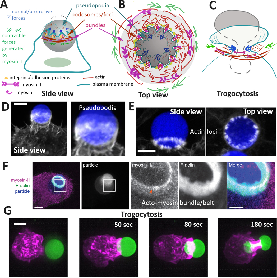
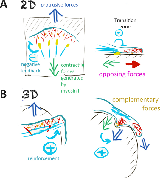
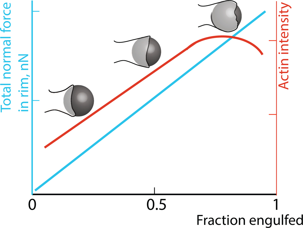
Cells ingest large particles, such as bacteria, viruses, or apoptotic cells, via the process of phagocytosis, which involves formation of an actin-rich structure known as the phagocytic cup. Phagocytic cup assembly and closure results from a concerted action of phagocytic receptors, regulators of actin polymerization, and myosin motors. Recent studies using advanced imaging approaches and biophysical techniques have revealed new information regarding phagocytic cup architecture, regulation of actin assembly, and the distribution, direction, and magnitude of the forces produced by the cytoskeletal elements that form the cup. These findings provide insights into the mechanisms leading to the assembly, expansion, and closure of phagocytic cups. The new data show that engulfment and internalization of phagocytic targets rely on several distinct yet complementary mechanisms that support the robust uptake of foreign objects and may be precisely tailored to the demands of specific phagocytic pathways.


Conflict of interest statement

Conflict of interest The authors declare that they have no known competing financial interests or personal relationships that could have appeared to influence the work reported in this paper.

Figures

Figure 1.
Figure 1.. Phagocytic cup architecture
(A, B) Phagocytic cup organization, shown in a side view (A) and a top-down view (B). The picture of phagocytic cup organization that arises from multiple studies includes the presence of ruffles or pseudopodia at the leading edge of the cup (nucleated primarily by Arp2/3 complex), a ring of Arp2/3-nucleated actin foci or podosomes located immediately behind the ruffling edge, and parallel, stress-fiber-like circumferential actin bundles collaboratively nucleated by Arp2/3 and formins. The ruffles, along with formin-nucleated filopodia, provide increased surface area to promote the initial contact between the phagocyte and the target. Podosomes/actin foci ensure the tight adhesion between the phagocyte and the target. Actin polymerization at foci is responsible for protrusive forces (blue arrows) that may serve a mechanosensory function and may also be involved in trogocytosis. Additional contractile forces (green double arrows) are generated via myosin II-mediated sliding of actin filaments. In complement-mediated phagocytosis, focal adhesion-like complexes containing integrin and talin promote receptor-cytoskeleton coupling needed for successful engulfment while in Fc-dependent phagocytosis, explosive actin polymerization along with the tight adhesion may be sufficient to enclose the phagocytic target. (C) Protrusive and contractile forces cooperate to promote “nibbling” during trogocytosis. (D, E) Phagocytic cup organization (D) and actin foci (E) in RAW macrophages expressing mEmerald-Lifeact (grey) and ingesting polyacrylamide microparticles (blue). D and left panel in E - side view, right panel in E - top-down view (adapted from [11]). (F) Constricting acto-myosin belt in a RAW macrophage ingesting a polyacrylamide particle (adapted from [11]). (G) An example of trogocytosis, with a RAW macrophage expressing mEmerald-Lifeact (magenta) ingesting a polyacrylamide microparticle (green). Scale bars, 5 um; zoom scale bar in F, 1 um.
Figure 2.
Figure 2.. Competition and cooperation between protrusive and contractile forces in 2D and 3D
(2D) During cell migration on a planar substrate, Arp2/3-nucleated actin polymerization drives leading edge protrusion and leads to assembly of focal complexes (orange), while myosin II-dependent contraction of stress fibers pulls at the lamellipodial actin network and promotes maturation of focal adhesions (yellow). Competition between the pulling and protrusive forces produces negative feedback that slows down forward movement of the leading edge. (3D) During phagocytic cup closure in 3D, both protrusive forces (generated by Arp2/3-driven actin assembly) and contractile forces (produced by the acto-myosin belt) are directed towards the center of the phagocytic target, reinforcing and complementing each other. Phagocytic podosomes are shown in orange.
Figure 3.
Figure 3.. Time course of actin assembly and force production during phagocytic cup closure
Constriction of the phagocytic cup and contraction of actin networks results in an increase in both actin intensity and normal forces.

Similar articles

Cited by

References

    1. Freeman SA, Grinstein S: Phagocytosis: receptors, signal integration, and the cytoskeleton. Immunol Rev 2014, 262:193–215. - PubMed
    1. Mattila PK, Lappalainen P: Filopodia: molecular architecture and cellular functions. Nat Rev Mol Cell Biol 2008, 9:446–454. - PubMed
    1. Young LE, Heimsath EG, Higgs HN: Cell type-dependent mechanisms for formin-mediated assembly of filopodia. Mol Biol Cell 2015, 26:4646–4659. - PMC - PubMed
    1. Mylvaganam S, Freeman SA, Grinstein S: The cytoskeleton in phagocytosis and macropinocytosis. Curr Biol 2021, 31:R619–R632. - PubMed
    1. Niedergang F, Grinstein S: How to build a phagosome: new concepts for an old process. Curr Opin Cell Biol 2018, 50:57–63. - PubMed

Publication types