. 2022 Jul 2;39(7):msac145.
doi: 10.1093/molbev/msac145.

Evolutionary History of Sexual Differentiation Mechanism in Insects

Affiliations

Evolutionary History of Sexual Differentiation Mechanism in Insects

Yasuhiko Chikami et al. Mol Biol Evol. .

Abstract

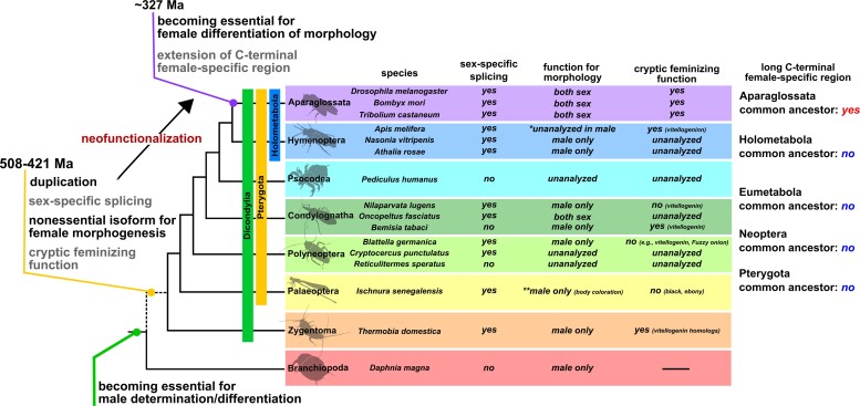
Alternative splicing underpins functional diversity in proteins and the complexity and diversity of eukaryotes. An example is the doublesex gene, the key transcriptional factor in arthropod sexual differentiation. doublesex is controlled by sex-specific splicing and promotes both male and female differentiation in holometabolan insects, whereas in hemimetabolan species, doublesex has sex-specific isoforms but is not required for female differentiation. How doublesex evolved to be essential for female development remains largely unknown. Here, we investigate ancestral states of doublesex using Thermobia domestica belonging to Zygentoma, the sister group of Pterygota, that is, winged insects. We find that, in T. domestica, doublesex expresses sex-specific isoforms but is only necessary for male differentiation of sexual morphology. This result supports the hypothesis that doublesex initially promoted male differentiation during insect evolution. However, T. domestica doublesex has a short female-specific region and upregulates the expression of vitellogenin homologs in females, suggesting that doublesex may already play some role in female morphogenesis of the common ancestor of Pterygota. Reconstruction of the ancestral sequence and prediction of protein structures show that the female-specific isoform of doublesex has an extended C-terminal disordered region in holometabolan insects but not in nonholometabolan species. We propose that doublesex acquired its function in female morphogenesis through a change in the protein motif structure rather than the emergence of the female-specific exon.

Keywords: doublesex; Zygentoma; alternative splicing; insect; sexual differentiation.


Figures

Fig. 1.
Fig. 1.
Molecular phylogeny of doublesex in Pancrustacea and Vertebrata. (A) Molecular phylogeny of doublesex and Mab-3-related transcriptional factors (DMRTs). Phylogenetic analysis was based on the amino acid sequences of DM domain and performed by IQ-TREE following multiple sequence alignment using MAFFT software. The maximum-likelihood method was applied. The 166 operational taxonomic units used for the phylogenetic analysis are listed in supplementary table S2, supplementary material online. Arrow heads indicate the DM domain-containing genes in Thermobia domestica. (B) Enlarged view of insect Dsx Clade2 (dsx-like clade). Numerical value on each node indicates SH-aLRT/UFBoot values. Values on nodes where both SH-aLRT and UFBoot are >80% are shown. Larger numbers are the supporting values on the branches relevant to this study. The full tree information can be found in a supplementary file, supplementary material online (nexus format).
Fig. 2.
Fig. 2.
Structural features of dsx and dsx-like in Thermobia domestica. (A) Exon–intron structures of dsx in Thermobia domestica. Upper and lower schematic images show the gene structure of dsx male-type and female-type, respectively. (B) Expression level of dsx in males and females of T. domestica. (C) Exon–intron structures of dsx-like in T. domestica. (D) Expression level of dsx-like in males and females. Exon–intron structure is determined by mapping the mRNA sequence of each gene to the genome of T. domestica. Expression level (B and D) was measured using RT-qPCR of dsx and dsx-like in the adult fat body and is indicated as relative values to expression of the reference gene, ribosomal protein 49 (rp49). Each plot signifies the mRNA expression level of each individual. Total N = 20 (dsx male-type), 23 (dsx female-type), and 24 (dsx-like). Results of Brunner–Munzel tests are indicated by asterisks: **P < 0.01; ***P < 0.001 and are described in supplementary table S3, supplementary material online.
Fig. 3.
Fig. 3.
Function of doublesex and doublesex-like in terms of body size, internal reproductive system, and gametogenesis of Thermobia domestica. (A) A pair of T. domestica. Female and male are somewhat similar in appearance. (B) Body size of RNAi treatment groups. Width of the pronotum (prothoracic tergum) was used as an indicator of body size. Graph shows mean ± SE (standard error). Results of the GLM analysis shown in supplementary table S5 (female) and S6 (male), supplementary material online. Any significant effect can be detected in the RNAi treatments. Total N = 49 in females and 36 in males. (C) Histology of gonads in the RNAi groups. Paraffin. Hematoxylin–Eosin staining. For ovary images, left and right panels in each treatment show germarium/previtellogenesis and vitellogenesis, respectively. (D) Effects of RNAi on male internal reproductive system. Upper photo shows the gross morphology of the reproductive systems in the nontreated male. Lower photos show the morphology of RNAi males (arrowheads show rounded seminal vesicle). Lowest photos are focused on the vas efferens (arrows show sperm clogged in the tubule). (E) Sperm of RNAi males. Upper photo and figure are sperm morphology in the nontreated male. The sperm forms doublets in the seminal vesicle. Lower figure shows sperm number of RNAi males. Results of the GLM analysis are shown in supplementary table S6, supplementary material online. Significant effect was detected in the dsx RNAi treatment (P = 0.00487). Total N = 29. (F) Effects of RNAi on the female internal reproductive system. (G) Effects of the RNAi on oocyte number. Upper photo shows the ovariole of the nontreated female. Lower figures show the number of oocytes in RNAi females along with oogenetic stages. Results of the GLM analysis are given in supplementary table S5, supplementary material online. The number of late vitellogenic oocytes was correlated with pronotum width, although any significant effect can be detected in RNAi treatments. Total N = 42 in each stage. In each panel, the egfp, dsx all, dsx-like and dsx + dsx-like represent the egfp dsRNA injected group (control), dsx sex-common region dsRNA injected group, dsx-like dsRNA injected group, and both dsx sex-common region and dsx-like dsRNAs injected group, respectively. Each plot in (B), (E), and (G) represent the value of each individual. cc, cystocyte; fc, follicle cell; gv, germinal vesicle; og, oogonia; ol, ovariole; pvo, previtellogenic oocyte; sc, spermatocyte; sp, sperm; st, spermatheca; sv, seminal vesicle; tf, testicular follicle; yg, yolk granule; ve, vas efferens; vd, vas deferens, vo, vitellogenic oocyte. Scales: 50 µm (C); 10 µm (E); 1,000 µm (D and F).
Fig. 4.
Fig. 4.
Function of doublesex and doublesex-like in the morphogenesis of genital organs in Thermobia domestica. (A) Sexually dimorphic traits of T. domestica: females possessing an ovipositor and males a penis. (B) Effects of RNAi treatments on male penial structure. Upper image: ventral side of male abdomen. Lower images focus on male penis. Arrowheads indicate ovipositor-like structure in dsx or both dsx and dsx-like RNAi groups. (C) SEM images of male penial structure. In dsx and dsx + dsx-like RNAi, the two photos are merged into one image. Left panel: ovipositor valvula II (inner sheath)-like structure. Right panel: ovipositor valvula I (outer sheath)-like structure. Details can be found in Supplementary material online. (D) Effects of RNAi treatments on female ovipositor. Upper images: ventral side of the female abdomen. Lower images: female ovipositor. (E) SEM images of female ovipositor structure. Left and right panels show the valvula II; middle panel shows the valvula I. The results of histological observations are shown in supplementary fig. S3, supplementary material online. Details can be found in Supplementary material online. (F) Schematic images of measured parts. (G) Effects of RNAi treatments on growth of ovipositor. Each plot indicates ovipositor length of each individual. Results of the GLM analysis are shown in supplementary table S5, supplementary material online. Ovipositor length correlated with prothoracic width (P = 2.00×10⁻¹⁶), although any significant effects can be seen in RNAi treatments. Total N = 38. In each panel, the egfp, dsx all, dsx-like, and dsx + dsx-like indicates egfp dsRNA injected group (control), dsx sex-common region dsRNA injected group, dsx-like dsRNA injected group, and both dsx sex-common region and dsx-like dsRNAs injected group, respectively. Scales: 1 cm (B and D); 50 µm (C and E).
Fig. 5.
Fig. 5.
Function of doublesex for vitellogenin expression in Thermobia domestica. (A) Vitellogenin expression level in RNAi males. (B) vitellogenin expression level in RNAi females. mRNA expression levels were measured through RT-qPCR. Shown are the log-scale relative values of expression levels of three vitellogenin homologs to the reference gene, ribosomal protein 49 (rp49). Each plot indicates mRNA expression levels of each individual. In each panel, egfp, dsx all, dsx-like, and dsx + dsx-like indicates egfp dsRNA injected group (control), dsx sex-common region dsRNA injected group, dsx-like dsRNA injected group, and both dsx sex-common region and dsx-like dsRNA injected group, respectively. Brunner–Munzel test was performed to ascertain the difference in mRNA expression level between control and dsx or dsx-like RNAi groups. P-values were adjusted with Holm’s method. *P < 0.05, **P < 0.001, ***P < 0.0001. P ≥ 0.05 is not shown. Statistical results are described in supplementary table S3, supplementary material online. Total N = 30 (vitellogenin-1), 24 (vitellogenin-2) and 39 (vitellogenin-3) in males and 33 (vitellogenin-1), 33 (vitellogenin-2), and 34 (vitellogenin-3) in females.
Fig. 6.
Fig. 6.
Evolution of C-terminal sequence of doublesex in insects. (A) ASs of dsx. AS were reconstructed from 49 dsx proteins of insects via maximum-likelihood methods of MEGA X. Details on species and proteins used for the AS reconstruction are found in supplementary table S8, supplementary material online. The most probable sequences were applied. Results of AS reconstruction are described in supplementary table S9, supplementary material online. Probabilities of sites of AS are listed in supplementary table S11, supplementary material online. All sites of Aparaglossata-specific region in AS other than Aparaglossata AS were gaps with probabilities >0.9. Details are given in tsection Materials and Methods. Upper scheme: dsx gene structure of D. melanogaster. Lower image: outcome of multiple sequence alignments (MSA) of dsx sequences by MAFFT. OD domain sequences at C-terminal side were used for MSA. White background in MSA result indicates the conserved sites that share residues in the 80% taxa. The Aparaglossata-specific motif is indicated by orange frame. (B) Predicted protein structures of dsx female-type in common ancestors of insect taxa. The phylogenetic relationship is based on topology from Misof et al. (2014). 3D images in the right panel indicate predicted structures of the OD domain including female-specific region of dsx. Protein structures were predicted using AlphaFold2-based algorism (ColabFold: Mirdita et al. 2021). Red in the 3D image indicates female-specific region, green indicates Aparaglossata-specific motif. Information on the reliable values, that is, predicted local distance difference test: plDDT, of the prediction is given in the Material and Methods section and supplementary fig. S11, supplementary material online.
Fig. 7.
Fig. 7.
Schematic diagram of the evolutionary history of doublesex proposed in this study and the feature of dsx in insects. In our hypothesis, the female isoform of dsx may not have been essential for female differentiation of morphological traits at least from the common ancestor of Dicondylia to the common ancestor of Aparaglossata. In contrast, this female isoform may have contributed to the expression of some genes in females (“cryptic feminizing function” in the figure). The phylogenetic relationship and divergence time are referenced in Misof et al. (2014). The dotted line in the phylogenetic relationship indicates that the taxa occurring from the common ancestor between Branchiopoda and Dicondylia to the common ancestor of Dicondylia are omitted. Here, we also show information on the current knowledge of dsx features in insects and a branchiopod. In Aparaglossata, we show only three representative species. This information was based on the following studies: Hildreth (1965), Burtis and Baker (1989) and Clough et al. (2014) in Drosophila melanogaster (Diptera), Ohbayashi et al. (2001), Suzuki et al. (2003), and Xu et al. (2017) in Bombyx mori (Lepidoptera), Shukla and Palli (2012) in Tribolium castaneum (Coleoptera), Roth et al. (2019) and Velasque et al. (2018) in Apis mellifera (Hymenoptera), Wang et al. (2020) in Nasonia vitripenis (Hymenoptera), Mine et al. (2017, 2021) in Athalia rosae (Hymenoptera), Wexler et al. (2019) in Pediculus humanus (Psocodea) and Blattella germanica (Dictyoptera), Zhuo et al. (2018) in Nilaparvata lugens (Hemiptera), Just et al. (2021) in Oncopeltus fasciatus (Hemiptera), Guo et al. (2018) in Bemisia tabaci (Hemiptera), Miyazaki et al. (2021) in the wood roach Cryptocercus punctulatus and Reticulitermes speratus (Dictyoptera), Takahashi et al. (2019, 2021) in Ischnura senegalensis (Odonata), this study in Thermobia domestica (Zygentoma), and Kato et al. (2011) in Daphnia magna (Branchiopoda). In Condylognatha, information on dsx in the blood-sucking bug Rhodnius prolixus is omitted. R. prolixus has sex-specific isoforms of dsx whose function has not been investigated (Wexler et al. 2014). The term “unanalyzed” means the functional analyses of dsx have not been performed in the relevant species. Information on the roles of dsx of some species in female morphogenesis is limited to some body parts: body coloration in I. senegalensis (Takahashi et al. 2021), leg pigmentation, pheromone synthesis, and wing morphology in Na. vitripenis (Wang, Rensink, et al. 2022; Wang, Sun, et al. 2022), and worker morphology in Ap. mellifera (Roth et al. 2019). The asterisk (*) in Ap. mellifera indicates that the functional analysis of dsx in males was not conducted although gonad differentiation of female workers was affected by dsx knockouts (Roth et al. 2019; see main text). The double-asterisk (**) in I. senegalensis shows that this species has polymorphic coloration in females, that is, gynomorph (normal female color) and andromorph (male-like color) and that dsx is involved in color formation of males and andromorphic females but not gynomorphic females (see Takahashi et al. 2021), suggesting that dsx is nonessential for the female color development. In our hypothesis, the essential roles of dsx for female development in O. fasciatus (Just et al. 2021) may have occurred in parallel with Aparaglossata.

Similar articles

Cited by

References

    1. R Core Team. 2020. R: A language and environment for statistical computing. R Foundation for Statistical Computing.
    1. An W, Wensink PC. 1995. Integrating sex-and tissue-specific regulation within a single Drosophila enhancer. Genes Dev. 9(2):256–266. - PubMed
    1. Bachtrog D, Mank JE, Peichel CL, Kirkpatrick M, Otto SP, Ashman TL, Hahn MW, Kitano J, Mayrose I, Ming R, et al. . 2014. Sex determination: why so many ways of doing it? PLoS Biol. 12(7):e1001899. - PMC - PubMed
    1. Baral S, Arumugam G, Deshmukh R, Kunte K. 2019. Genetic architecture and sex–specific selection govern modular, male–biased evolution of doublesex. Sci Adv. 5(5):eaau3753. - PMC - PubMed
    1. Bear A, Monteiro A. 2013. Both cell-autonomous mechanisms and hormones contribute to sexual development in vertebrates and insects. Bioessays 35(8):725–732. - PubMed

Publication types