. 2022 Aug;298(8):102248.
doi: 10.1016/j.jbc.2022.102248. Epub 2022 Jul 9.

A new paradigm for regulation of protein phosphatase 2A function via Src and Fyn kinase-mediated tyrosine phosphorylation

Affiliations

A new paradigm for regulation of protein phosphatase 2A function via Src and Fyn kinase-mediated tyrosine phosphorylation

Jean-Marie Sontag et al. J Biol Chem. 2022 Aug.

Abstract

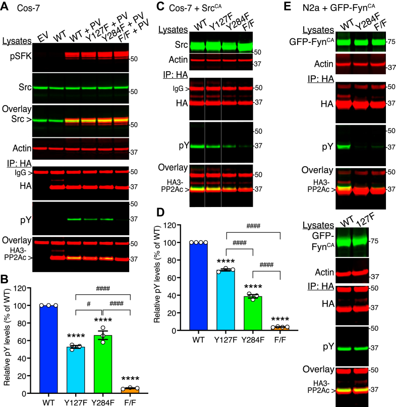
Protein phosphatase 2A (PP2A) is a major phospho-Ser/Thr phosphatase and a key regulator of cellular signal transduction pathways. While PP2A dysfunction has been linked to human cancer and neurodegenerative disorders such as Alzheimer's disease (AD), PP2A regulation remains relatively poorly understood. It has been reported that the PP2A catalytic subunit (PP2Ac) is inactivated by a single phosphorylation at the Tyr307 residue by tyrosine kinases such as v-Src. However, multiple mass spectrometry studies have revealed the existence of other putative PP2Ac phosphorylation sites in response to activation of Src and Fyn, two major Src family kinases (SFKs). Here, using PP2Ac phosphomutants and novel phosphosite-specific PP2Ac antibodies, we show that cellular pools of PP2Ac are instead phosphorylated on both Tyr127 and Tyr284 upon Src activation, and on Tyr284 following Fyn activation. We found these phosphorylation events enhanced the interaction of PP2Ac with SFKs. In addition, we reveal SFK-mediated phosphorylation of PP2Ac at Y284 promotes dissociation of the regulatory Bα subunit, altering PP2A substrate specificity; the phosphodeficient Y127/284F and Y284F PP2Ac mutants prevented SFK-mediated phosphorylation of Tau at the CP13 (pSer202) epitope, a pathological hallmark of AD, and SFK-dependent activation of ERK, a major growth regulatory kinase upregulated in many cancers. Our findings demonstrate a novel PP2A regulatory mechanism that challenges the existing dogma on the inhibition of PP2A catalytic activity by Tyr307 phosphorylation. We propose dysregulation of SFK signaling in cancer and AD can lead to alterations in PP2A phosphorylation and subsequent deregulation of key PP2A substrates, including ERK and Tau.

Keywords: ERK; Fyn; PP2A; Src; Tau; protein phosphatase 2A; tyrosine phosphorylation.


Conflict of interest statement

Conflicts of interest B. E. W. and B. W. S. are cofounders of Turkey Creek Biotechnology LLC and have equity ownership. The authors declare no other competing conflicts of interest with the contents of this article.

Figures

Figure 1
Figure 1
Expression of constitutively active Src or Fyn kinase induces tyrosine phosphorylation of endogenous PP2Ac and HA-tagged WT and Y307F PP2Ac.A, lysates from control (−) and SrcCA-transformed (+) MEFs were subjected to microcystin-Sepharose pull downs (MPDs). Total cell lysates and MPDs were analyzed by Western with antibodies recognizing phosphotyrosine (pY) and endogenous PP2Ac. Representative Western blots of total cell lysates and HA-immunoprecipitates prepared from SYF (B), N2a (C), or Cos-7 (D and E) cells transfected with either empty vector (EV), HA-/HA3-tagged wildtype PP2Ac (WT), or the Y307F phosphomutant of PP2Ac (Y307F) together with plasmids encoding SrcCA, GFP-tagged FynCA, or EV. Where indicated, a subset of these cells was treated with pervanadate (+PV) prior to harvesting. Well-characterized anti-pY antibodies were used to assess tyrosine phosphorylation of proteins in response to SFKCA and pervanadate. Tubulin antibodies were used to control for protein loading. Similar results were observed in at least four separate experiments.
Figure 2
Figure 2
Distinct effects of pervanadate, ScrCA, and FynCAon PP2Ac WT, Y127F, Y284F, and Y127/284F tyrosine phosphorylation. Lysates and HA-immunoprecipitates were prepared from control and pervanadate-treated (+PV) Cos-7 cells (A and B), SrcCA-transfected Cos-7 cells (C and D), and GFP-FynCA-transfected N2a cells (E) that were cotransfected with plasmids encoding HA3-tagged PP2Ac wildtype (WT), Y127F (Y127F), Y284F (Y284F), Y127/284F (F/F), or the empty vector (EV). Representative Western blots with the indicated antibodies are shown in (A), (C), and (E). Note: the pSFK antibody recognizes Src only when phosphorylated at Y416. B, relative levels of pY signal in HA3-PP2Ac immunoprecipitates from (A). Data (mean ± SEM from n = 3 separate experiments) were appraised using one-way ANOVA (F = 236; p < 0.0001) with Tukey’s post hoc multiple comparisons test. ∗∗∗∗p < 0.0001, compared with WT; ####p < 0.0001, #p < 0.05. D, relative levels of pY signal in HA3-PP2Ac immunoprecipitates from (C). Data (mean ± SEM from n = 3–4 separate experiments) were appraised using one-way ANOVA (F = 2067; p < 0.0001) with Tukey’s post hoc multiple comparisons test. ∗∗∗∗p < 0.0001, compared with WT; ####p < 0.0001. (E) similar results were observed in three separate experiments.
Figure 3
Figure 3
Western analyses using newly generated phosphosite-specific antibodies reveal that PP2Ac is differentially phosphorylated by FynCAand SrcCAat the Y284 and Y127 epitopes.A, representative Western blots of HA-immunoprecipitates prepared from N2a cells coexpressing HA3-tagged PP2Ac and either GFP- FynCA or an empty vector (EV) and probed with mouse anti-HA, alpaca anti-pY284 (pY284), or rabbit anti-pY127 (pY127) PP2Ac antibodies. Where indicated, a subset of these cells were treated with pervanadate (+PV) prior to harvesting. B, Western analysis of HA-immunoprecipitates prepared from EV- or SrcCA-transfected Cos-7 cells that were cotransfected with plasmids encoding HA3-tagged PP2Ac wildtype (WT), Y127F (Y127F), Y284F (Y284F), or Y127/284F (F/F). C and D, Western analyses of total cell lysates and FLAG-immunoprecipitates prepared from Cos-7 cells that were cotransfected with plasmids encoding SrcCA, FLAG-tagged Bα regulatory subunit (FLAG-Bα), and/or the empty vector (-). Blots were probed with anti-PP2Ac, anti-PP2A B subunit, anti-pY (C), or the novel PP2Ac phosphospecific Y284 and Y127 (D) antibodies. Where indicated, a subset of these cells was treated with pervanadate (PV) prior to harvesting. E, Western analyses of HA-immunoprecipitates prepared from Cos-7 cells that were cotransfected with plasmids encoding SrcCA and HA-tagged PP2Ac WT or the L309Δ, D88N, and H59Q mutants. Expression of SrcCA in the lysates was verified using the pY416-SFK antibody.
Figure 4
Figure 4
SFK-mediated tyrosine phosphorylation of PP2Ac decreases its binding to the regulatory Bα subunit.A, total lysates and HA-immunoprecipitates were prepared from empty vector (EV)- or SrcCA-transfected Cos-7 cells that were cotransfected with plasmids encoding either HA3-tagged PP2Ac wildtype (WT), Y127F (Y127F), Y284F (Y284F), or Y127/284F (F/F). Blots were analyzed using anti-Bα, anti-PP2Ac, or anti-HA antibodies. B, relative levels of Bα were quantified in HA3-PP2Ac immunoprecipitates. Data are mean ± SEM from at least n = 4 separate experiments and were appraised using unpaired t-tests as indicated. ∗p < 0.05; ∗∗∗∗p < 0.0001, SrcCAversus EV; ns, not significant. C, Western blots of total lysates and HA-immunoprecipitates prepared from empty vector (EV)- or GFP-FynCA-transfected N2a cells that were cotransfected with plasmids encoding either HA3-tagged PP2Ac wildtype (WT), Y284F (Y284F), or Y127F (Y127F). Levels of GFP-Fyn and Bα in the input used for immunoprecipitation (lysates) are shown for reference. D, relative levels of Bα binding in HA3-PP2Ac immunoprecipitates. Data are mean ± SEM from n = 3 separate experiments and were appraised using one-way ANOVA (F = 29.02; p = 0.0001) with Tukey’s post hoc multiple comparisons test. ∗∗p < 0.01, ∗∗∗p < 0.001; ns, not significant.
Figure 5
Figure 5
PP2Ac phosphomutants exhibit decreased interactions with SFKCA.A, duplicate HA3-PP2Ac immunoprecipitates from the same transfected Cos-7 cells analyzed in Figure 2C were subjected to Western analysis using anti-pY416 SFK and anti-HA antibodies. B, relative levels of SrcCA in HA3-PP2Ac immunoprecipitates. Data are mean ± SEM from n = 4 separate experiments and were appraised using one-way ANOVA (F= 97.17; p < 0.0001) with Tukey’s post hoc multiple comparisons test. ∗p < 0.05, ∗∗∗∗p < 0.0001, compared with WT; #p < 0.05. C, representative Western blots of total lysates and HA-immunoprecipitates prepared from empty vector (EV)- or GFP-FynCA-expressing N2a cells that were cotransfected with plasmids encoding either HA3-tagged PP2Ac wildtype (WT), Y284F (Y284F), Y127F (Y127F), or Y127/284F (F/F). D, relative levels of FynCA in HA3-PP2Ac immunoprecipitates. Data are mean ± SEM from n = 3 separate experiments and were appraised using one-way ANOVA (F = 59.07; p < 0.0001) with Tukey’s post hoc multiple comparisons test. ∗∗∗∗p < 0.0001, compared to WT; ns, not significant.
Figure 6
Figure 6
The Y127/284F and Y284F phosphoincompetent mutants inhibit SrcCA- and FynCA-mediated ERK activation, respectively.A, Cos-7 cells were transfected with plasmids encoding HA3-tagged PP2Ac WT, Y127F, Y284F, or the empty vector (EV). Total cell homogenates from serum-starved cells were analyzed by immunoblotting for active ERK using validated antibodies recognizing the phosphorylated forms of ERK2 (or p42-MAPK, lower band) and ERK1 (or p44-MAPK, upper band), and total ERK1/2. PP2Ac expression levels in the cells are shown as reference. Pervanadate-treated control cells and WT-transfected cells incubated with the SFK inhibitor, PP2, were used as controls. B, Western analyses of active ERK in Cos-7 cells that were cotransfected with plasmids encoding SrcCA and HA3-tagged WT, Y127F, Y284F, F/F or Y307F PP2Ac, or the empty vector (EV). Extracts from EV-transfected cells served as controls. HA3-PP2Ac and Src expression levels in these cells are shown as reference. C, relative phosphorylated ERK (pERK) levels in Cos-7 cells transfected as in B. Data are mean ± SEM from at least three separate experiments and were appraised using one-way ANOVA (F = 45.13; p < 0.0001) with Tukey’s post hoc multiple comparisons test. ∗∗p < 0.01, ∗∗∗∗p < 0.0001, compared to WT + SrcCA; #p < 0.05; ns, not significant. D, representative Western blots of cell homogenates prepared from control SrcCA-, or FynCA-transfected N2a cells that were cotransfected with an empty vector (EV), HA3-tagged PP2Ac wildtype (WT), Y127F, Y284F, or F/F. Duplicate aliquots of the cell lysates were analyzed for active ERK, HA-PP2Ac, Src, and GFP-Fyn expression levels. Note that top ERK panels were assembled from the same blot. E, relative phosphorylated ERK (pERK) levels in N2a cells transfected as in D. Data are mean ± SEM from n = 3 separate experiments and were appraised using one-way ANOVA (F = 66.78; p < 0.0001) with Tukey’s post hoc multiple comparisons test. ∗∗∗∗p < 0.0001, compared with WT + SrcCA; ####p < 0.0001; ns, not significant.
Figure 7
Figure 7
PP2Ac phosphomutants inhibit SrcCA- and FynCA-mediated Tau phosphorylation.A, the same total cell homogenates from control (Fig. 6D, left panel), SrcCA- (Fig. 6D, center panel), or FynCA- (Fig. 5C, right panel) transfected N2a cells that were cotransfected with EV or HA-PP2Ac species were reanalyzed by Western for endogenous Tau phosphorylation at the CP13 phosphoepitope (pSer202) and for total Tau. B, relative Ser 202-phosphorylated Tau (p-Tau) levels in N2a cells transfected as in A. Data are mean ± SEM from n = 3 separate experiments and were appraised using one-way ANOVA (F = 61.67; p < 0.0001) with Tukey’s post hoc multiple comparisons test. ∗p < 0.05, ∗∗∗∗p < 0.0001, compared with WT + SFKCA; ns, not significant. C, representative immunoblots of total and Ser 9-phosphorylated GSK-3β from the same cell lysates analyzed in A. Similar results were obtained in two other experiments.

References

    1. Parsons S.J., Parsons J.T. Src family kinases, key regulators of signal transduction. Oncogene. 2004;23:7906–7909. - PubMed
    1. Elias D., Ditzel H.J. Fyn is an important molecule in cancer pathogenesis and drug resistance. Pharmacol. Res. 2015;100:250–254. - PubMed
    1. Roskoski R., Jr. Src protein-tyrosine kinase structure, mechanism, and small molecule inhibitors. Pharmacol. Res. 2015;94:9–25. - PubMed
    1. Lee G. Tau and src family tyrosine kinases. Biochim. Biophys. Acta. 2005;1739:323–330. - PubMed
    1. Ittner A., Ittner L.M. Dendritic tau in Alzheimer's disease. Neuron. 2018;99:13–27. - PubMed

Publication types

MeSH terms

LinkOut - more resources