Review
. 2022 Oct 1:172:105815.
doi: 10.1016/j.nbd.2022.105815. Epub 2022 Jul 9.

Dendritic involvement in inhibition and disinhibition of vulnerable dopaminergic neurons in healthy and pathological conditions

Affiliations
Review

Dendritic involvement in inhibition and disinhibition of vulnerable dopaminergic neurons in healthy and pathological conditions

R C Evans. Neurobiol Dis. .

Abstract

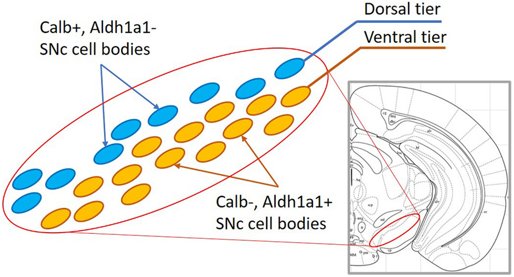
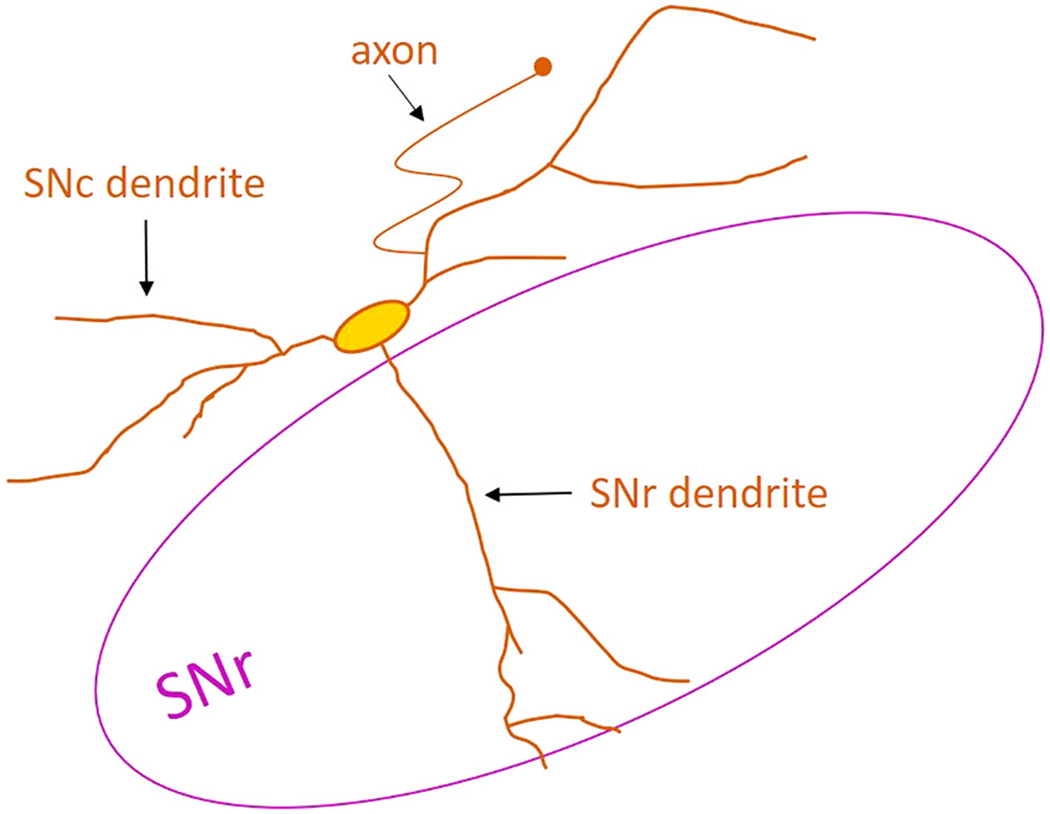
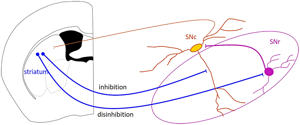
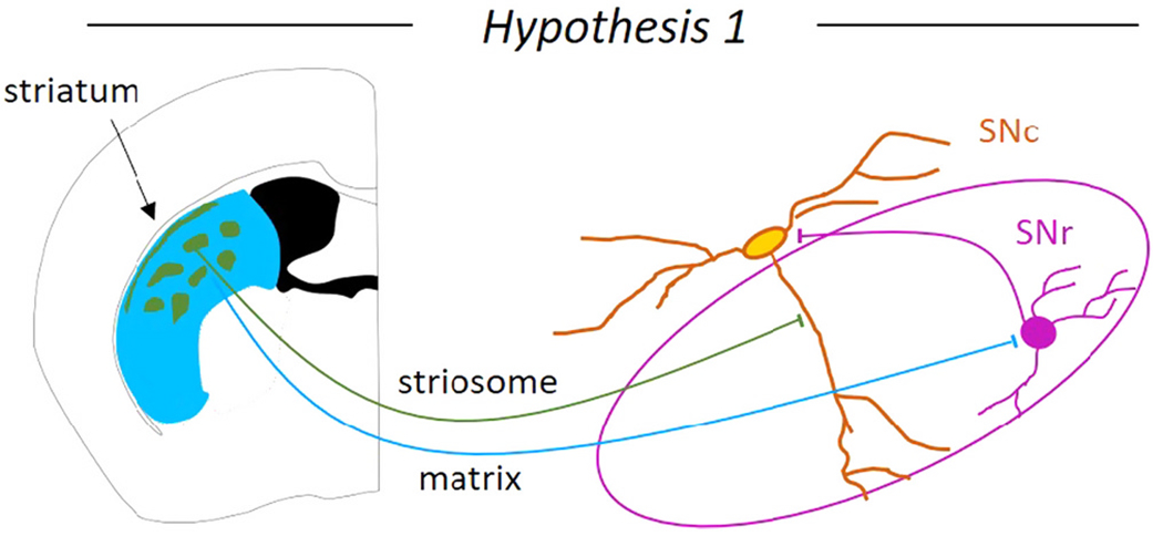
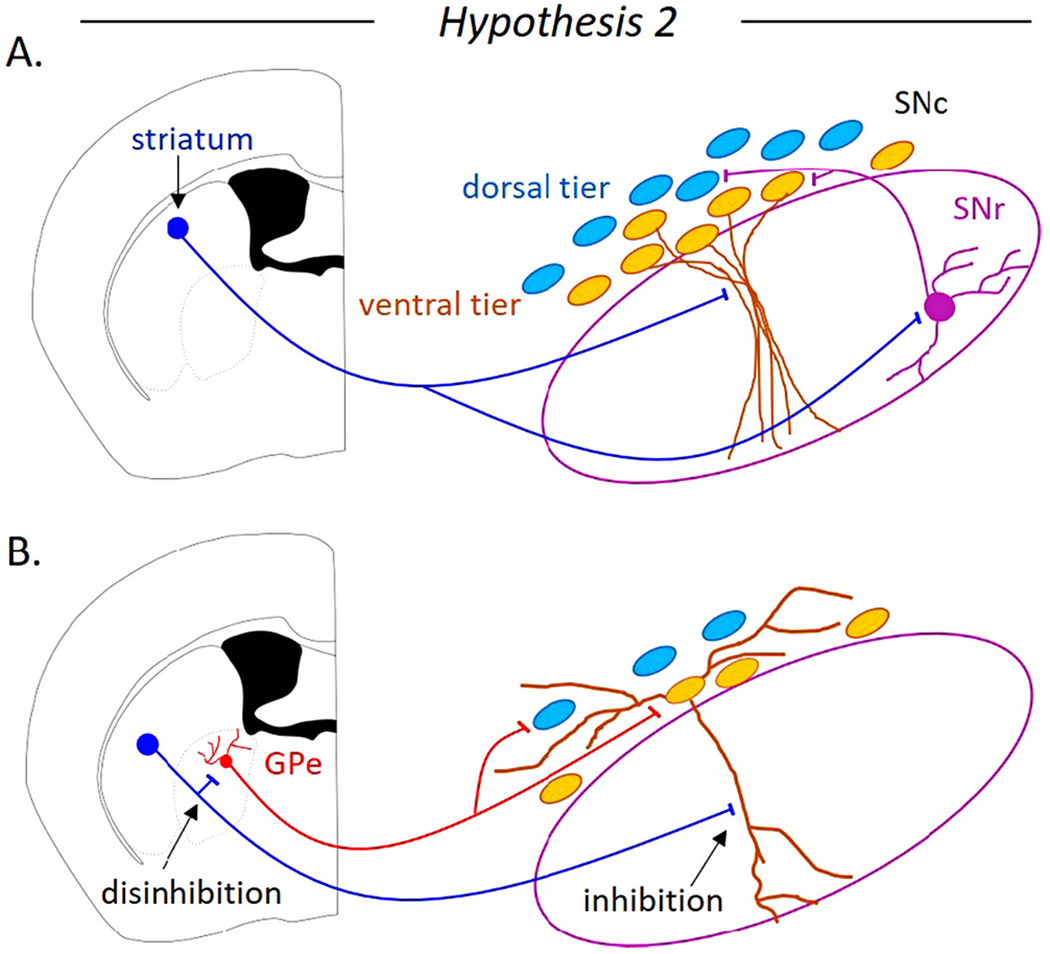
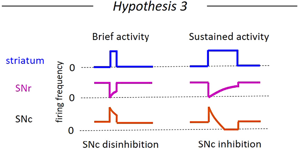
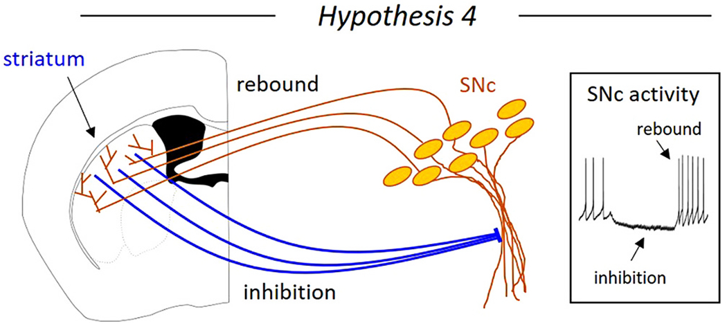
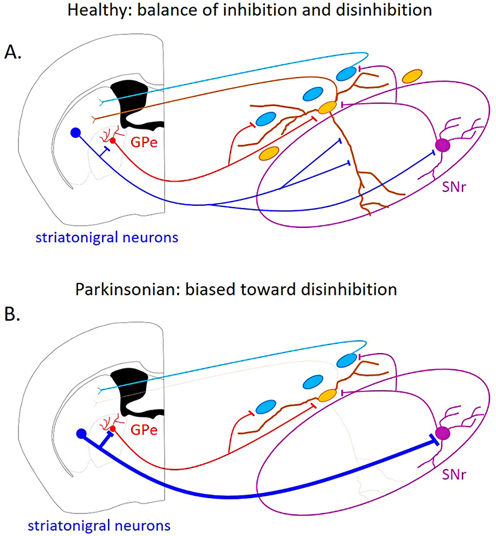
Dopaminergic neurons in the substantia nigra pars compacta (SNc) differentially degenerate in Parkinson's Disease, with the ventral region degenerating more severely than the dorsal region. Compared with the dorsal neurons, the ventral neurons in the SNc have distinct dendritic morphology, electrophysiological characteristics, and circuit connections with the basal ganglia. These characteristics shape information processing in the ventral SNc and structure the balance of inhibition and disinhibition in the striatonigral circuitry. In this paper, I review foundational studies and recent work comparing the circuitry of the ventral and dorsal SNc neurons and discuss how loss of the ventral neurons early in Parkinson's Disease could affect the overall balance of inhibition and disinhibition of dopamine signals.

Keywords: Basal ganglia; Circuit mapping; Dendrites; Dendritic morphology; Disinhibition; Dopamine; Inhibition; Nigrostriatal; Parkinson's disease; SNc; Striatonigral; Striatum; Substantia nigra.


Conflict of interest statement

Declaration of Competing Interest

None.

Figures

Fig. 1.
Fig. 1.. Diagram of the ventral and dorsal tier of the substantia nigra pars compacta (SNc).
Yellow ovals represent the calbindin-negative, Aldh1a1-positive ventral tier dopaminergic neurons that are more vulnerable to degeneration. Blue ovals represent the dorsal tier calbindin-positive, Aldh1a1-negative dopaminergic neurons that are more resilient. (For interpretation of the references to colour in this figure legend, the reader is referred to the web version of this article.)
Fig. 2.
Fig. 2.. Dendritic morphology of ventral SNc neurons.
The ventrally-located SNc dopaminergic neurons have two distinct dendrite types: SNc dendrites projecting along the substantia nigra pars compacta cell body layer and SNr dendrites projecting ventrally into the GABAergic substantia nigra pars reticulata. The axon-bearing dendrite is most often an SNc dendrite.
Fig. 3.
Fig. 3.. Canonical pathway for striatonigral inhibition and disinhibition.
Striatal projection neurons can directly inhibit substantia nigra pars compacta (SNc) dopaminergic neurons, and can disinhibit them by inhibiting substantia nigra pars reticulata (SNr) GABAergic neurons. The inhibition and disinhibition of the SNc neurons will shape the dopamine signal sent to the striatum through SNc dopaminergic axons.
Fig. 4.
Fig. 4.. Hypothesis 1: Striosome inhibits and matrix disinhibits dopaminergic neurons of the substantia nigra pars compacta (SNc).
The two striatal compartments, the striosomes and the matrix may exert opposing control of the SNc dopaminergic neurons. The striosomes are thought to directly inhibit the SNc dopaminergic neurons on the SNr dendrite, while the matrix is thought to disinhibit the SNc dopaminergic neurons through inhibition of substantia nigra pars reticulata (SNr) GABAergic neurons.
Fig. 5.
Fig. 5.. Hypothesis 2: the ventral tier neurons of the substantia nigra pars compacta (SNc) are inhibited by the striatum, while the dorsal tier neurons of the SNc are disinhibited.
A. The striatal direct pathway may inhibit the ventrally-located SNc neurons and disinhibit the dorsally-located SNc neurons through the GABAergic neurons of the substantia nigra pars reticulata (SNr). B. A non-canonical pathway for striatonigral disinhibition is through the globus pallidus external segment (GPe). The striatal direct pathway spiny projection neurons inhibit a subpopulation of GPe neurons known to directly inhibit dopaminergic SNc neurons.
Fig. 6.
Fig. 6.. Hypothesis 3: The timing of the striatal activity determines whether the dopamine neurons of the substantia nigra pars compacta (SNc) are inhibited or disinhibited.
Brief increases in striatal projection neuron firing frequency briefly inhibits the substantia nigra pars reticulata (SNr) GABAergic neurons to disinhibit SNc dopaminergic neurons (left). Sustained increases in striatal projection neuron firing could result in short term synaptic depression between the striatum and the SNr neurons or between the SNr neurons and the SNc, resulting in a short disinhibition followed by a period of inhibition in the SNc dopaminergic neurons.
Fig. 7.
Fig. 7.. Hypothesis 4: Striatal inhibition of substantia nigra pars compacta (SNc) dopaminergic neurons generates rebound activity.
Striatal inhibition of SNc neurons recruits intrinsic rebound mechanisms in the ventral SNc neurons, causing increased dopaminergic neuron activity when inhibition is released.
Fig. 8.
Fig. 8.. Hypothetical imbalance between inhibition and disinhibition of SNc dopaminergic neurons under pathological conditions.
A. In the healthy condition, the balance of inhibition and disinhibition of the substantia nigra pars compacta (SNc) dopaminergic neurons is intact. B. Under Parkinsonian conditions, when the ventral SNc neurons have degenerated, direct striatonigral inhibition onto the SNc neurons is reduced, while disinhibitory pathways may be strengthened.

References

    1. Abecassis ZA, Berceau BL, Win PH, García D, Xenias HS, Cui Q, Pamukcu A, Cherian S, Hernández VM, Chon U, et al., 2020. Npas1+-Nkx2.1+ neurons are an integral part of the Cortico-pallido-cortical loop. J. Neurosci 40, 743–768. 10.1523/JNEUROSCI.1199-19.2019. - DOI - PMC - PubMed
    1. Aguila J, Cheng S, Kee N, Cao M, Wang M, Deng Q, Hedlund E, 2021. Spatial RNA sequencing identifies robust markers of vulnerable and resistant human midbrain dopamine neurons and their expression in Parkinson’s disease. Front. Mol. Neurosci 14, 699562 10.3389/fnmol.2021.699562. - DOI - PMC - PubMed
    1. Alberico SL, Cassell MD, Narayanan NS, 2015. The vulnerable ventral tegmental area in Parkinson’s disease. Basal Ganglia 5, 51–55. 10.1016/j.baga.2015.06.001. - DOI - PMC - PubMed
    1. Ambrosi P, Lerner TN, 2021. Striatonigrostriatal circuit architecture for disinhibition of dopamine signaling. BioRxiv. 10.1101/2021.06.22.449416, 2021.06.22.449416. - DOI - PMC - PubMed
    1. Beckstead MJ, Williams JT, 2007. Long-term depression of a dopamine IPSC. J. Neurosci 27, 2074–2080. 10.1523/JNEUROSCI.3251-06.2007. - DOI - PMC - PubMed

Publication types