Multicenter Study
. 2023 Feb;72(2):392-403.
doi: 10.1136/gutjnl-2022-327498. Epub 2022 Jul 12.

Accurate liquid biopsy for the diagnosis of non-alcoholic steatohepatitis and liver fibrosis

Affiliations
Multicenter Study

Accurate liquid biopsy for the diagnosis of non-alcoholic steatohepatitis and liver fibrosis

Giulia Angelini et al. Gut. 2023 Feb.

Abstract

Objective: Clinical diagnosis and approval of new medications for non-alcoholic steatohepatitis (NASH) require invasive liver biopsies. The aim of our study was to identify non-invasive biomarkers of NASH and/or liver fibrosis.

Design: This multicentre study includes 250 patients (discovery cohort, n=100 subjects (Bariatric Surgery Versus Non-alcoholic Steato-hepatitis - BRAVES trial); validation cohort, n=150 (Liquid Biopsy for NASH and Liver Fibrosis - LIBRA trial)) with histologically proven non-alcoholic fatty liver (NAFL) or NASH with or without fibrosis. Proteomics was performed in monocytes and hepatic stellate cells (HSCs) with iTRAQ-nano- Liquid Chromatography - Mass Spectrometry/Mass Spectrometry (LC-MS/MS), while flow cytometry measured perilipin-2 (PLIN2) and RAB14 in peripheral blood CD14+CD16- monocytes. Neural network classifiers were used to predict presence/absence of NASH and NASH stages. Logistic bootstrap-based regression was used to measure the accuracy of predicting liver fibrosis.

Results: The algorithm for NASH using PLIN2 mean florescence intensity (MFI) combined with waist circumference, triglyceride, alanine aminotransferase (ALT) and presence/absence of diabetes as covariates had an accuracy of 93% in the discovery cohort and of 92% in the validation cohort. Sensitivity and specificity were 95% and 90% in the discovery cohort and 88% and 100% in the validation cohort, respectively.The area under the receiver operating characteristic (AUROC) for NAS level prediction ranged from 83.7% (CI 75.6% to 91.8%) in the discovery cohort to 97.8% (CI 95.8% to 99.8%) in the validation cohort.The algorithm including RAB14 MFI, age, waist circumference, high-density lipoprotein cholesterol, plasma glucose and ALT levels as covariates to predict the presence of liver fibrosis yielded an AUROC of 95.9% (CI 87.9% to 100%) in the discovery cohort and 99.3% (CI 98.1% to 100%) in the validation cohort, respectively. Accuracy was 99.25%, sensitivity 100% and specificity 95.8% in the discovery cohort and 97.6%, 99% and 89.6% in the validation cohort. This novel biomarker was superior to currently used FIB4, non-alcoholic fatty liver disease fibrosis score and aspartate aminotransferase (AST)-to-platelet ratio and was comparable to ultrasound two-dimensional shear wave elastography.

Conclusions: The proposed novel liquid biopsy is accurate, sensitive and specific in diagnosing the presence and severity of NASH or liver fibrosis and is more reliable than currently used biomarkers.

Clinical trials: Discovery multicentre cohort: Bariatric Surgery versus Non-Alcoholic Steatohepatitis, BRAVES, ClinicalTrials.gov identifier: NCT03524365.Validation multicentre cohort: Liquid Biopsy for NASH and Fibrosis, LIBRA, ClinicalTrials.gov identifier: NCT04677101.

Keywords: HEPATIC FIBROSIS; NONALCOHOLIC STEATOHEPATITIS.


Conflict of interest statement

Competing interests: GM reports consulting fees from Novo Nordisk,_Fractyl Inc and Recor Inc; she is also scientific current advisor and consultant of Metadeq Limited, and current advisor and consultant of Keyron Limited, GHP Scientific Limited, and Jemyll Limited. FR reports receiving research grants from Ethicon and Medtronic; consulting fees from Novo Nordisk, Ethicon and Medtronic; serving on scientific advisory boards for GI Dynamics; and is former director and current stock option holder of Metadeq Limited and former director and current advisor of Keyron Limited and GHP Scientific Limited. CWlR reports grants from the Irish Research Council, Science Foundation Ireland, Anabio and the Health Research Board; serves on advisory boards of Novo Nordisk, Herbalife, GI Dynamics, Eli Lilly, Johnson & Johnson, Sanofi Aventis, AstraZeneca, Janssen, Bristol-Myers Squibb, Glia and Boehringer Ingelheim. ClR is a member of the Irish Society for Nutrition and Metabolism outside the area of work commented on here, and is the chief medical officer and director of the Medical Device Division of Keyron since January 2011; both of these are unremunerated positions. CWlR is also current director, shareholder and stock option holder of Metadeq Limited, current director of GHP Scientific Limited, was a previous investor in Keyron, which develops endoscopically implantable medical devices intended to mimic the surgical procedures of sleeve gastrectomy and gastric bypass. The product has only been tested in rodents and none of Keyron’s products are currently licensed. They do not have any contracts with other companies to put their products into clinical practice. No patients have been included in any of Keyron’s studies and they are not listed on the stock market. He continues to provide scientific advice to Keyron for no remuneration. All other authors declare no competing interests.

Figures

Figure 1
Figure 1
Laser scanning confocal immunofluorescence of monocytes, HSCs and hepatocytes from a representative subject with NASH/fibrosis (upper panels) and a representative subjects with negative histology for NASH/fibrosis (lower panels). (A–D) RAB14 staining in monocytes (A, B) and in HSCs (C, D). (E) Quantification of RAB14 protein expression by flow cytometry, in monocytes and HSCs of 20 subjects with NASH/fibrosis and 20 subjects with negative histology for NASH/fibrosis. (F) Spearman correlation analysis and linear regression line fitting of RAB14 expression in monocytes and HSCs. (G–J) PLIN2 staining in monocytes (G, H) and in hepatocytes (I, J). (K) Quantification of PLIN2 protein expression by flow cytometry in monocytes and hepatocytes of 20 subjects with NASH/fibrosis and 20 subjects with negative histology for NASH/fibrosis. (L) Spearman correlation analysis and linear regression line fitting of PLIN2 expression in monocytes and hepatocytes. (M–P) LDs staining with Nile Red in monocytes (M, N) and in hepatocytes (O, P). The mean NAS was 4.05±0.15 in the 20 subjects with NASH and liver fibrosis and 0.65±0.11 in the 20 subjects study without NASH and liver fibrosis from LIBRA. SAF-F mean was 1.95±0.17. Data are expressed as mean±SEM for linear regression analysis; Spearman rank correlation coefficients (R2) and p values are shown. Magnification ×60. Scale bar: 50 µm. DAPI, 4', 6-Diamidino-2-Phenylindole; HSC, hepatic stellate cell; LD, lipid droplet; MFI, mean florescence intensity; NAS, Non-alcoholic Fatty Liver Disease Activity Score; NASH, non-alcoholic steatohepatitis; PLIN2, perilipin-2.
Figure 2
Figure 2
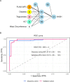
PLIN2 diagnostic performance for predicting presence/absence of NASH. (A) Architecture of the NN used. Each input node represents a biomarker, while edges represent the weights between layers. The thickness of the edge is proportional to the magnitude of each weight. Positive weights are plotted as black lines; negative weights as grey lines. In the NN, there are five input nodes: PLIN2, presence of diabetes, plasma triglycerides and ALT levels and waist circumference. There are three hidden nodes and one output node for presence of NASH. Two biases nodes are included. The bias node covers the same function of the intercept in a regression model. (B) ROC curves for predicting the presence of NASH (NAS ≥3) in the discovery and in the validation cohorts. FPR=1–specificity. AUC, area under the curve; B, bias weight; FPR, false-positive rate; H, hidden node; I, input node; MFI, mean florescence intensity; NAS, Non-alcoholic Fatty Liver Disease Activity Score; NASH, non-alcoholic steatohepatitis; NN, neural network; O, output node; PLIN2, perilipin-2; ROC, receiver operating characteristic; TPR, true-positive rate.
Figure 3
Figure 3
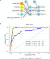
PLIN2 diagnostic performance for predicting NASH severity. (A) PLIN2 biomarker network for diagnosing of NASH severity through NAS levels. Architecture of the NN used. Each input node represents a biomarker, while edges represent the weights between layers. The thickness of the edge is proportional to the magnitude of each weight. Positive weights are plotted as black lines; negative weights as grey lines. In the NN, there are five input nodes: PLIN2, presence of diabetes, plasma triglycerides and ALT levels and waist circumference. There are three hidden nodes and three output nodes for NAS level=0 for total NAS score <3, NAS level=1 for total NAS score=3, NAS level=2 for total NAS score ≥4. Two bias nodes are included. The bias node covers the same function of the intercept in a regression model. (B) ROC curves for each level of NASH severity as identified by the NAS value (see aforementioned) in the validation cohort. FPR=1–specificity. AUC, area under the curve; B, bias weight; FPR, false-positive rate; H, hidden node; I, input node; MFI, mean fluorescence intensity; NAS, Non-alcoholic Fatty Liver Disease Activity Score; NASH, non-alcoholic steatohepatitis; NN, neural network; O, output node; PLIN2, perilipin-2; ROC, receiver operating characteristic; TPR, true-positive rate.
Figure 4
Figure 4
Whisker plots of monocyte PLIN2 levels (MFI) measured by flow cytometry in the training and validation cohorts in different NASH stages: histological NAS <3, NAS=3 and NAS ≥4. MFI, mean fluorescence intensity; NAS, Non-alcoholic Fatty Liver Disease Activity Score; NASH, non-alcoholic steatohepatitis; PLIN2, perilipin-2.
Figure 5
Figure 5
RAB14 (A) and elastography (C) diagnostic performance for presence/absence of NASH. The model estimates ±SE (A) (RAB14) are intercept −62.25±22.15, RAB14 −0.87±0.81, waist circumference 12.03±4.28, plasma glucose 1.09±3.66, age 3.33±2.40, HDL cholesterol −1.80±4.64 and ALT −0.23±1.25. The model estimates ±SE (C) (elastography) are intercept −61.69±22.84, elastography 2.49±2.48, waist circumference 10.1±4.84, plasma glucose 1.64±3.74, age 2.97±2.49, HDL cholesterol −1.43±1.70 and ALT −0.16±1.22. Monocyte RAB14 levels (MFI) measured by flow cytometry at Steatosis, Activity, Fibrosis-Fibrosis (SAF-F) scores 0 (NO Fibrosis), ≥1 (YES Fibrosis) in the training and validation cohorts (B). Ultrasound transient elastography (kPa) at SAF-F scores 0 (NO Fibrosis), ≥1 (YES Fibrosis) in the training and validation cohorts (D). AUC, area under the curve; FPR, false-positive rate; HDL, high-density lipoprotein; MFI, mean fluorescence intensity; ROC, receiver operating characteristic; TPR, true-positive rate.

Comment in

References

    1. Wong VW-S, Chitturi S, Wong GL-H, et al. . Pathogenesis and novel treatment options for non-alcoholic steatohepatitis. Lancet Gastroenterol Hepatol 2016;1:56–67. 10.1016/S2468-1253(16)30011-5 - DOI - PubMed
    1. Pioglitazone vs vitamin E vs placebo for treatment of non-diabetic patients with nonalcoholic steatohepatitis (PIVENS). Available: https://clinicaltrials.gov/ct2/show/NCT00063622
    1. Friedman SL, Ratziu V, Harrison SA, et al. . A randomized, placebo-controlled trial of cenicriviroc for treatment of nonalcoholic steatohepatitis with fibrosis. Hepatology 2018;67:1754–67. 10.1002/hep.29477 - DOI - PMC - PubMed
    1. Haukeland JW, Damås JK, Konopski Z, et al. . Systemic inflammation in nonalcoholic fatty liver disease is characterized by elevated levels of CCL2. J Hepatol 2006;44:1167–74. 10.1016/j.jhep.2006.02.011 - DOI - PubMed
    1. Smits MM, van Geenen EJM. The clinical significance of pancreatic steatosis. Nat Rev Gastroenterol Hepatol 2011;8:169–77. 10.1038/nrgastro.2011.4 - DOI - PubMed

Publication types

Associated data