. 2022 Nov;136(6):1841-1850.
doi: 10.1007/s00414-022-02863-5. Epub 2022 Jul 12.

Density of TMEM119-positive microglial cells in postmortem cerebrospinal fluid as a surrogate marker for assessing complex neuropathological processes in the CNS

Affiliations

Density of TMEM119-positive microglial cells in postmortem cerebrospinal fluid as a surrogate marker for assessing complex neuropathological processes in the CNS

Simone Bohnert et al. Int J Legal Med. 2022 Nov.

Abstract

Routine coronal paraffin-sections through the dorsal frontal and parieto-occipital cortex of a total of sixty cases with divergent causes of death were immunohistochemically (IHC) stained with an antibody against TMEM119. Samples of cerebrospinal fluid (CSF) of the same cases were collected by suboccipital needle-puncture, subjected to centrifugation and processed as cytospin preparations stained with TMEM119. Both, cytospin preparations and sections were subjected to computer-assisted density measurements. The density of microglial TMEM119-positive cortical profiles correlated with that of cytospin results and with the density of TMEM119-positive microglial profiles in the medullary layer. There was no statistically significant correlation between the density of medullary TMEM119-positive profiles and the cytospin data. Cortical microglial cells were primarily encountered in supragranular layers I, II, and IIIa and in infragranular layers V and VI, the region of U-fibers and in circumscribed foci or spread in a diffuse manner and high density over the white matter. We have evidence that cortical microglia directly migrate into CSF without using the glympathic pathway. Microglia in the medullary layer shows a strong affinity to the adventitia of deep vessels in the myelin layer. Selected rapidly fatal cases including myocardial infarcts and drowning let us conclude that microglia in cortex and myelin layer can react rapidly and its reaction and migration is subject to pre-existing external and internal factors. Cytospin preparations proved to be a simple tool to analyze and assess complex changes in the CNS after rapid fatal damage. There is no statistically significant correlation between cytospin and postmortem interval. Therefore, the quantitative analyses of postmortem cytospins obviously reflect the neuropathology of the complete central nervous system. Cytospins provide forensic pathologists a rather simple and easy to perform method for the global assessment of CNS affliction.

Keywords: Biomarker; Cerebrospinal fluid; Forensic neuropathology; Forensic neurotraumatology; Immunocytochemistry; Immunohistochemistry.


Conflict of interest statement

The authors declare that they have no potential conflict of interest.

Research involving human participants and/or animals

None

Figures
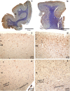
Fig. 1
Fig. 1
Low resolution overview of coronal sections through the isocortex of the parietal lobe (a, b). Female aged 91 years, TBI, PMI 125 h (a), and female aged 28 years, carbon monoxide intoxication, postmortem interval 10 h (b) In (a) IHC-staining is confined mainly to the crest of the gyri and weak or absent in the wall and fundus of sulci. Status cribrosus in the white matter of both gyri and perivascular loss of staining. Arrow points to prominent internal granular layer characteristic of cortical areae in parieto-occipital regions. Only few TMEM119-positive microglial cells with thin processes were seen in the supragranular layers I–III and in the deeper layers V–VI including U-fibers and the central medullary ray of gyri (c, e). Many processes of microglial cells appear blurred. This could be an effect of the long postmortem interval. Increase of TMEM119 microglial cells in the supragranular layer after intoxication in layers I–III (d) and infragranular layers V–VI (f) together with U-fibers and fibers coursing in the medullary ray of this gyrus. At the border between lamina II–I, microglial cells appear crowded and seem to loose their processes and microglial pericarya with few or without processes seem to head to the outer pia-covered region of layer I
Fig. 2
Fig. 2
a Strongly positive cytospin preparation, female aged 48 years, sepsis, postmortem interval 83 h. Arrow points to grouped TMEM119-positive microglia (b) Same case as in a. The high-power microscopic image depicts microglial cells on their way to (big arrows) and through the pia (thin arrows) into the subarachnoid CSF. c Female aged 72 years, suffocation after aspiration, post-mortem-interval 106 h. Layer I at the fundus of a sulcus. The outer parts of the molecular layer appeared oedematous and groups of microglial cells and fragments of processes were interspersed in this spongy part of the molecular layer (thin arrow). The pia mater in this case is obviously detached and glia cells and fragments seemed to enter the CSF (thick arrow) but they were also attached to arachnoid fibers beyond the pia mater (thin arrow). d Same case as in a, subcortical myelin layer with tangentially cut vascular profile with TMEM119-positive profiles closely attached to the vascular adventitia. Shrinkage artifacts do not unequivocally disclose the mode of association or transition of numerous TMEM119-positive microglial cells from myelin layer to perivascular adventitia
Fig. 3
Fig. 3
Correlation between cortical TMEM119-positive microglial density and CSF TMEM119-positive microglial density (cytospin preparations)
Fig. 4
Fig. 4
Correlation between cortical and white matter TMEM119-positive microglial density
Fig. 5
Fig. 5
Correlation between white matter TMEM119-positive microglial cell density and CSF TMEM119-positive microglial density (cytospin preparations)
Fig. 6
Fig. 6
Heatmap. Correlations between cause of death, sex, age, postmortem interval (PMI), cardiopulmonal resuscitation (CPR), intensity of staining in CSF, and brain weight

References

    1. Daneman R, Prat A. The blood-brain barrier. Cold Spring Harb Perspect Biol. 2015;7:a020412. doi: 10.1101/cshperspect.a020412. - DOI - PMC - PubMed
    1. Ballabh P, Braun A, Nedergaard M. The blood-brain barrier: an overview structure, regulation, and clinical implications. Neurobiol Dis. 2004;16:1–13. doi: 10.1016/j.nbd.2003.12.016. - DOI - PubMed
    1. Benveniste H, Lee H, Volkow N. The glymphatic pathway: waste removal from CNS via cerebrospinal fluid transport. Neuroscientist. 2017;23:454–465. doi: 10.1177/1073858417691030. - DOI - PMC - PubMed
    1. Agoston DV, Shutes-David A, Peskind E. Biofluid biomarkers of traumatic brain injury. Brain Inj. 2017;31:1195–1203. doi: 10.1080/02699052.2017.1357836. - DOI - PubMed
    1. Harel A, Kvist M, Nuutinen S, Välimaa L. Biomarkers of traumatic brain injury temporal changes in body fluids. eNeuro. 2016;3:1–13. - PMC - PubMed

LinkOut - more resources