

Pediatric pineal region tumors: institutional experience of surgical managements with posterior interhemispheric transtentorial approach
- PMID: 35821434
- PMCID: PMC10432319
- DOI: 10.1007/s00381-022-05595-4
Pediatric pineal region tumors: institutional experience of surgical managements with posterior interhemispheric transtentorial approach
Abstract
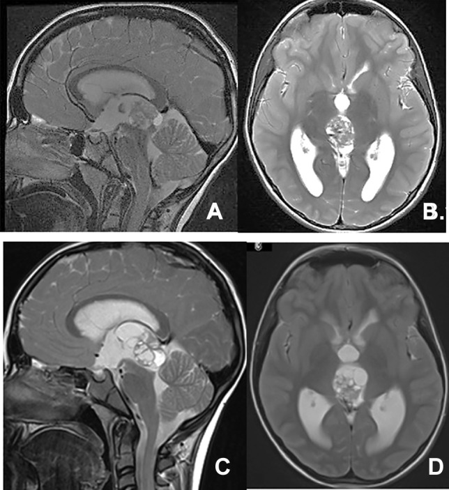
Purpose: Resecting pineal region tumors in children is often challenging. Several approaches have been proposed and practiced. A personal series of pediatric pineal region tumors resected through craniotomy with posterior interhemispheric occipital transtentorial (OT) approach are reviewed. We present the surgical techniques, pitfalls, and their results.
Material and methods: Eighty patients ranging in age from 3 months to 21 years old, and treated over 3 decades were reviewed. Hydrocephalus caused the main presenting symptoms and was noted in 74 patients. It was treated prior to the craniotomy for tumor resection with endoscopic third ventriculostomy (ETV) in 33, external ventricular drainage in 26, and precraniotomy shunt in 15. Nine patients had ETV together with endoscopic biopsy. All patients had a parieto-occipital craniotomy in a prone position. Through a tentorial section, a gross total resection of the tumor was attempted except for germinomas.
Results: The tumor pathology showed 32 germ cell tumors (GCT), 22 benign astrocytomas, 13 pineal parenchymal tumors, 5 ATRTs, 3 papillary tumors, and 5 others. Of GCTs, 18 were teratomas. The extent of resection consisted of 55 gross total resections, 13 subtotal resections, 10 partial, and 2 biopsies with one postoperative death. Hemiparesis in 2, cerebellar ataxia in another 2, and hemiballismus in 1 were transient and improved over time. One had permanent hemisensory loss and another patient had bilateral oculomotor palsy. Postoperative homonymous hemianopia occurred in 2 patients but subsided over a short period of time. Parinaud's sign was noted in 24 patients, of which 16 were transient.
Conclusion: The posterior interhemispheric OT approach provides a safe route and comfortable access to the pineal region in children. A great majority of postoperative neurological complications are the results of direct manipulations of the midbrain at tumor resection. Identification and preservation of the tumor-brain interface are of paramount importance. GCTs other than teratomas are treated with neoadjuvant chemotherapy and may eliminate the need for craniotomy. Exophytic midbrain JPAs are amenable to resection.
Keywords: Germ cell tumor; Hydrocephalus headings: pineal region tumors; Midbrain glioma; Neuroendoscopy; Occipital transtentorial resection; Pineal tumor; Pineoblastoma; Superior medullary velum.
© 2022. The Author(s).
Conflict of interest statement
The authors declare no competing interests.
Figures


Similar articles
-
Endoscopic-assisted interhemispheric parieto-occipital transtentorial approach for microsurgical resection of a pineal region tumor: operative video and technical nuances.Neurosurg Focus. 2016 Jan;40 Video Suppl 1:2016.1.FocusVid.15450. doi: 10.3171/2016.1.FocusVid.15450. Neurosurg Focus. 2016. PMID: 26722692
-
Simultaneous single-trajectory endoscopic biopsy and third ventriculostomy in pediatric pineal region tumors.Acta Neurol Belg. 2021 Dec;121(6):1535-1542. doi: 10.1007/s13760-020-01387-2. Epub 2020 Jun 6. Acta Neurol Belg. 2021. PMID: 32506355
-
Second-look surgery for pineal region tumors.Childs Nerv Syst. 2023 Sep;39(9):2349-2352. doi: 10.1007/s00381-022-05676-4. Epub 2022 Oct 1. Childs Nerv Syst. 2023. PMID: 36181520
-
Pineal/germ cell tumors and pineal parenchymal tumors.Childs Nerv Syst. 2023 Oct;39(10):2649-2665. doi: 10.1007/s00381-023-06081-1. Epub 2023 Oct 13. Childs Nerv Syst. 2023. PMID: 37831207 Review.
-
An update on the surgical treatment of malignant pineal region tumors.Clin Neurosurg. 1985;32:397-428. Clin Neurosurg. 1985. PMID: 2415282 Review.
Cited by
-
Understanding and Managing Pineal Parenchymal Tumors of Intermediate Differentiation: An In-Depth Exploration from Pathology to Adjuvant Therapies.J Clin Med. 2024 Feb 23;13(5):1266. doi: 10.3390/jcm13051266. J Clin Med. 2024. PMID: 38592098 Free PMC article. Review.
-
Supracerebellar highway-fast and safe road to pediatric pineal tumor resection.Childs Nerv Syst. 2025 Aug 13;41(1):260. doi: 10.1007/s00381-025-06919-w. Childs Nerv Syst. 2025. PMID: 40802062
-
Diagnosis and Management of Pineal Germinoma: From Eye to Brain.Eye Brain. 2023 Apr 13;15:45-61. doi: 10.2147/EB.S389631. eCollection 2023. Eye Brain. 2023. PMID: 37077304 Free PMC article. Review.
-
A rare case of pineal astrocytoma with hemorrhagic changes: Diagnostic and surgical challenges.Radiol Case Rep. 2025 Apr 10;20(6):3153-3158. doi: 10.1016/j.radcr.2025.03.003. eCollection 2025 Jun. Radiol Case Rep. 2025. PMID: 40247959 Free PMC article.
References
-
- Jouvet A, Fauchon F, Liberski P, Saint-Pierre G, Didier-Bazes M, Heitzmann A, et al. Papillary tumor of the pineal region. Am J Surg Pathol. 2003;27:505–512. - PubMed
-
- Thomas C, Wefers A, Bens S, Nemes K, Agaimy A, Oyen F, Vogelgesang S, Rodriguez FJ, Brett FM, McLendon R, Bodi I, Burel-Vandenbos F, Keyvani K, Tippelt S, Poulsen FR, Lipp ES, Giannini C, Reifenberger G, Kuchelmeister K, Pietsch T, Kordes U, Siebert R, Frühwald MC, Johann D, Sill M, Kool M, von Deimling A, Paulus W, Hasselblatt M. Desmoplastic myxoid tumor, SMARCB1-mutant: clinical, histopathological and molecular characterization of a pineal region tumor encountered in adolescents and adults. Acta Neuropathol. 2019;139:277–286. - PubMed
-
- Fangusaro J, Wu S, MacDonald S, Murphy E, Shaw D, Bartels U, Soumen Khatua S, Souweidane M, Lu H-M, Morris D, Panigrahy A, Onar-Thomas A, Fouladi M, Gajjar A, Dhall G. Phase II trial of response-based radiation therapy for patients with localized CNS nongerminomatous germ cell tumors: a children’s oncology group study. J Clin Oncol. 2019;37:3283–3290. - PMC - PubMed
-
- Goldman S, Bouffet E, Fisher PG, Allen JC, Robertson PL, Chuba PI, Donahue B, Kretschmar CS, Zhou T, Buxton AB, Pollack IF. Phase II trial assessing the ability of neoadjuvant chemotherapy with or without second-look surgery to eliminate measurable disease for nongerminomatous germ cell tumors: a children’s oncology group study. J Clin Oncol. 2015;33:2464–2471. - PMC - PubMed
MeSH terms
LinkOut - more resources
Full Text Sources
Medical
