

Micro-Raman spectroscopy study of blood samples from myocardial infarction patients
- PMID: 35821543
- PMCID: PMC9708773
- DOI: 10.1007/s10103-022-03604-1
Micro-Raman spectroscopy study of blood samples from myocardial infarction patients
Abstract
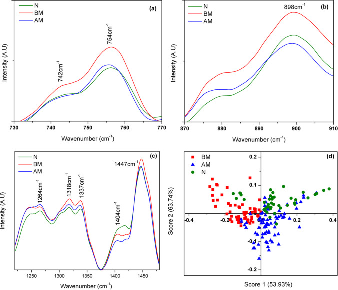
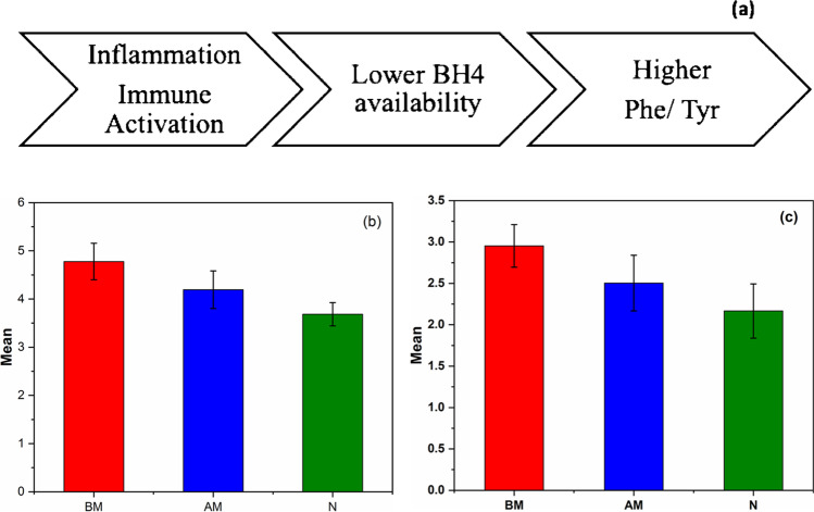
Acute myocardial infarction (MI) is found to be a major causative factor for global mortality and morbidity. This situation demands necessity of developing efficient and rapid diagnostic tools to detect acute MI. Raman spectroscopy is a non-destructive optical diagnostic technique, which has high potential in probing biochemical changes in clinical samples during initiation and progress of diseases. In this work, blood was taken as the sample to examine inflammation in acute MI patients using Raman spectroscopy. Ratio of Raman peak intensities that corresponds to phenylalanine (1000 cm-1) and tyrosine (825 cm-1) can facilitate indirect information about tetrahydrobiopterin (BH4) availability, which can indicate inflammatory status in patients. This ratio obtained was higher for MI patients in comparison with control subjects. The decrease in phenylalanine and tyrosine ratio (Phe-Tyr ratio) is attributed to the prognosis of standard of care (medications like antiplatelets including aspirin, statin and revascularisation) leading to inflammation reduction. Phe-Tyr ratio estimated from the Raman spectra of blood can be exploited as a reliable method to probe inflammation due to MI. The method is highly objective, require only microliters of sample and minimal sample preparation, signifying its clinical utility.
Keywords: BH4; Inflammation; Laser Raman spectroscopy; Myocardial infarction; Optical diagnostics.
© 2022. The Author(s).
Conflict of interest statement
The authors declare no competing interests.
Figures


References
-
- World Health Organization (2013) Health topics: cardiovascular diseases. http://www.Who.Int/Topics/Cardiovascular_diseases/En/. Accessed June 2021
MeSH terms
Substances
LinkOut - more resources
Full Text Sources
Medical
