. 2022 Sep;67(5):2040-2047.
doi: 10.1111/1556-4029.15093. Epub 2022 Jul 12.

A method for the development of cranial fracture histology slides

Affiliations

A method for the development of cranial fracture histology slides

Carolyn V Isaac et al. J Forensic Sci. 2022 Sep.

Abstract

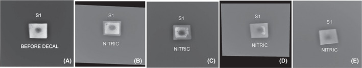
Cranial vault fractures are of medicolegal interest as they have long-term impacts to someone's health and may contribute to an individual's death. The ability to distinguish antemortem from perimortem fractures and to assess the age of the injury is increasingly dependent on histology. Despite the increasing role of histology in assessing the microanatomy of osseous fractures, there are no methods currently available which account for the nuances and difficulties in creating high-quality histologic slides of cranial vault fractures that allow visualization of cellular features associated with healing bone. The authors present a modified method specific to slide development of human cranial vault fractures derived from the trial-and-error process of creating 730 such slides over a 3-year period which are suitable for the evaluation of the tissues, cells, and nuclei involved in fracture healing. This method adapts and troubleshoots typical histological procedures including sample excision, fixation, decalcification, dehydrating, clearing, embedding, microtomy, and staining, and introduces new procedures including preprocessing photography and cassette placement. By implementing these modifications, the number of poor-quality slides that required a new section to be sent to the histology laboratory was greatly reduced. Proactively implementing this new method into cranial fracture histologic slide development significantly reduces the number of slide rejections due to common issues like folding, chatter, or insufficient staining, saving both time and financial resources for forensic practitioners, researchers, and histotechnologists.

Keywords: bone histology; cranial fractures; decalcified bone; forensic anthropology; forensic pathology; fracture dating; fracture histology; histology method; histotechnology.


Conflict of interest statement

The authors declare there are no conflicts of interest.

Figures

FIGURE 1
FIGURE 1
Recommended preprocessing photographic series: (A) fracture in situ, (B) excised fracture sample from an ectocranial view, (C) excised fracture sample from an endocranial view, (D) thick section of the fracture sample from the ectocranial view with forceps indicating the section used for slide development, (E) cross‐sectional view of the thick section of the fracture sample, (F) trimmed fracture sample in cross‐section. The resulting histologic slide: (G) photomicrograph of the fracture sample stained with Masson's trichrome (scanned with Aperio CS2 digital slide scanner). Note how the fracture margin morphology seen in the slide is also visible in (E,F) as is the curvature of the inner table. The preprocessing photography can help to orient the slide and identify the outer and inner tables. [Color figure can be viewed at wileyonlinelibrary.com]
FIGURE 2
FIGURE 2
Radiographic decalcification assessment of a burr hole defect immersed in 5% nitric acid (A) before decalcification, (B) 5 days immersed, (C) 7 days immersed, (D) 10 days immersed, and (E) complete decalcification at 14 days. A setting of 50 kVp and 2.5 mAs was used for each radiograph.
FIGURE 3
FIGURE 3
Fractured infant bone braced in a biopsy sponge within a cassette. Bracing with the sponge prevents sample movement during processing and ensures the fracture margins remain approximated. [Color figure can be viewed at wileyonlinelibrary.com]
FIGURE 4
FIGURE 4
Orientation of fracture sample relative to microtome blade. Arrow indicates direction of blade travel with potential poor blade interaction sites marked in red. (A) Orienting the sample parallel to the blade is not recommended. (B) Overall bone is oriented at an angle, but the fracture edge could be problematic. (C) A balanced angle of orientation for the overall bone and fracture edges. [Color figure can be viewed at wileyonlinelibrary.com]
FIGURE 5
FIGURE 5
Common artifacts resulting from microtome sectioning. (A) H&E slide with folding along the fracture margins. (B) Trichrome slide with knife marks indicated by oblique striations. (C) AB/OG slide of a fracture in advanced healing which shows extensive wrinkling of the sample along inferior margin. [Color figure can be viewed at wileyonlinelibrary.com]
FIGURE 6
FIGURE 6
The variable results of AB/OG staining. (A) A washed‐out AB/OG slide due to old or exhausted staining reagents. (B) A properly stained AB/OG slide with fresh reagents. [Color figure can be viewed at wileyonlinelibrary.com]

Similar articles

References

    1. Kleinman PK, Marks SC, Richmond JM, Blackbourne BD. Inflicted skeletal injury: a postmortem radiologic‐histopathologic study in 31 infants. Am J Roentgenol. 1995;165(3):647–50. 10.2214/ajr.165.3.7645487 - DOI - PubMed
    1. Klotzbach H, Delling G, Richter E, Sperhake J, Püschel K. Post‐mortem diagnosis and age estimation of infants' fractures. Int J Leg Med. 2003;117(2):82–9. 10.1007/s00414-002-0338-3 - DOI - PubMed
    1. Cappella A, de Boer HH, Cammilli P, De Angelis D, Messina C, Sconfienza LM, et al. Histologic and radiological analysis on bone fractures: estimation of posttraumatic survival time in skeletal trauma. Forensic Sci Int. 2019;302:109909. 10.1016/j.forsciint.2019.109909 - DOI - PubMed
    1. Delabarde T, Reynolds M, Decourcelle M, Pascaretti‐Grizon F, Ludes B. Skull fractures in forensic putrefied/skeletonised cases: the challenge of estimating the post‐traumatic interval. Morphologie. 2020;104(344):27–37. 10.1016/j.morpho.2020.01.002 - DOI - PubMed
    1. Naqvi A, Raynor E, Freemont AJ. Histological ageing of fractures in infants: a practical algorithm for assessing infants suspected of accidental or non‐accidental injury. Histopathology. 2019;75:74–80. 10.1111/his.13850 - DOI - PMC - PubMed