Review
. 2022 May 26;4(1):vdac080.
doi: 10.1093/noajnl/vdac080. eCollection 2022 Jan-Dec.

Standard clinical approaches and emerging modalities for glioblastoma imaging

Affiliations
Review

Standard clinical approaches and emerging modalities for glioblastoma imaging

Joshua D Bernstock et al. Neurooncol Adv. .

Abstract

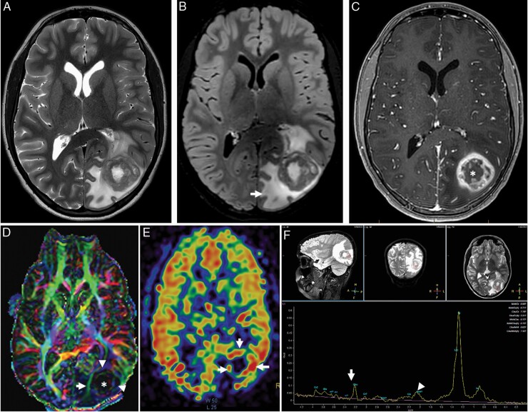
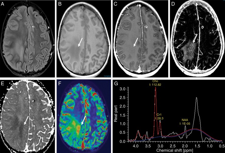
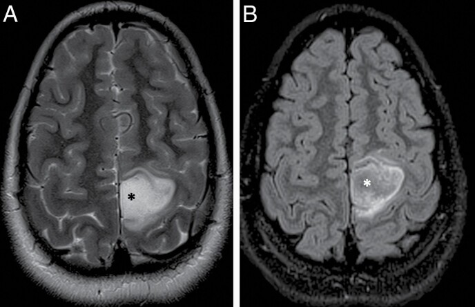
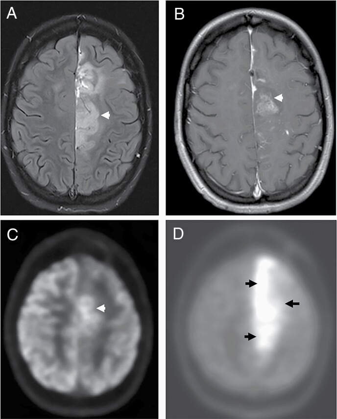
Glioblastoma (GBM) is the most common primary adult intracranial malignancy and carries a dismal prognosis despite an aggressive multimodal treatment regimen that consists of surgical resection, radiation, and adjuvant chemotherapy. Radiographic evaluation, largely informed by magnetic resonance imaging (MRI), is a critical component of initial diagnosis, surgical planning, and post-treatment monitoring. However, conventional MRI does not provide information regarding tumor microvasculature, necrosis, or neoangiogenesis. In addition, traditional MRI imaging can be further confounded by treatment-related effects such as pseudoprogression, radiation necrosis, and/or pseudoresponse(s) that preclude clinicians from making fully informed decisions when structuring a therapeutic approach. A myriad of novel imaging modalities have been developed to address these deficits. Herein, we provide a clinically oriented review of standard techniques for imaging GBM and highlight emerging technologies utilized in disease characterization and therapeutic development.

Keywords: MRI; PET; glioblastoma (GBM); mass spectrometry; radiographic progression; tumor progression.


Figures

Figure 1.
Figure 1.
Typical imaging appearance of glioblastoma in a 12 year old male subject. Axial T2-weighted (A) and fluid attenuated inversion recovery (FLAIR) (B) images demonstrate a heterogeneously T2 hyperintense tumor centered at the left occipital lobe with extensive peritumoral edema (arrow). Axial post contrast T1 weighted imaging demonstrating classic heterogenous, rim enhancing lesion (C). The color coded fractional anisotropy image (D) through the tumor demonstrates distrupted brain architecture at the necrotic tumor core (*). The left inferior longitudinal fasciculus is thinned out and displaced due to the mass effect of the tumor. The arrowheads at the tumor periphery demonstrate low intensity compared to the contralateral hemisphere suggesting, lower FA values. Cerebral blood flow (CBF) from 3D PCASL arterial spin labelling perfusion imaging (E) demonstrates high blood flow at the peripheral enhancing component of the tumor (arrows). The single voxel MR spectroscopy (F) demonstrates severely truncated choline (arrow) and N-Acetyl Aspartate (NAA) peaks (arrowhead) as well as a dominant lipid/lactate peak. This is a typical imaging feature when a spectroscopy voxel includes both tumor and necrotic tissue.
Figure 2.
Figure 2.
Importance of advanced imaging in the evaluation of GBM. Axial T2 FLAIR images (A) through the level of the centrum semiovale demonstrate patchy areas of FLAIR hyperintensity in the deep cerebral white matter that are hypointense (arrow) on the pre-contrast T1-weighted sequence (B) with imperceptible enhancement (arrow) on the post contrast T1-weighted sequence (C). The enhancement is vivid (arrow) on the delta T1 map (D) that also demonstrates low (arrow) ADC value (E) suggesting hypercellularity; high (arrow) cerebral blood volume (CBV) (F) and a tall choline peak compared to NAA on MR spectroscopic imaging (G).
Figure 3.
Figure 3.
T2-FLAIR mismatch sign in an 11 year old female patient with a diagnosis of IDH-mutant, 1p/19q non-codeleted and p53-muted anaplastic astrocytoma in the left parietal lobe. (A) Axial T2-weighted image demonstrating an ill-defined tumor in the left paracentral lobule region with almost homogenous hyperintense T2 signal at the center of the tumor (*) that is mostly hypointense on the corresponding FLAIR image (B).
Figure 4.
Figure 4.
Metabolic imaging of glioblastoma at recurrence. (A) Axial T2 FLAIR images through the level of the centrum semiovale demonstrate a large, ill-defined, heterogenous tumor involving the body of the left cingulate gyrus that demonstrates patchy enhancement on the post contrast T1-weighted sequence. (B)18FDG PET (C) demonstrates hypermetabolism involving only the anterior aspect of the tumor, whereas the 11C-Methionine PET and (D) depicts the entirety of the tumor more conspicuously.
Figure 5.
Figure 5.
Fluorescence guided surgery using 5-ALA induced PpIX. Intraoperative images of a GBM patient undergoing 5-ALA induced PpIX fluorescence guided surgery. (A) Conventional white light image and (B) fluorescence image of the same surgical field of view as (A), demonstrating a region with red-pink fluorescence corresponding to a tumor tissue with significant accumulation of the tumor biomarker, PpIX.
Figure 6.
Figure 6.
Imaging appearance of a glioblastoma following partial resection. Axial T2-weighted images (A) demonstrate a large hematoma (white asterisk) at the anterior aspect of the larger resection cavity (red asterisk) in the right parietal lobe. The arrow points to a residual T2 heterogenous component that demonstrates nodular enhancement (arrow) on the axial post contrast T1 weighted image (B) at the same level. The enhancing tissue demonstrates low ADC values (arrow) (C) suggesting hypercellularity; high cerebral blood volume (CBV) (D) (arrow) suggesting hypervascularity; tall choline peak compared to NAA (E) and a high choline/NAA ratio (F) on MR spectroscopic imaging.
Figure 7.
Figure 7.
Imaging appearances of a glioblastoma following treatment with bevacizumab. Axial diffusion weighted image (A) through the level of centrum semiovale demonstrates a confluent area of high signal (arrowheads) surrounding the cystic resection cavity associated with low values (arrowheads) on the corresponding ADC map suggestive of diffusion restriction developed after treatment with bevacizumab. The precontrast axial T1-weighted image (C) at the same level demonstrates irregular T1 hyperintensities. Enhancement, if any, is hard to appreciate on the post contrast T1 weighted image (D) at the same level, demonstrating the importance of the delta T1 map (not shown).
Figure 8.
Figure 8.
Pseudoresponse following bevacizumab therapy in a 15 year old male patient with recurrent high grade glioma with histone H3.3 G34 mutation in the left temporal lobe. Extensive heterogenous enhancement (A, post-contrast T1-weighted image) and edema with mass effects (D, FLAIR image) have been significantly reduced 3 weeks after the start of bevacizumab therapy (B, E, post-contrast T1-weighted image and FLAIR image, respectively); however, there is evidence of worsening of enhancement (C, motion degraded post-contrast T1-weighted image) and infiltrative tumor component (F, FLAIR image), on a follow-up MRI obtained 8 weeks after the start of bevacizumab therapy.

References

    1. Ostrom QT, Gittleman H, Farah P, et al. . CBTRUS statistical report: primary brain and central nervous system tumors diagnosed in the United States in 2006-2010. Neuro Oncol. 2013;15(Suppl 2):ii1–i56. - PMC - PubMed
    1. Stupp R, Mason WP, van den Bent MJ, et al. . Radiotherapy plus concomitant and adjuvant temozolomide for glioblastoma. N Engl J Med. 2005;352(10):987–996. - PubMed
    1. Herrlinger U, Tzaridis T, Mack F, et al. . Lomustine-temozolomide combination therapy versus standard temozolomide therapy in patients with newly diagnosed glioblastoma with methylated MGMT promoter (CeTeG/NOA-09): a randomised, open-label, phase 3 trial. Lancet. 2019;393(10172):678–688. - PubMed
    1. Stupp R, Taillibert S, Kanner A, et al. . Effect of tumor-treating fields plus maintenance temozolomide vs maintenance temozolomide alone on survival in patients with glioblastoma: a randomized clinical trial. JAMA. 2017;318(23):2306–2316. - PMC - PubMed
    1. Lara-Velazquez M, Al-Kharboosh R, Jeanneret S, et al. . Advances in brain tumor surgery for Glioblastoma in adults. Brain Sci. 2017;7(12). - PMC - PubMed

LinkOut - more resources