. 2022 Jan 13:2:828697.
doi: 10.3389/fragi.2021.828697. eCollection 2021.

Obesity Accelerates Age-Associated Defects in Human B Cells Through a Metabolic Reprogramming Induced by the Fatty Acid Palmitate

Affiliations

Obesity Accelerates Age-Associated Defects in Human B Cells Through a Metabolic Reprogramming Induced by the Fatty Acid Palmitate

Daniela Frasca et al. Front Aging. .

Abstract

We have measured the secretion of autoimmune antibodies in plasma samples and in culture supernatants of blood-derived B cells from four groups of individuals: young lean (YL), elderly lean (EL), young obese (YO) and elderly obese (EO). We found secretion comparable in YO and EL individuals, suggesting that obesity accelerates age-associated defects in circulating B cells. To define at least one possible molecular pathway involved, we used an in vitro model in which B cells from YL and EL individuals have been stimulated with the Fatty Acid (FA) palmitate, the most common saturated FA in the human body. The rationale to use palmitate is that there is a chronic increase in circulating levels of palmitate, due to increased spontaneous lipolysis occurring during aging and obesity, and this may induce autoimmune B cells. Results herein show that in vitro incubation of B cells from YL and EL individuals with the FA palmitate induces mRNA expression of T-bet, the transcription factor for autoimmune antibodies, as well as secretion of autoimmune IgG antibodies, with B cells from YL individuals looking similar to B cells from EL individuals, confirming our initial hypothesis. The generation of autoimmune B cells in the presence of the FA palmitate was found to be associated with a metabolic reprogramming of B cells from both YL and EL individuals. These results altogether show the critical role of the FA palmitate in inducing human B cell immunosenescence and show for the first time the importance of metabolic pathways in this process.

Keywords: B cells; aging; autoimmunity; metabolism; obesity.


Conflict of interest statement

The authors declare that the research was conducted in the absence of any commercial or financial relationships that could be construed as a potential conflict of interest.

Figures

FIGURE 1
FIGURE 1
Effect of aging and obesity on the secretion of autoimmune IgG antibodies. Plasma samples were isolated from YL, EL, YO and EO individuals and analyzed by ELISA for the presence of AD-specific (A) or MDA-specific (D) IgG. B cells, isolated from the same individuals in A using magnetic beads, were stimulated for 8 days with CpG, supernatants were collected after 8 days and AD-specific (B) or MDA-specific (E) IgG were measured by ELISA. Correlations of AD-specific (C) and MDA-specific (F) IgG in plasma and culture supernatants. Mean comparisons between groups were performed by two-way ANOVA: *p < 0.05, **p < 0.01, ***p < 0.001, ****p < 0.0001. Correlations were calculated by bivariate Pearson’s correlation analyses.
FIGURE 2
FIGURE 2
Effect of aging and obesity on lipid accumulation by B cells. PBMC were isolated from YL, EL, YO and EO individuals and stained first with the Deep Red Neutral Lipid Stain LipidTOX and then with anti-CD45, anti-CD19, and the Live/Dead detection kit. (A) MFI profiles of one representative donor/group. MFI values for negative controls (without the addition of LipidTOX are, respectively: 915 (YL), 1214 (YO), 837 (EL), 1025 (EO). (B) MFI data from all donors. Mean comparisons between groups were performed by two-way ANOVA: **p < 0.01, ****p < 0.0001.
FIGURE 3
FIGURE 3
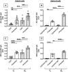
Palmitate in vitro stimulates the secretion of AD-specific autoimmune antibodies in B cells from both YL and EL individuals. B cells, isolated from YL and EL individuals using magnetic beads, were stimulated with CpG, alone or in the presence of palmitate, for 8 days. Then supernatants were collected and analyzed by ELISA for the presence of AD-specific (A) or MDA-specific (C) IgG. Naïve B cells, sorted from the blood of tha same YL and EL individuals, were stimulated with CpG/anti-Ig, alone or in the presence of palmitate, for 8 days. Culture supernatants were tested by ELISA for the presence of AD-specific (B) or MDA-specific (D) IgG. Mean comparisons between groups were performed by two-way ANOVA: **p < 0.01, ***p < 0.001, ****p < 0.0001.
FIGURE 4
FIGURE 4
Palmitate in vitro increases mRNA expression of T-bet, the transcription factor for autoimmune antibody production, in B cells from both YL and EL individuals. Total B cells (A) and naïve B cells (B), isolated from the same YL and EL individuals in Figure 3, were stimulated with CpG or CpG/anti-Ig respectively, alone or in the presence of palmitate, for 1 day. Then the mRNA was extracted and qPCR reactions run to evaluate the expression of tbx21. Results show qPCR values (2−ΔCt) of tbx21, normalized to GAPDH. Mean comparisons between groups were performed by two-way ANOVA: ***p < 0.001, ****p < 0.0001.
FIGURE 5
FIGURE 5
Palmitate in vitro induces a hyper-metabolic phenotype in B cells from both YL and EL individuals. Total B cells from the same YL and EL individuals in Figures 3, 4 were stimulated with CpG, alone or in the presence of palmitate for 6 h, and evaluated for the mRNA expression of HK-2, LDHA, PDHX and ACACB. Heatmap shows qPCR values (2−ΔCt) of metabolic markers, normalized to GAPDH, from four individuals/group.
FIGURE 6
FIGURE 6
Palmitate in vitro induces higher ECAR in B cells from both YL and EL individuals. Total B cells from the same YL and EL individuals in Figures 3, 4 were stimulated with CpG, alone or in the presence of palmitate for 6 h, and seeded into the wells of an extracellular flux analyzer at the concentration of 2 × 10⁵/well in triplicate and run in a mitostress test. Bottom two groups refer to: B cells from YL individuals. Top two groups refer to: B cells from EL individuals.

Similar articles

Cited by

References

    1. Apovian C. M., Gokce N. (2012). Obesity and Cardiovascular Disease. Circulation 125 (9), 1178–1182. 10.1161/circulationaha.111.022541 - DOI - PMC - PubMed
    1. Bernasconi N. L., Traggiai E., Lanzavecchia A. (2002). Maintenance of Serological Memory by Polyclonal Activation of Human Memory B Cells. Science 298 (5601), 2199–2202. 10.1126/science.1076071 - DOI - PubMed
    1. Bonadonna R. C., Groop L. C., Simonson D. C., DeFronzo R. A. (1994). Free Fatty Acid and Glucose Metabolism in Human Aging: Evidence for Operation of the Randle Cycle. Am. J. Physiol. 266 (3 Pt 1), E501–E509. 10.1152/ajpendo.1994.266.3.E501 - DOI - PubMed
    1. Bradford M. M. (1976). A Rapid and Sensitive Method for the Quantitation of Microgram Quantities of Protein Utilizing the Principle of Protein-Dye Binding. Anal. Biochem. 72, 248–254. 10.1016/0003-2697(76)90527-3 - DOI - PubMed
    1. Buffa S., Pellicanò M., Bulati M., Martorana A., Goldeck D., Caruso C., et al. (2013). A Novel B Cell Population Revealed by a CD38/CD24 Gating Strategy: CD38(−)CD24(−) B Cells in Centenarian Offspring and Elderly People. Age 35 (5), 2009–2024. 10.1007/s11357-012-9488-5 - DOI - PMC - PubMed

LinkOut - more resources