. 2022 Aug 1;128(2):339-349.
doi: 10.1152/jn.00063.2022. Epub 2022 Jul 13.

Oropharyngeal capsaicin exposure improves infant feeding performance in an animal model of superior laryngeal nerve damage

Affiliations

Oropharyngeal capsaicin exposure improves infant feeding performance in an animal model of superior laryngeal nerve damage

Chloe E Edmonds et al. J Neurophysiol. .

Abstract

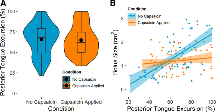
Sensorimotor feedback is critical to safe and effective swallowing. Because of this, sensory interventions have the potential to treat dysphagia. One such treatment may be found in capsaicin, which activates the internal branch of the superior laryngeal nerve (iSLN). The iSLN initiates the pharyngeal swallow, and a more sensitive iSLN should more readily elicit swallowing and improve swallow safety. We explored the neurophysiological mechanism by which capsaicin improves swallow performance using an infant pig model with a unilateral iSLN lesion. Using high-speed videofluoroscopy, we collected oropharyngeal kinematic data while pigs suckled on bottles, before and after applying capsaicin to the posterior tongue and valleculae. We found that capsaicin application decreased maximal bolus sizes, which improved swallow safety. Furthermore, capsaicin improved performance when infant pigs swallowed more moderately sized boluses. However, capsaicin did not change swallow frequency, the number of sucks prior to each swallow, nor total pharyngeal transit time (TPT). Similarly, excursions of the hyoid, thyroid, and posterior tongue were unchanged. TPT and hyoid and thyroid excursions maintained relationships with bolus size post-capsaicin, suggesting that these variables are less sensitive to sensory intervention. The timing and extent of posterior tongue movement were only correlated with bolus size pre-capsaicin, which could imply that capsaicin fundamentally changes in relationships between tongue movements and bolus size. Our results provide insight into the neural control of swallowing and capsaicin's mechanism of action, and suggest that capsaicin may be beneficial in treating acute infant dysphagia.NEW & NOTEWORTHY Chemical sensory interventions alter swallow physiology, which is well-documented in adults but relatively unexplored in infants. Using videofluoroscopy, we found that capsaicin exposure limited infant pigs' bolus sizes to improve swallow performance without changing swallow frequency. Capsaicin increased the likelihood of safe swallowing with more moderately sized boluses and changed relationships between bolus size and tongue movements, which may impact performance. This work highlights the potential role of capsaicin in treating acute infant dysphagia.

Keywords: capsaicin; infant swallowing; kinematics; pediatrics; sensorimotor feedback.


Conflict of interest statement

No conflicts of interest, financial or otherwise, are declared by the authors.

Figures

None
Graphical abstract
Figure 1.
Figure 1.
Frequency of swallowing in infant pigs prior to (left, blue) and following (right, orange) the application of capsaicin to the soft palate and valleculae (pre-capsaicin n = 76 swallows, post-capsaicin n = 85). No significant changes in swallow frequency were observed following capsaicin application (χ2 = 0.926, P = 0.336). Large black circles represent means; box and whisker plots show median and interquartile range; width of each plot indicates the frequency distribution of the data along the y-axis.
Figure 2.
Figure 2.
Total pharyngeal transit (TPT) time (A) as it is affected by capsaicin application and (B) as it relates to bolus size in both experimental conditions (pre-capsaicin n = 81 swallows, post-capsaicin n = 90 swallows). In both panels, pre-capsaicin data are shown in blue, and post-capsaicin data are shown in orange. There is a significant decrease in TPT following capsaicin application (χ2 = 6.86, P = 0.009), and TPT is related to bolus size both prior to (R2 = 0.123, P = 7.70 × 10⁻⁴) and following (R2 = 0.640, P = 2.20 × 10⁻¹⁶) capsaicin application. In A, large black circles represent means; box and whisker plots show median and interquartile range; width of each plot indicates the frequency distribution of the data along the y-axis; brackets connecting plots show significant differences identified by linear mixed effects models.
Figure 3.
Figure 3.
The effect of capsaicin on hyoid excursion during the swallow (A), timing of peak hyoid elevation as a percentage of swallow duration (B), thyroid excursion during the swallow (C), and timing of peak thyroid elevation as a percentage of swallow duration (D). In all panels, pre-capsaicin data are shown in blue, while post-capsaicin data are shown in orange (pre-capsaicin n = 81 swallows, post-capsaicin n = 89 swallows). Hyoid excursion and thyroid excursion significantly decreased (χ2 = 15.3, P = <0.001 and χ2 = 5.85, P = 0.016 respectively) following capsaicin application, while the timing of peak hyoid and thyroid elevation were consistent (χ2 = 0.042, P = 0.837 and χ2 = 0.508, P = 0.476 respectively) across treatments. Large black circles represent means; box and whisker plots show median and interquartile range; width of each plot indicates the frequency distribution of the data along the y-axis; and brackets connecting plots show significant differences identified by linear mixed effects models.
Figure 4.
Figure 4.
Excursion of the posterior tongue, given as a percentage of each infant pig’s maximum posterior tongue excursion (pre-capsaicin n = 62 swallows, post-capsaicin n = 67 swallows). A: the effect of capsaicin application on scaled posterior tongue excursion. B: scaled posterior tongue excursion as it relates to bolus size in both experimental conditions. In both panels, pre-capsaicin data are shown in blue, and post-capsaicin data are shown in orange. Scaled posterior tongue excursion did not change (χ2 = 0.007, P = 0.936) following capsaicin application. The relationship between scaled posterior tongue excursion and bolus size was significant prior to (R2 = 0.431, P = 4.25 × 10⁻⁹) but not following (R2 = 0.010, P = 0.201) capsaicin application. In A, large black circles represent means; box and whisker plots show median and interquartile range; width of each plot indicates the frequency distribution of the data along the y-axis.
Figure 5.
Figure 5.
Posterior tongue ratio (PTR) as it is affected by capsaicin application (A) and as it relates to bolus size in both experimental conditions (B) (pre-capsaicin n = 62 swallows, post-capsaicin n = 67 swallows). In both panels, pre-capsaicin data are shown in blue, and post-capsaicin data are shown in orange. PTR significantly decreased (χ2 = 4.24, P = 0.040) following capsaicin application. The relationship between PTR and bolus size was significant prior to (R2 = 0.218, P = 7.62 × 10⁻⁵) but not following (R2 = −0.015, P = 0.966) capsaicin application. In A, large black circles represent means; box and whisker plots show median and interquartile range; width of each plot indicates the frequency distribution of the data along the y-axis; brackets connecting plots show significant differences identified by linear mixed effects models.
Figure 6.
Figure 6.
The relationship between capsaicin application and bolus size in cm2 (pre-capsaicin n = 92, post-capsaicin n = 95). The dots shown on the left represent swallows made pre-capsaicin, while those on the right represent swallows made post-capsaicin. The color of each dot indicates the degree of airway compromise during each swallow: safe swallows shown in green, swallows with airway penetration shown in yellow, and swallows with aspiration shown in red.

References

    1. Kandel ER, Schwartz JH. The organization and planning of movement. In: Principles of Neural Science, edited by Kandel ER, Schwartz JH, Jessell TM, Siegelbaum SA, Hudspeth AJ. New York: McGraw Hill Medical, 2013, p. 743–768.
    1. Wolpert DM, Goodbody SJ, Husain M. Maintaining internal representations: the role of the human superior parietal lobe. Nat Neurosci 1: 529–533, 1998. doi:10.1038/2245. - DOI - PubMed
    1. Humbert IA, German RZ. New directions for understanding neural control in swallowing: the potential and promise of motor learning. Dysphagia 28: 1–10, 2013. doi:10.1007/s00455-012-9432-y. - DOI - PMC - PubMed
    1. Steele CM, Miller AJ. Sensory input pathways and mechanisms in swallowing: a review. Dysphagia 25: 323–333, 2010. doi:10.1007/s00455-010-9301-5. - DOI - PMC - PubMed
    1. Altman KW, Richards A, Goldberg L, Frucht S, McCabe DJ. Dysphagia in stroke, neurodegenerative disease, and advanced dementia. Otolaryngol Clin North Am 46: 1137–1149, 2013. doi:10.1016/j.otc.2013.08.005. - DOI - PubMed

Publication types