Review
. 2022 Sep;158(3):253-260.
doi: 10.1007/s00418-022-02124-x. Epub 2022 Jul 12.

Megapinosomes and homologous structures in hematopoietic cells

Affiliations
Review

Megapinosomes and homologous structures in hematopoietic cells

Andrea Bauer et al. Histochem Cell Biol. 2022 Sep.

Abstract

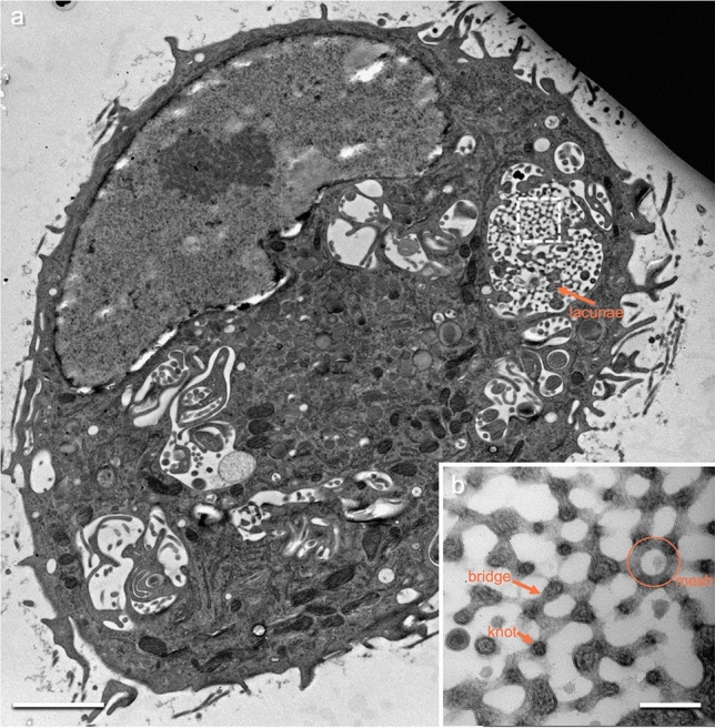
Megapinosomes are endocytic organelles found in human macrophage colony-stimulating factor (M-CSF) monocyte-derived M macrophages. They are large (several microns) and have a complex internal structure that is connected with the cytosol and consists of interconnected knots and concave bridges with sizes in the range of 100 nm. We called this structure trabecular meshwork. The luminal part of the megapinosome can be connected with luminal tubules and cisterns that form the megapinosome complex. The structures are especially well visible in scanning electron tomography when macrophages are prepared by high-pressure freezing and freeze substitution. Our research received a new impulse after studying the literature on hematopoietic cells, where very similar, most likely homologous, structures have been published in peritoneal macrophages as well as in megakaryocytes and blood platelets. In platelets, they serve as membrane storage that is used for structural changes of platelets during activation.

Keywords: Blood platelets; Electron microscopy; High-pressure freezing; M macrophages; Megakaryocytes; Megapinosomes; STEM tomography.


Conflict of interest statement

The authors declare no competing interests.

The authors declare no conflicts of interest.

Figures

Fig. 1
Fig. 1
M macrophage high-pressure frozen, freeze-substituted, and imaged in TEM, showing a megapinosome and elements of megapinosome complexes. b Higher magnification of the megapinosome in a, showing the dense trabecular meshwork topologically equivalent to the cytosol and the bright luminal part. The two parts are separated by a membrane. The trabecular meshwork consists of interconnected knots and concave bridges. In the 2D TEM image, one knot connects via bridges with three adjacent knots. Usually, five to six bridges form a mesh surrounding a lumen with an average diameter of 110 nm. Another cytosolic structure within the megapinosome are the lacunae. The scale bars represent 2 µm in a and 200 nm in b
Fig. 2
Fig. 2
Megapinosome complex in 3D, obtained from STEM tomography data. a Virtual section showing the trabecular meshwork on top. b The same virtual section with the trabecular meshwork that is part of the cytosol in red, and the luminal part in blue. c The segmented (red) trabecular meshwork in 3D. d The corresponding highly branched channel system of the luminal part of the megapinosome in 3D. It is connected with cisterns with a continuous 30-nm gap, as well as with tubules with an average diameter of 165 nm. The trabecular meshwork as well as the luminal part are continuous, meaning that, from each point of the trabecular meshwork (in red), each other point in the trabecular meshwork can be reached without passing through a membrane, and the same is true for the luminal part in blue. The scale bar represents 2 µm; all images at the same magnification. A movie of these data is available in the Supplementary Material
Fig. 3
Fig. 3
A large portion of a megapinosome complex segmented from a total of 17 a and 9 c STEM tomograms from two adjoint 600-nm-thick sections. The bright-blue parts form a continuum. The dark-blue parts are not connected with the bright-blue parts in the segmented area. They may, however, be connected in another section plane. In brown, the structures are segmented that are endocytic but not connected to the megapinosome. Many of these structures most likely represent canonical endosomes. b is an enlarged portion of the encircled area in a. The blue luminal part as well as the brown endosomes are transparent so that the yellow endocytosed gold particles become visible. The gold particles are markers for endocytic structures. The yellow arrows depict the gold particles in the luminal part of the megapinosome. The scale bars represent 2 µm
Fig. 4
Fig. 4
Several megapinosomes connected by tubules. Some of these tubules reach into the extracellular space. a is an overview of a M macrophage with the segmentation of a portion of the megapinosome complex. b Higher magnification of the encircled area in a, showing the connection of two megapinosomes with a tubule. c Virtual section of a tomogram where a tubule reaches the extracellular space (blue arrow). d Virtual section with the segmented luminal part of the megapinosome complex. The scale bars represent 2 µm
Fig. 5
Fig. 5
Portion of the megapinosome complex in Fig. 3. Beside the luminal part of the megapinosome (in blue), we found additional luminal structures that are not connected to the megapinosome lumen. They did not contain gold particles and are darker than the luminal parts of the megapinosome segmented in blue. Since we found two categories of these structures, we segmented the structures in the trabecular meshwork in yellow and the part adjacent to the trabecular meshwork in green. Whereas the green structure looks as expected for small tubular and vesicular structures, the yellow part shows polymorph shapes. Whether these structures are homologous to the “dense tubular system” in blood platelets needs to be further investigated. The scale bars represent 500 nm
Fig. 6
Fig. 6
Hypothetical schematic representation of the topological relationship of megapinosomes and homologous structures. Only a portion of the structure is shown, similar to Fig. 1a. The cytosolic parts are red, the endocytic luminal parts are blue, and the most likely endoplasmic reticulum-derived parts are yellow. The membranes to endocytic luminal structure are black, and the membranes to the luminal structure probably homologous to the DTS are grey. a An M macrophage’s megapinosome with the cytosolic trabecular meshwork. b Homologous structures in peritoneal macrophages from guinea pigs. In contrast to a, the endoplasmic reticulum-derived luminal part forms a continuous structure. The same is true for blood platelets in c, with the cytosolic parts in red, the OCS in blue, and the DTS in yellow. The scale bar represents 200 nm

References

    1. Alberts B, Johnson A, Lewis J, Morgan D, Raff M, Roberts K, Walter P. Molecular biology of the cell. 6. New York: Garland; 2014.
    1. Bauer A, Subramanian N, Villinger C, Frascaroli G, Mertens T, Walther P. Megapinocytosis: a novel endocytic pathway. Histochem Cell Biol. 2016;145:617–627. doi: 10.1007/s00418-015-1395-22016. - DOI - PubMed
    1. Bauer A, Frascaroli G, Walther P. Megapinosome: morphological description of a novel organelle. Struct Biol. 2020;210:107505. doi: 10.1016/j.jsb.2020.107505. - DOI - PubMed
    1. Behnke O. Electron microscopic observations on the membrane systems of the rat blood platelet. Anat Rec. 1967;158(2):121–137. doi: 10.1002/AR.1091580203. - DOI - PubMed
    1. Behnke O. An electron microscope study of the megacaryocyte of the rat bone marrow. I. The development of the demarcation membrane system and the platelet surface coat. J Ultrastruct Res. 1968;24(5):412–433. doi: 10.1016/S0022-5320(68)80046-2. - DOI - PubMed