Review
. 2022 Sep;119(3):470-479.
doi: 10.36660/abc.20210052.

Critical Analysis and Limitations of the Diagnosis of Heart Failure with Preserved Ejection Fraction (HFpEF)

[Article in English, Portuguese]
Affiliations
Review

Critical Analysis and Limitations of the Diagnosis of Heart Failure with Preserved Ejection Fraction (HFpEF)

[Article in English, Portuguese]
Viviane Tiemi Hotta et al. Arq Bras Cardiol. 2022 Sep.

Abstract

With the increase in the population's life expectancy and the higher frequency of risk factors such as obesity, hypertension and diabetes, an increase in the prevalence of heart failure with preserved ejection fraction (HFpEF) is expected. However, to date, the diagnosis and treatment of patients with HFpEF remain challenging. The syndromic diagnosis of HFpEF includes several etiologies and diseases with specific treatments but has points in common regarding the clinical presentation, laboratory evaluation related to biomarkers, such as BNP and NT-ProBNP, and echocardiographic evaluation of cardiac remodeling and left ventricular diastolic filling pressures. Extensive randomized clinical trials involving the treatment of this condition have failed to demonstrate benefits to the patient, making it necessary to reflect on the diagnosis, mechanisms of morbidity, mortality and reversibility in this syndrome. In this review, the current concepts, controversies and challenges, especially regarding diagnosis, will be addressed, critically analyzing the European Heart Failure Association score for the diagnosis of HFpEF.

Com o aumento da expectativa de vida da população e a maior frequência de fatores de risco como obesidade, hipertensão arterial e diabetes, espera-se um aumento na prevalência de insuficiência cardíaca com fração de ejeção preservada (ICFEp). Entretanto, no momento, o diagnóstico e o tratamento de pacientes com ICFEp permanecem desafiadores. O diagnóstico sindrômico de ICFEp inclui diversas etiologias e doenças com tratamentos específicos, mas que apresentam pontos em comum em relação à apresentação clínica e à avaliação laboratorial no que diz respeito aos biomarcadores como BNP e NT-ProBNP, à avaliação ecocardiográfica do remodelamento cardíaco e às pressões de enchimento diastólico ventricular esquerdo. Extensos ensaios clínicos randomizados envolvendo a terapia nesta síndrome falharam na demonstração de benefícios para o paciente, fazendo-se necessária uma reflexão acerca do diagnóstico, dos mecanismos de morbidade, da taxa de mortalidade e da reversibilidade. Na revisão, serão abordados os conceitos atuais, as controvérsias e, especialmente, os desafios no diagnóstico da ICFEp através de uma análise crítica do escore da European Heart Failure Association.


Conflict of interest statement

Potencial Conflito de Interesse

Declaro não haver conflito de interesses pertinentes.

Figures
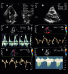
Figura 2
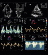
Figura 2. Critérios ecocardiográficos morfológicos (A e B) e funcionais (C a F) para aplicação do algoritmo diagnóstico para pacientes com suspeita de ICFEp. Os critérios morfológicos incluem a medida do volume indexado do átrio esquerdo (A), e o cálculo do índice de massa miocárdica e a espessura relativa de parede (B). Os critérios funcionais incluem a relação E/e’, calculada a partir da medida de onda E ao Doppler mitral (C) (v = 73,7 cm/s) e ondas e’ ao Doppler tecidual septal (D) (v = 8,6 cm/s) e lateral (E) (v = 16,1 cm/s), além da velocidade de pico do jato de regurgitação tricúspide (v = 2,07 cm/s) para estimar a medida da pressão sistólica de artéria pulmonar (F). v: velocidade.
Figura 1
Figura 1. Fluxograma para avaliação clínica, integrando fatores de risco, exame físico, avaliação de biomarcadores e análise ecocardiográfica. DM: diabetes mellitus; ECG: eletrocardiograma; FA: fibrilação atrial; IT: insuficiência tricúspide; PSAP: pressão sistólica em artéria pulmonar; VolAEi: volume do átrio esquerdo indexado; IMVE: índice de massa do ventrículo esquerdo; ERP: espessura relativa da parede ventricular; BNP: peptídeo natriurético atrial; GLS: strain global longitudinal; ICFEp: insuficiência cardíaca com fração de ejeção preservada.* Critério menor não deve ser contabilizado dentro do mesmo domínio.
Figure 1
Figure 1. Clinical evaluation flowchart integrating risk factors, physical examination, evaluation of biomarkers and echocardiographic analysis. AF: atrial fibrillation;DM: diabetes mellitur; TR: tricuspid regurgitation; LAVI: left atrial volume index; LVMI: left ventricular mass index; RWT: left ventricular relative wall thickness; BNP: B-type natriuretic peptide; GLS: global longitudinal strain; PASP: pulmonary artery systolic pressure; HFpEF: heart failure with preserved ejection fraction. * Minor criterion should not be counted within the same domain.
Figure 2
Figure 2. Morphological (A and B) and functional (C to F) echocardiographic criteria for the diagnostic algorithm application in patients with suspected HFpEF. The morphological criteria included the measurement of the left atrial volume index (A) and the calculation of the myocardial mass index and relative wall thickness (B). The functional criteria include the E/e´ ratio calculated from the measurement of the E wave on mitral Doppler (C) (v = 73.7 cm/s) and e´ wave septal (D) (v = 8.6 cm/s) and lateral (E) (v = 16.1 cm/s) velocities on tissue Doppler, in addition to the tricuspid regurgitation jet peak velocity (v = 2.07 cm/s) to measure the pulmonary artery systolic pressure (F). v: velocity.
Figura 3
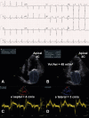
Figura 3. Exemplo ilustrativo de aplicação do escore diagnóstico em indivíduo com suspeita de ICFEp. Paciente do sexo feminino, 64 anos, com antecedentes de síndrome metabólica (obesidade grau III – IMC: 35,6, HAS e DM) e queixa de dispneia aos mínimos esforços (CF III NYHA). Ao ECG (acima), observa-se sinais de hipertrofia ventricular esquerda pelos critérios de Sokolow-Lyon. Apresenta ao ETT espessura do septo interventricular e parede posterior de 12 mm e IMVE: 105 g/m2 (1 ponto). Volume indexado do átrio esquerdo estimado ao corte apical 4C (acima à esquerda) e apical 2C (acima à direita) em 48 ml/m2 (2 pontos). Doppler tecidual evidencia onda e’ septal = 4 cm/s (abaixo à esquerda) e onda e’ lateral = 3 cm/s (abaixo à direita) (2 pontos). Assim, aplicando-se o escore para o diagnóstico de ICFEp, a paciente apresenta 5 pontos e, portanto, diagnóstico de ICFEp. VolAEi: volume do átrio esquerdo indexado.
Figura 4
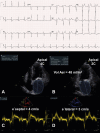
Figura 4. Paciente de 78 anos com obesidade, hipertensão arterial sistêmica, diabetes melito tipo 2 e fibrilação atrial paroxística em CF II NYHA. Ao ETT, apresenta AE = 50 mm, volume indexado de AE = 38 ml/m2 (2 pontos), índice de massa VE: 89 g/m2 e espessura relativa de parede = 0,47 e PSAP não analisável, relação E/e’ = 8,8 e BNP = 367 pg/ml (2 pontos). Aplicando-se o escore para o diagnóstico de ICFEp, paciente apresenta 4 pontos e, portanto, diagnóstico inconclusivo de ICFEp.
Figure 3
Figure 3. Illustrative example of the diagnostic score application in a patient with suspected HFpEF. A 64-year-old female patient had a history of metabolic syndrome (obesity grade III – Body Mass Index: 35.6, systemic arterial hypertension and diabetes mellitus) and complaints of dyspnea on minimal effort (FC III NYHA). The ECG (above) showed signs of left ventricular hypertrophy according to the Sokolow-Lyon criteria. The TTE displays an interventricular septum and posterior wall thickness of 12 mm and LVMI: 105 g/m2 (1 point). The left atrial volume index estimated at the apical 4C (top left) and apical 2C (top right) views was 48 mL/m2 (2 points). Tissue Doppler shows e’ wave septal velocity = 4 cm/s (bottom left) and lateral e´ wave velocity = 3 cm/s (bottom right) (2 points). Thus, by applying the score for the diagnosis of HFpEF, the patient attained 5 points and, therefore, the HFpEF diagnosis was confirmed.
Figure 4
Figure 4. A 78-year-old patient with obesity, systemic arterial hypertension, type 2 diabetes mellitus, paroxysmal atrial fibrillation, in NYHA FC II. On the TTE, LA = 50 mm, LAVI = 38 mL/m2 (2 points), LV mass index: 89 g/m2, LV relative wall thickness = 0.47, non-analyzable PASP and E/e´ ratio = 8.8. BNP = 367 pg/mL (2 points). After applying the score for the diagnosis of HFpEF, the patient attained 4 points and, therefore, had an inconclusive diagnosis of HFpEF.

References

    1. Pieske B, Tschöpe C, de Boer RA, Fraser AG, Anker SD, Donal E. How to diagnose heart failure with preserved ejection fraction: the HFA-PEFF diagnostic algorithm: a consensus recommendation from the Heart Failure Association (HFA) of the European Society of Cardiology (ESC) 10.1093/eurheartj/ehz641Eur Heart J. 2019;40(40):3297–3317. - PubMed
    1. Van Riet EES, Hoes AW, Wagenaar KP, Limburg A, Landman MA, Rutten FH, et al. Epidemiology of heart failure: the prevalence of heart failure and ventricular dysfunction in older adults over time. A systematic review. 10.1002/ejhf.483Eur J Heart Fail. 2016;18(3):242–252. - PubMed
    1. Seferovic PM, Petrie MC, Filippatos GS, Anker SD, Rosano G, Bauersachs J, et al. Type 2 diabetes mellitus and heart failure: a position statement from the Heart Failure Association of the European Society of Cardiology. 10.1002/ejhf.1170Eur J Heart Fail. 2018;20(5):853–872. - PubMed
    1. Ponikowski P, Voors AA, Anker SD, Bueno H, Cleland JG, Coats AJ, et al. 2016 ESC Guidelines for the diagnosis and treatment of acute and chronic heart failure: the task force for the diagnosis and treatment of acute and chronic heart failure of the European Society of Cardiology (ESC). Developed with the special contribution of the Heart Failure Association (HFA) of the ESC. 10.1002/ejhf.592Eur J Heart Fail. 2016;18(8):891–975. - PubMed
    1. Fernandes SL, Carvalho RR, Santos LG, Sá FM, Ruivo C, Mendes SL, et al. Pathophysiology and treatment of heart failure with preserved ejection fraction: state of the art and prospects for the future. 10.36660/abc.20190111Arq Bras Cardiol. 2020;114(1):120–129. - PMC - PubMed