. 2022 Aug 1;128(2):405-417.
doi: 10.1152/jn.00469.2021. Epub 2022 Jul 13.

Laryngeal and swallow dysregulation following acute cervical spinal cord injury

Affiliations

Laryngeal and swallow dysregulation following acute cervical spinal cord injury

Teresa Pitts et al. J Neurophysiol. .

Abstract

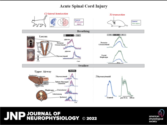
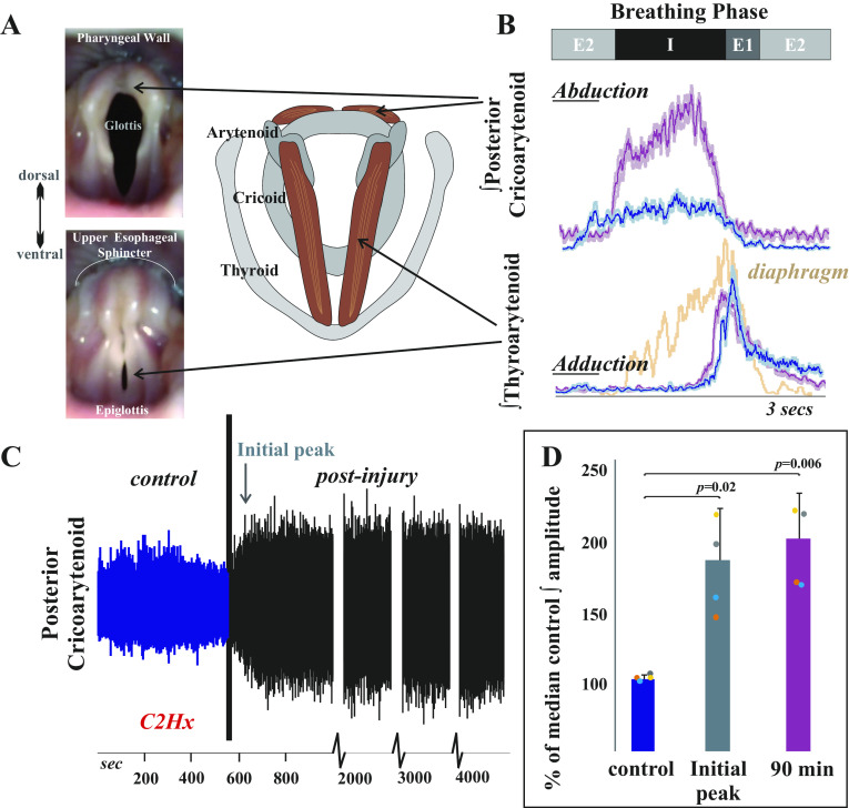
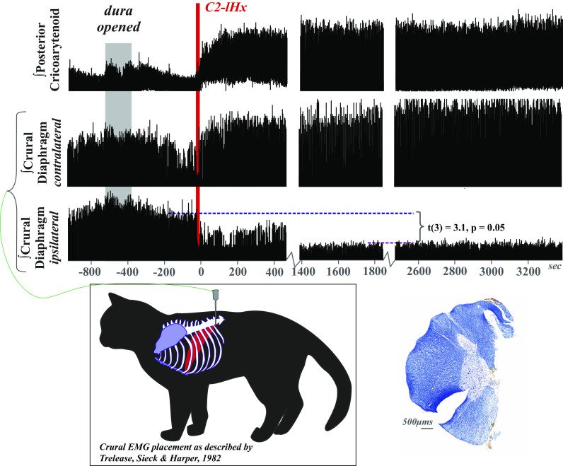
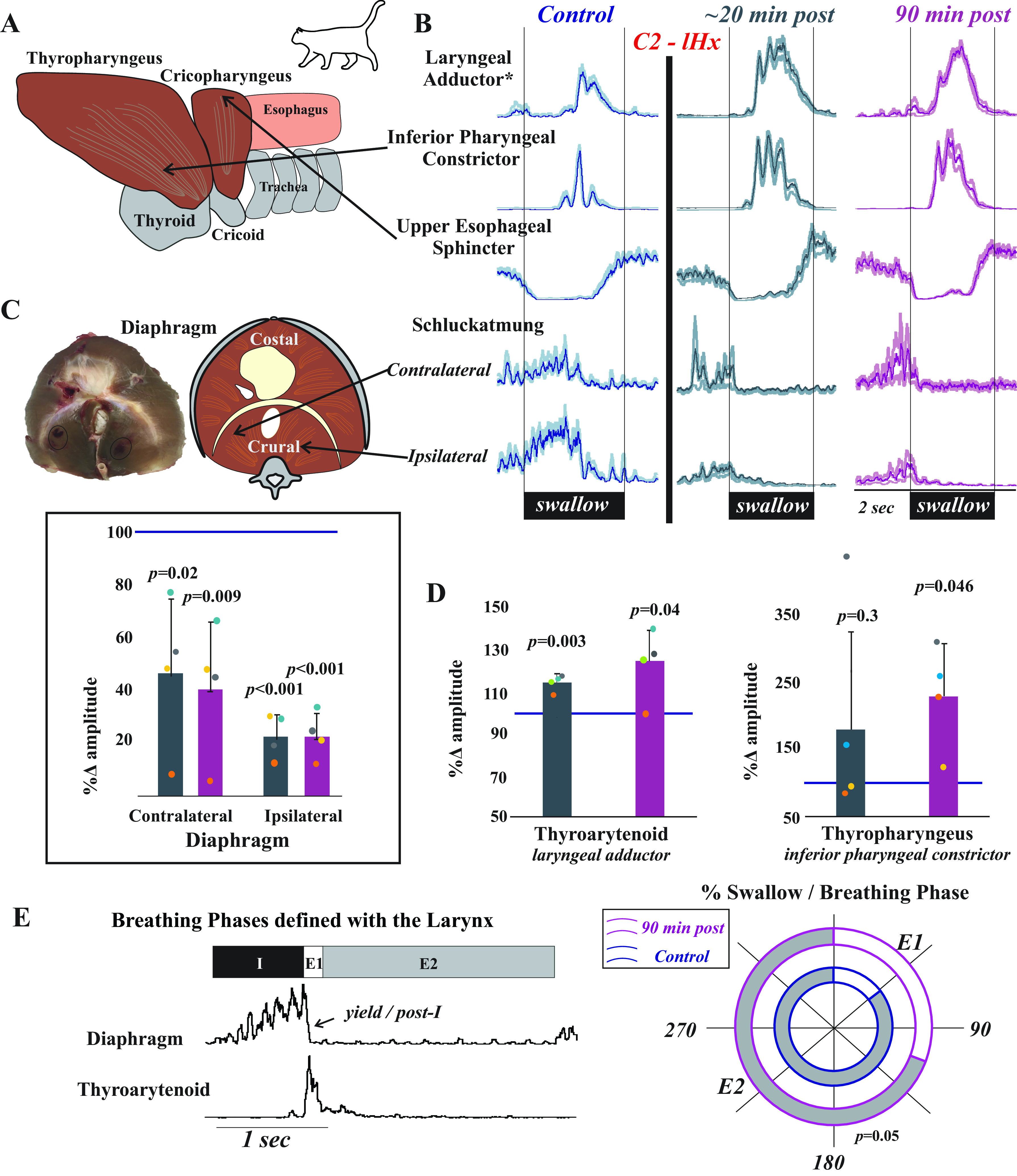
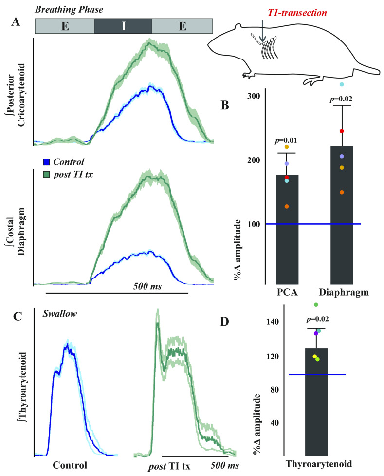
Laryngeal function is vital to airway protection. Although swallow is mediated by the brainstem, the mechanism underlying the increased risk of dysphagia after cervical spinal cord injury (SCI) is unknown. We hypothesized that: 1) loss of descending phrenic drive affects swallow and breathing differently, and 2) loss of ascending spinal afferent information alters swallow regulation. We recorded electromyograms (EMGs) from upper airway and chest wall muscles in freely breathing pentobarbital-anesthetized cats and rats. Laryngeal abductor activity during inspiration increased about twofold following C2 lateral hemisection. Ipsilateral to the injury, the crural diaphragm EMG amplitude was reduced during breathing (62 ± 25% change postinjury), but no animal had complete termination of activity; 75% of animals had increased contralateral diaphragm recruitment, but this did not reach significance. During swallow, laryngeal adductor and pharyngeal constrictor muscles increased activity, and diaphragm activity was bilaterally suppressed. This was unexpected because of the ipsilateral-specific response during breathing. Swallow-breathing coordination was disrupted by injury, and more swallows occurred during early expiration. Finally, to determine if the chest wall is a major source of feedback for laryngeal regulation, we performed T1 total transections in rats. As in the C2 lateral hemisection, inspiratory laryngeal recruitment was the first feature noted after injury. In contrast to the C2 lateral hemisection, diaphragmatic drive increased after T1 transection. Overall, we found that SCI alters laryngeal drive during swallow and breathing, and alters swallow-related diaphragm activity. Our results show behavior-specific effects, suggesting that swallow is affected more than breathing is by SCI, and emphasizing the need for additional studies on the effect of ascending afferents from the spinal cord on laryngeal function.NEW & NOTEWORTHY This is the first manuscript to determine the impact of cSCI on laryngeal and swallow function, and to describe a possible mechanism for dysphagia and altered airway protection after injury.

Keywords: breathing; cervical; larynx; spinal cord injury; swallow.


Conflict of interest statement

No conflicts of interest, financial or otherwise, are declared by the authors.

Figures

None
Graphical abstract
Figure 1.
Figure 1.
Inspiratory laryngeal drive increases following cervical spinal cord injury. A: endoscopic laryngeal images of a spontaneously breathing, spinally intact cat (isoflurane anesthesia) show abduction (opening) and adduction (closing) of the vocal folds. Muscular attachments (posterior cricoarytenoid and thyroarytenoid) to cartilages in the larynx (arytenoid, cricoid, and thyroid) are illustrated to the right. The vocal folds are white bands of specialized vibratory tissue that are necessary for voice production, and the space between the vocal folds is the glottis. B: laryngeal valving is regulated across the respiratory cycle, with contraction of the posterior cricoarytenoid opening the glottic space during inspiration, and the thyroarytenoid narrowing the glottic space during early expiration (E1). This narrowing increases early-expiratory subglottic pressure and therefore reduces the initial flow of expired gases. Representative traces of electromyogram (EMG) activity recorded from posterior cricoarytenoid (top) and thyroarytenoid (bottom) muscles during eupnea prior to (blue traces) and 90 min after (purple traces) injury. Diaphragm trace (orange) is underlaid on bottom thyroarytenoid panel for reference. There is a substantial increase in laryngeal inspiratory EMG activity after C2 lateral hemisection in cats. Traces are waveform averages of the rectified and smoothed (50 ms) EMGs across 1 min of stable eupneic activity. C: an unrectified raw trace of the posterior cricoarytenoid EMG shows increased inspiratory motor drive immediately following C2 lateral hemisection in spontaneously breathing cats (intravenous pentobarbital anesthesia), that was sustained >90 min after injury. D: the plotted mean integrated posterior cricoarytenoid EMG amplitudes illustrate a significant increase from control (preinjury) in the periods immediately after the injury [t(3) = −5.1, P = 0.02] and 90 min postinjury [t(3) = −6.3, P = 0.008; paired t tests]. There was no significant change in thyroarytenoid EMG amplitude immediately after injury [t(3) = −0.4, P = 0.68] or at 90 min postinjury [t(3) = 1.98, P = 0.19].
Figure 2.
Figure 2.
Respiratory drive is altered following cervical spinal cord injury. Electromyogram (EMG) activity was recorded from posterior cricoarytenoid and bilateral crural diaphragm muscles prior to, during, and after C2 lateral hemisection (C2-lHx) in freely breathing (pentobarbital anesthetized) cats with intact vagi. There was a reduction in peak EMG amplitude, but no evidence of paralysis of the ipsilateral crural diaphragm after lateral hemisection. In three of the four animals, drive of the contralateral diaphragm increased qualitatively, but the effect was not significant as a group t(3) = −1.05, P = 0.3. EMG was rectified and smoothed at 50 ms. One minute of stable eupneic breathing before and at 1 h postinjury was analyzed, and to limit the potential confound of cycle-by-cycle variability, the percent change in peak EMG amplitude was compared with the median peak amplitude during the control period. Crural diaphragm EMG placement is illustrated (postmortem confirmation example in Fig. 3C). A representative example of the C2 lateral hemisection counterstained for Nissl and myelin shows an accurate and complete right side hemisection (left side slit to maintain orientation throughout lesion block during histological processing). Supplemental Table S1 contains additional cardiorespiratory measures.
Figure 3.
Figure 3.
Laryngeal and pharyngeal drive during swallow increases after cervical spinal cord injury. A: while both of the lower pharyngeal muscles (thyropharyngeus and cricopharyngeus) are involved in pharyngeal constriction and maintaining tone in the upper esophageal sphincter, we record from the thyropharyngeus to assess inferior pharyngeal constriction and from the cricopharyngeus to assess upper esophageal sphincter activity. B: representative traces of electromyogram (EMG) activity recorded from muscles during swallow prior to (blue) and ∼20 min (gray) and 90 min (purple) after C2 lateral hemisection (C2-lHx) in cats show changes after injury. The laryngeal adductor is the thyroarytenoid muscle, and traces are waveform averages of the rectified and smoothed (50 ms) EMGs. Swallows were induced with infusion of 3 mL of water into the oropharynx. The control waveform (blue) demonstrates the actuation of the diaphragm during swallow (termed “Schluckatmung”) during expiration. Oral infusion of water reliably elicits swallow, and peak EMG amplitude during swallow for laryngeal closure and pharyngeal constriction significantly increased after injury; swallow-related activity of the crural diaphragm significantly decreased on both sides. C: postmortem and illustrated images of the diaphragm show locations of the EMG placements. The plot shows decreases in waveform average amplitudes for swallow-related diaphragm activity at ∼20 (gray) and 90 (purple) min postinjury as a percent of the control amplitude. Contralateral crural diaphragm amplitude was decreased compared with control at ∼20-min [t(3) = 3.082, P = 0.03] and 90-min postinjury [t(3) = 4.7, P = 0.02]; ipsilateral crural diaphragm amplitude was also decreased at ∼20-min [t(3) = 18.5, P < 0.001] and 90-min postinjury [t(3) = 17.6, P < 0.001] (paired t tests). D: laryngeal adductor (thyroarytenoid) EMG activity during swallow was significantly increased at ∼20-min [t(3) = −6.9, P = 0.003] and 90-min postinjury [t(3) = −2.1, P = 0.04] compared with control. Inferior pharyngeal constrictor (thyropharyngeus) activity during swallow was significantly increased at 90-min postinjury [t(3) = −3.3, P = 0.045]. E: representative EMG example of breathing are shown with breathing phases defined using laryngeal drive. The percentages of swallow in each phase were plotted across a 180° circle plot (white indicates swallows in E1; gray indicates swallows in E2; inner circle with blue outline indicates early time point; outer circle with pink outline indicates 90-min time point). A Wilcoxon signed-rank test detected a significant change in swallow breathing coordination, with significantly greater number of swallows occurring during E1 after injury (Z = −1.9, P = 0.05).
Figure 4.
Figure 4.
Respiratory and swallow drive is altered after T1 spinal cord transection in the rat. To test the hypothesis that thoracic afferent feedback may be a significant contributor to laryngeal regulation, we performed T1 total transections in freely breathing (vagi intact) pentobarbital anesthetized female Sprague Dawley rats. A: representative traces of electromyogram (EMG) activity recorded from posterior cricoarytenoid (top) and costal diaphragm (bottom) muscles during eupnea prior to (blue traces) and ∼20 min after (green traces) total transection at the T1 spinal level in rats show increased activity after injury. Traces are waveform averages of the rectified and smoothed (50 ms) EMGs from 1 min of stable eupneic activity. B: EMG amplitudes 20 min after transection were compared with the median amplitude during the control period to control for cycle-by-cycle variability and plotted as a percent of control. As assessed by paired t tests, there were significant increases in posterior cricoarytenoid [t(4) = −4.6, P = 0.01] and costal diaphragm [t(4) = −4.04, P = 0.02] amplitudes posttransection. C: representative waveform averages of thyroarytenoid EMG swallow activity during control and post-T1 total transection (∼20 min) illustrate an increase in thyroarytenoid EMG activity after transection. D: this effect was significant [t(4) = −3.43, P = 0.02].

Similar articles

Cited by

References

    1. Pitts T, Rose MJ, Mortensen AN, Poliacek I, Sapienza CM, Lindsey BG, Morris KF, Davenport PW, Bolser DC. Coordination of cough and swallow: a meta-behavioral response to aspiration. Respir Physiol Neurobiol 189: 543–551, 2013. doi:10.1016/j.resp.2013.08.009. - DOI - PMC - PubMed
    1. Cukier-Blaj S, Bewley A, Aviv JE, Murry T. Paradoxical vocal fold motion: a sensory-motor laryngeal disorder. Laryngoscope 118: 367–370, 2008. doi:10.1097/MLG.0b013e31815988b0. - DOI - PubMed
    1. Allegretto M, Morrison M, Rammage L, Lau DP. Selective denervation: reinnervation for the control of adductor spasmodic dysphonia. J Otolaryngol 32: 185–189, 2003. doi:10.2310/7070.2003.40431. - DOI - PubMed
    1. Barton RT. Treatment of spastic dysphonia by recurrent laryngeal nerve section. Laryngoscope 89: 244–249, 1979. doi:10.1288/00005537-197902000-00007. - DOI - PubMed
    1. Neri G, Castiello F, Vitullo F, De Rosa M, Ciammetti G, Croce A. Post-thyroidectomy dysphonia in patients with bilateral resection of the superior laryngeal nerve: a comparative spectrographic study. Acta Otorhinolaryngol Ital 31: 228–234, 2011. - PMC - PubMed

Publication types