. 2022 Jul 12;40(2):111064.
doi: 10.1016/j.celrep.2022.111064.

CaMKII binds both substrates and activators at the active site

Affiliations

CaMKII binds both substrates and activators at the active site

Can Özden et al. Cell Rep. .

Abstract

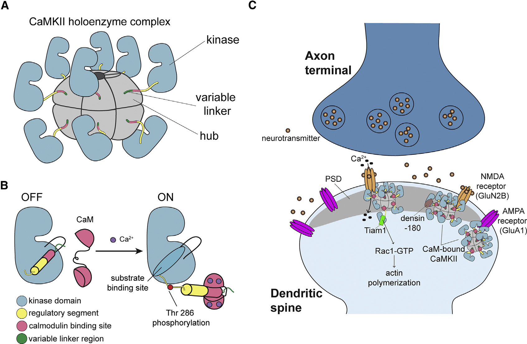
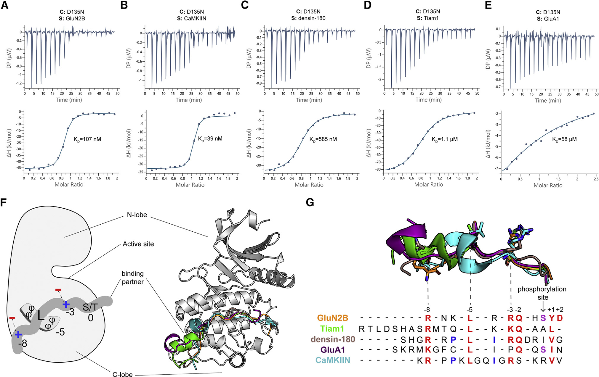
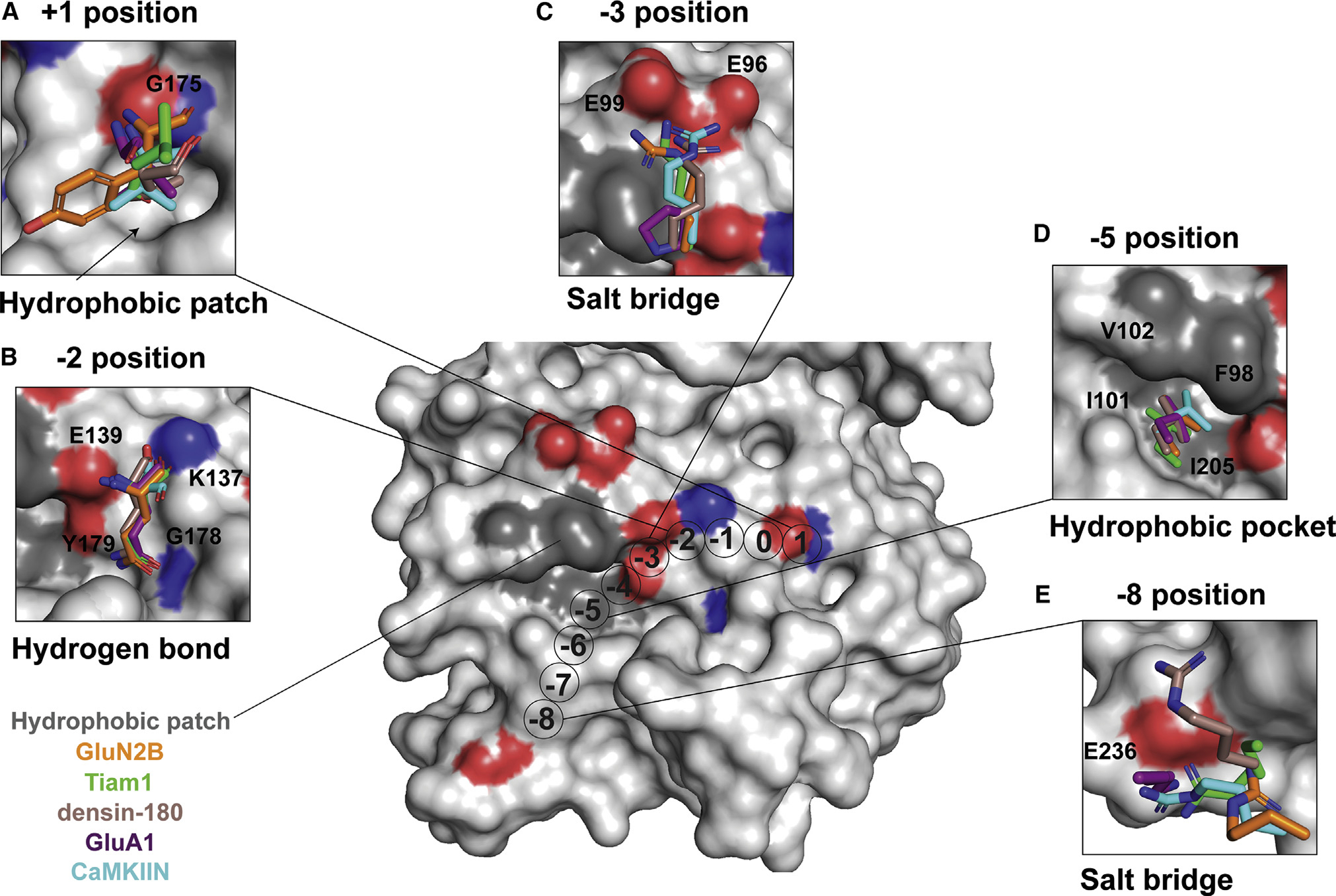
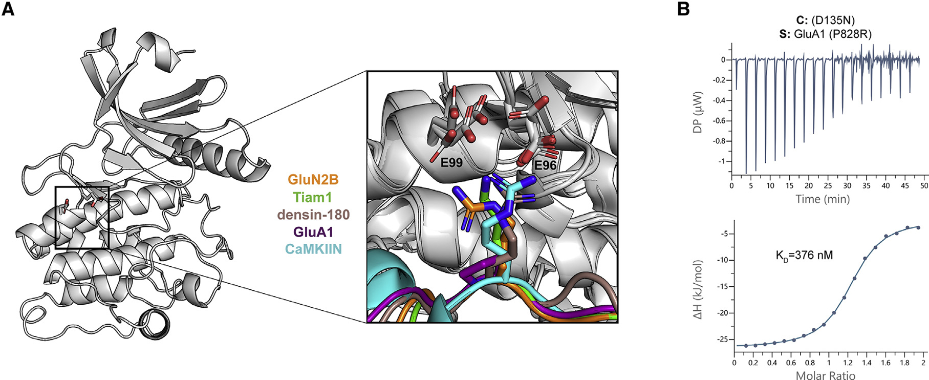
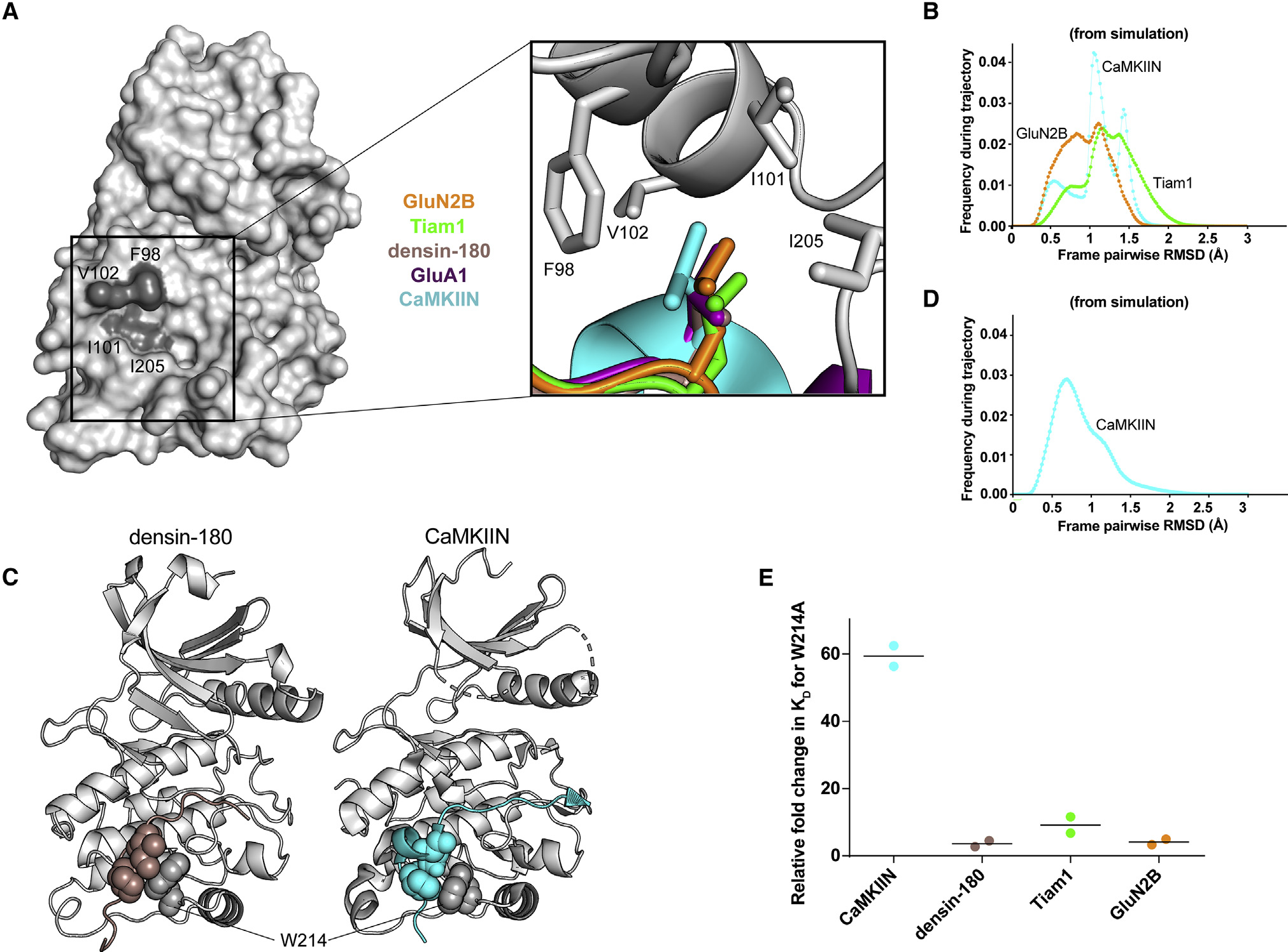
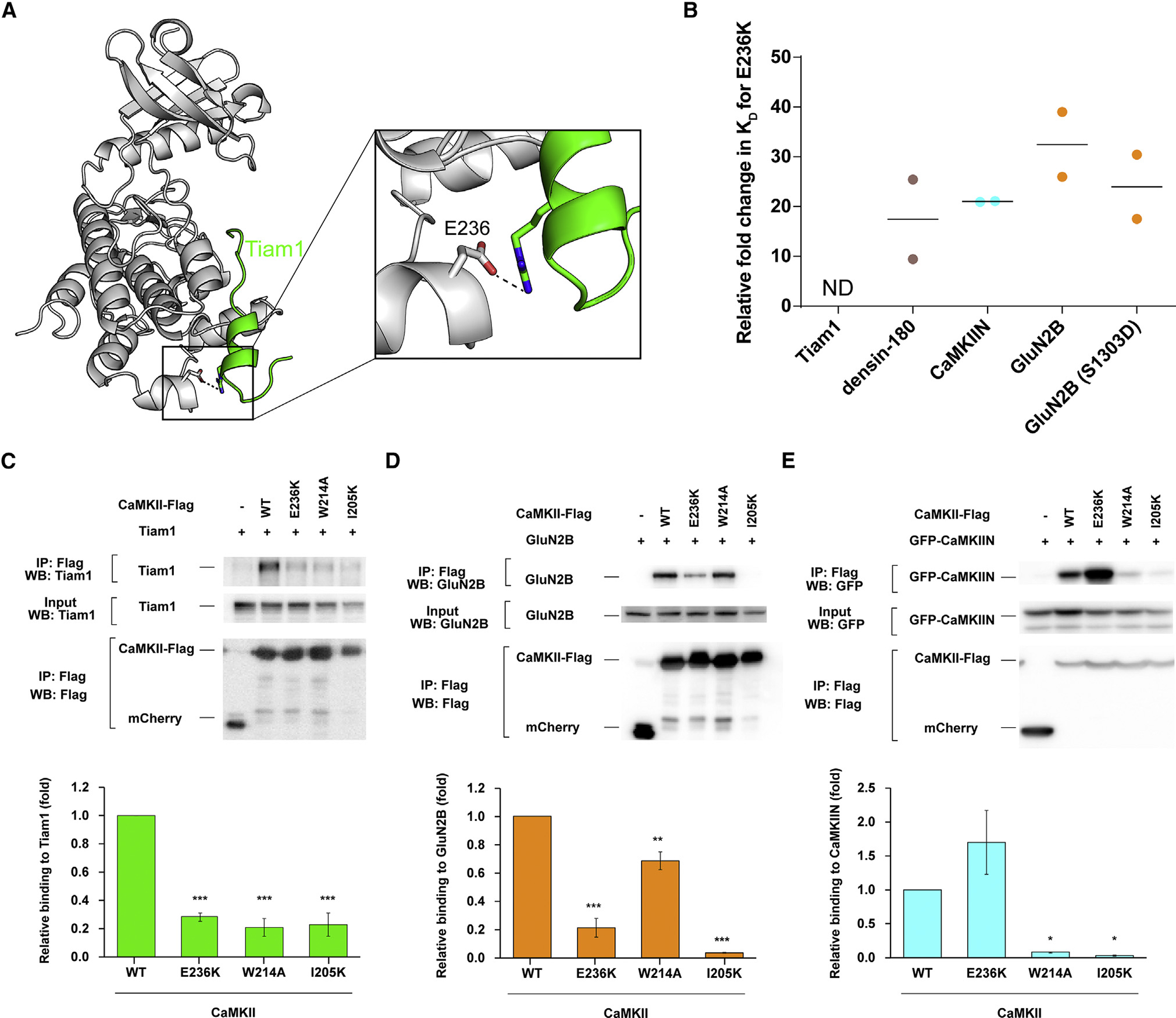
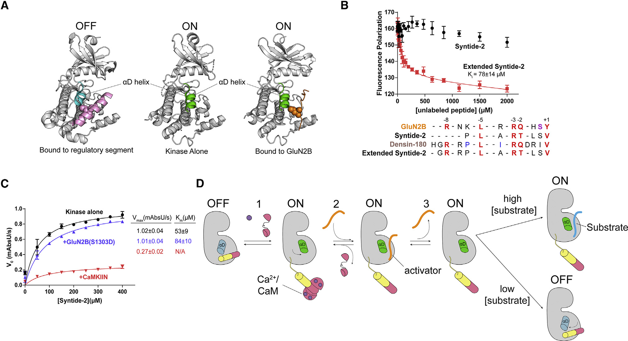
Ca2+/calmodulin-dependent protein kinase II (CaMKII) is a signaling protein required for long-term memory. When activated by Ca2+/CaM, it sustains activity even after the Ca2+ dissipates. In addition to the well-known autophosphorylation-mediated mechanism, interaction with specific binding partners also persistently activates CaMKII. A long-standing model invokes two distinct S and T sites. If an interactor binds at the T-site, then it will preclude autoinhibition and allow substrates to be phosphorylated at the S site. Here, we specifically test this model with X-ray crystallography, molecular dynamics simulations, and biochemistry. Our data are inconsistent with this model. Co-crystal structures of four different activators or substrates show that they all bind to a single continuous site across the kinase domain. We propose a mechanistic model where persistent CaMKII activity is facilitated by high-affinity binding partners that kinetically compete with autoinhibition by the regulatory segment to allow substrate phosphorylation.

Keywords: AMPA-type glutamate receptor; CP: Molecular biology; Ca(2+)/calmodulin dependent protein kinase II; LTP; NMDA-type glutamate receptor; Tiam1; X-ray crystallography.


Conflict of interest statement

Declaration of interests Y.H. received research funds from Fujitsu Laboratories and Dwango.

Figures

Figure 1.
Figure 1.. CaMKII architecture and the interaction partners at excitatory synapses
(A) The architecture of a dodecameric CaMKII holoenzyme. (B) Ca2+/CaM binding activates CaMKII by competitively binding the regulatory segment, thereby freeing the substrate binding site. Active CaMKII autophosphorylates at Thr 286. (C) CaMKII interactions at the excitatory postsynaptic structure, mostly in the postsynaptic density (PSD), of the dendritic spine.
Figure 2.
Figure 2.. There is a single binding site on the CaMKII kinase domain
(A–E) Representative ITC measurements of D135N kinase domain binding to peptide partners. Contents of the cell (C) and syringe (S) are indicated. The mean Kd value from two independent measurements is labeled. (F) Left: schematic of the interactions with binding partners. The phi symbol indicates hydrophobic residues. Right: overlay of five cocrystal structures, peptides shown as cartoon in corresponding colors seen in key to the right. (G) The sequence alignment of CaMKII binding partners. Binding partner position numbering is based on the prototypical GluN2B substrate with the phosphorylation site set to zero. Aligned peptide structures are shown above for reference. Conserved residues are colored red. Phosphorylatable residues at the phosphorylation site are colored purple. Residues involved in a docking event with W214 are colored blue.
Figure 3.
Figure 3.. Conserved binding motifs on the catalytic domain surface
Shown is a surface representation of the CaMKII kinase domain, highlighting residues that mediate interactions with binding partners. The color code matches previous figures (GluN2B, orange; Tiam1, green; densin-180, brown; GluA1, purple; CaMKIIN, cyan). (A) At the +1 position, a hydrophobic patch (arrow) is formed by F173, P177, L185, and Y222. GluA1, CaMKIIN, and densin-180 have Val or Ile at the +1 position, which are buried in this hydrophobic groove. Backbone atoms of the +1 residue hydrogen bond with the backbone of G175 (highlighted red and blue on the structure). (B) At the −2 position, all interactors have a glutamine except for CaMKIIN, which has a serine. The glutamine side-chain amide oxygen forms a hydrogen bond with the backbone of G178, and the amino group interacts with the side chain of Y179. The backbone carbonyl interacts with the side chain of K137, and the backbone amino group interacts with E139. (C) At the −3 position, lysine or arginine interacts with E96 and E99. The basic residues of interaction partners are positioned 2.4–4.2 Å between E96 and E99. GluA1 has a proline at the −3 position, which is flipped away from E96/99. (D) At the −5 position, a conserved leucine across all interactors nestles into a hydrophobic pocket formed by F98, I101, V102, and I205. (E) Lysine or arginine at the −8 position forms a salt bridge with E236.
Figure 4.
Figure 4.. Electrostatic interactions with a basic residue at the −3 position facilitate high-affinity binding
(A) CaMKII kinase domain shown as a cartoon; the E96 and E99 residues are shown as sticks. A magnified view of all five co-crystal structures is overlaid to highlight the basic residue at the −3 position (except for GluA1 in purple) interacting with the two glutamic acids in the kinase domain. (B) ITC data for the D135N CaMKII kinase domain (cell) and GluA1 with P828R mutation (syringe). The mean Kd value is from two independent measurements.
Figure 5.
Figure 5.. Hydrophobic interactions mediate binding
(A) Surface representation of the CaMKII kinase domain, with residues forming the hydrophobic pocket labeled (dark gray). Inset: overlay of leucine residues from all co-crystal structures bound in the hydrophobic pocket. (B) Histograms of RMSD from MD simulations between every pair of trajectory frames for F98, I101, V102, and I205 with −5 peptide leucine. (C) Crystal structures with sphere representation of the isoleucine and proline or leucine residues of densin-180 (brown) and CaMKIIN (cyan) docking onto W214 (gray) of the kinase domain. (D) RMSD histogram from MD simulations for W214 interacting with isoleucine and proline of CaMKIIN. (E) Kd values were extracted from ITC data, and relative fold changes were calculated by dividing the observed Kd from the mutant (W214A) by the D135N kinase domain. Individual data points are shown, and the line indicates the average.
Figure 6.
Figure 6.. Electrostatic interaction with E236 facilitates binding
(A) View of the interaction between CaMKII E236 (gray) and Tiam1 R1549 (green). (B) Kd values were extracted from ITC data, and relative fold changes were calculated by dividing the observed Kd from the mutant (E236K) by the D135N kinase domain. Individual data points are shown, and the line indicates the average. (C–E) Effects of CaMKII mutations (E236K, W214A, and I205K) on interactions with Tiam1, GluN2B, and CaMKIIN. HEK293T cells were co-transfected with FLAG-tagged CaMKII variants and Tiam1-mGFP (modified GFP), GluN2B, or EGFP-CaMKIIN. Cell lysates were immunoprecipitated with FLAG antibody, and samples were immunoblotted with Tiam1, GluN2B, GFP, and FLAG antibodies. Representative blots are shown in the top panels. Quantification of the co-immunoprecipitation from 3 or 4 independent experiments is shown in a graph of (C) Tiam1 (n = 3), (D) GluN2B (n = 3), and (E) CaMKIIN (n = 4). Error bars indicate standard error of the means. The amount of co-immunoprecipitated Tiam1, GluN2B, or CaMKIIN was normalized by the amount in cell lysate and immunoprecipitated CaMKII. *p < 0.05, **p < 0.01, and ***p < 0.001 compared with WT CaMKII; one-way ANOVA with Shaffer’s post hoc test comparisons. WT, wild type; IP, immunoprecipitation.
Figure 7.
Figure 7.. GluN2B binding interferes with autoinhibition
(A) Comparison of the αD helix between crystal structures of the autoinhibited with regulatory segment (pink) bound (left; PDB: 2VZ6), uninhibited kinase with no regulatory segment (center; PDB: 6VZK), and active kinase with GluN2B (orange) bound (right; PDB: 7UIS). The αD helix goes from rotated inward in the off-state (blue) to rotated outward 45° in the on state (green). (B) Competition assay against GluN2B using Syntide-2 (black) and the extended version of Syntide-2 (red). Error bars indicate standard deviation from triplicate at each data point. Sequence alignment of GluN2B, Syntide-2, densin-180, and the extended Syntide-2 is shown below the graph. For clarity, the alignments start at the +1 position (C-terminal end) and contain the −8 basic residue (N-terminal end). Full sequences used are listed in the STAR Methods. (C) Coupled kinase assay results with kinase alone and in the presence of GluN2B and CaMKIIN. The significance of the change in Km values is significant when using unpaired t test with Welch’s correction (p = 0.0208). (D) Proposed model of maintaining CaMKII activity by binding to a high-affinity activator. CaMKII binds Ca2+/CaM and is activated, and the αD helix rotates out (Rxn 1). A high-affinity activator binds to the substrate binding site (Rxn 2). When the Ca2+ signal dissipates, CaM dissociates from CaMKII, but the activator remains bound, competing with the regulatory segment. The activator dissociates, but the αD helix remains in the active conformation (Rxn 3). In high [substrate], the substrate will bind and be phosphorylated. In low [substrate], the activator will rebind or the αD helix will rotate back in to accommodate regulatory segment binding.

References

    1. Bakan A, Meireles LM, and Bahar I (2011). ProDy: protein dynamics inferred from theory and experiments. Bioinformatics 27, 1575–1577. 10.1093/bioinformatics/btr168. - DOI - PMC - PubMed
    1. Barker SC, Kassel DB, Weigl D, Huang X, Luther MA, and Knight WB (1995). Characterization of pp60c-src tyrosine kinase activities using a continuous assay: autoactivation of the enzyme is an intermolecular autophosphorylation process. Biochemistry 34, 14843–14851. 10.1021/bi00045a027. - DOI - PubMed
    1. Barria A, Derkach V, and Soderling T (1997a). Identification of the Ca2+/calmodulin-dependent protein kinase II regulatory phosphorylation site in the alpha-amino-3-hydroxyl-5-methyl-4-isoxazole-propionate-type glutamate receptor. J. Biol. Chem. 272, 32727–32730. 10.1074/jbc.272.52.32727. - DOI - PubMed
    1. Barria A, Muller D, Derkach V, Griffith LC, and Soderling TR (1997b). Regulatory phosphorylation of AMPA-type glutamate receptors by CaM-KII during long-term potentiation. Science 276, 2042–2045. 10.1126/science.276.5321.2042. - DOI - PubMed
    1. Bayer KU, De Koninck P, Leonard AS, Hell JW, and Schulman H (2001). Interaction with the NMDA receptor locks CaMKII in an active conformation. Nature 411, 801–805. 10.1038/35081080. - DOI - PubMed

Publication types

MeSH terms

Substances

LinkOut - more resources