. 2022 Oct-Dec:262:110777.
doi: 10.1016/j.cbpb.2022.110777. Epub 2022 Jul 10.

Gene expression and functional analysis of Aha1a and Aha1b in stress response in zebrafish

Affiliations

Gene expression and functional analysis of Aha1a and Aha1b in stress response in zebrafish

Huanhuan Xiao et al. Comp Biochem Physiol B Biochem Mol Biol. 2022 Oct-Dec.

Abstract

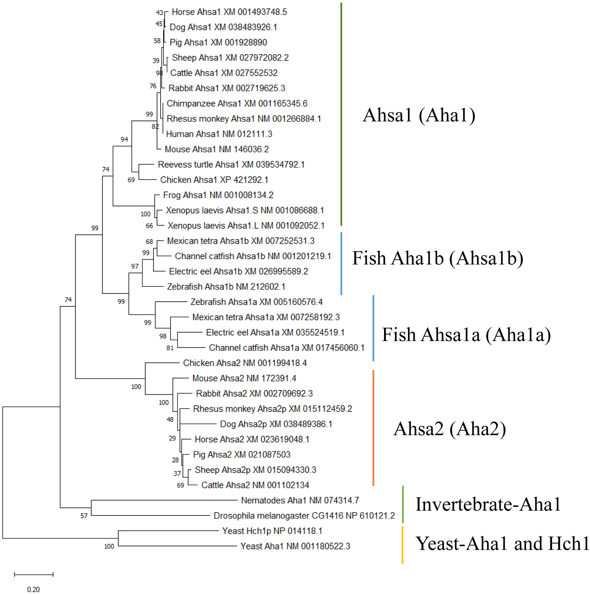
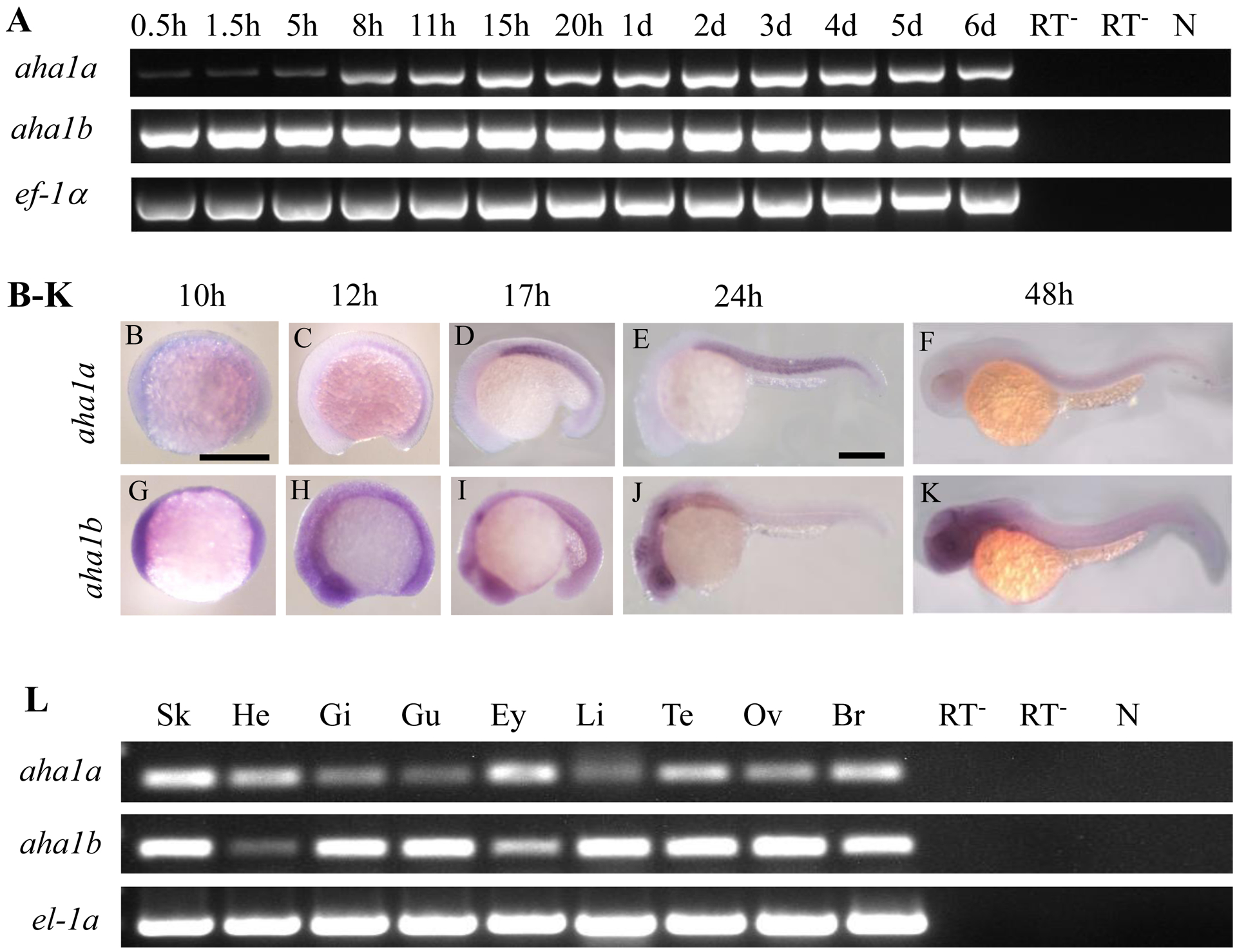
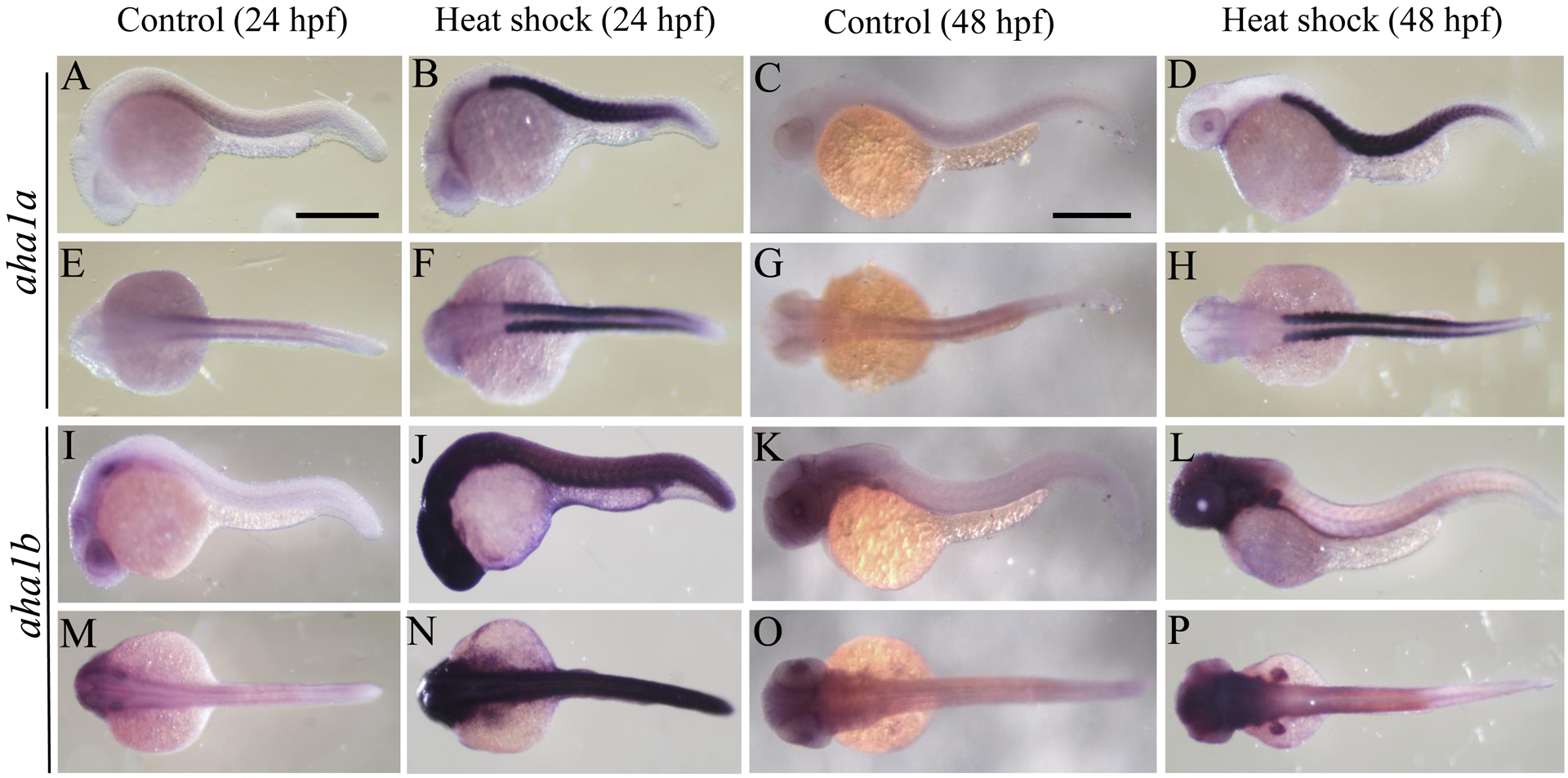
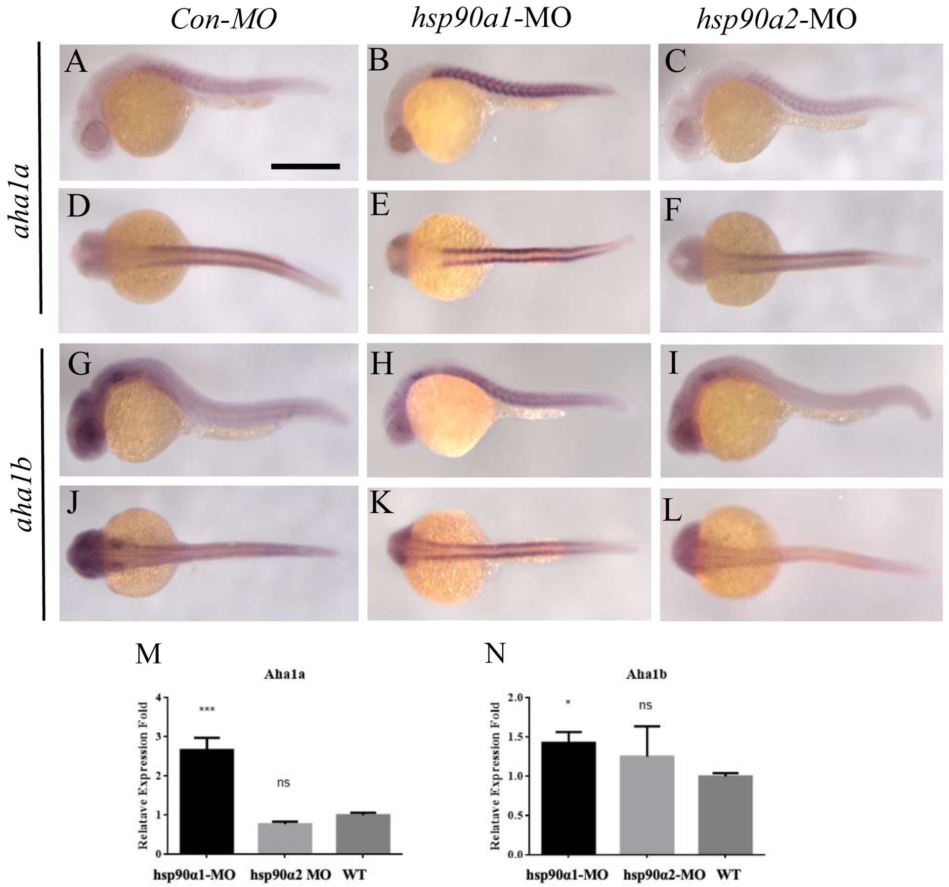
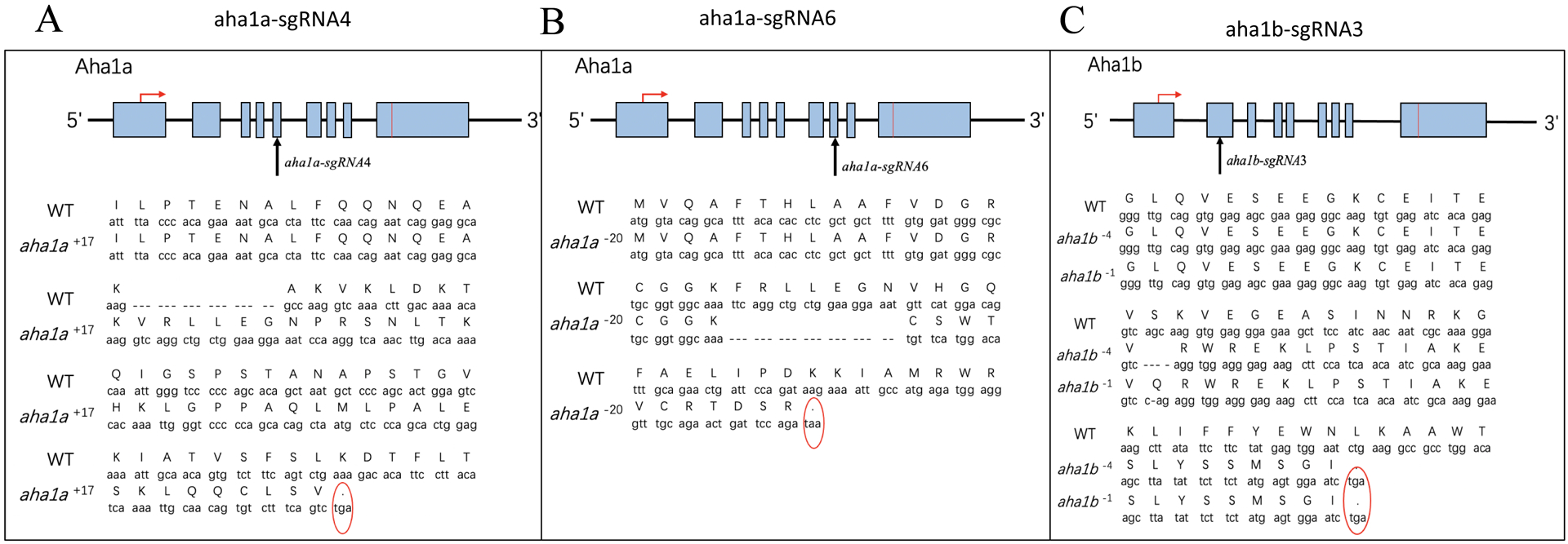
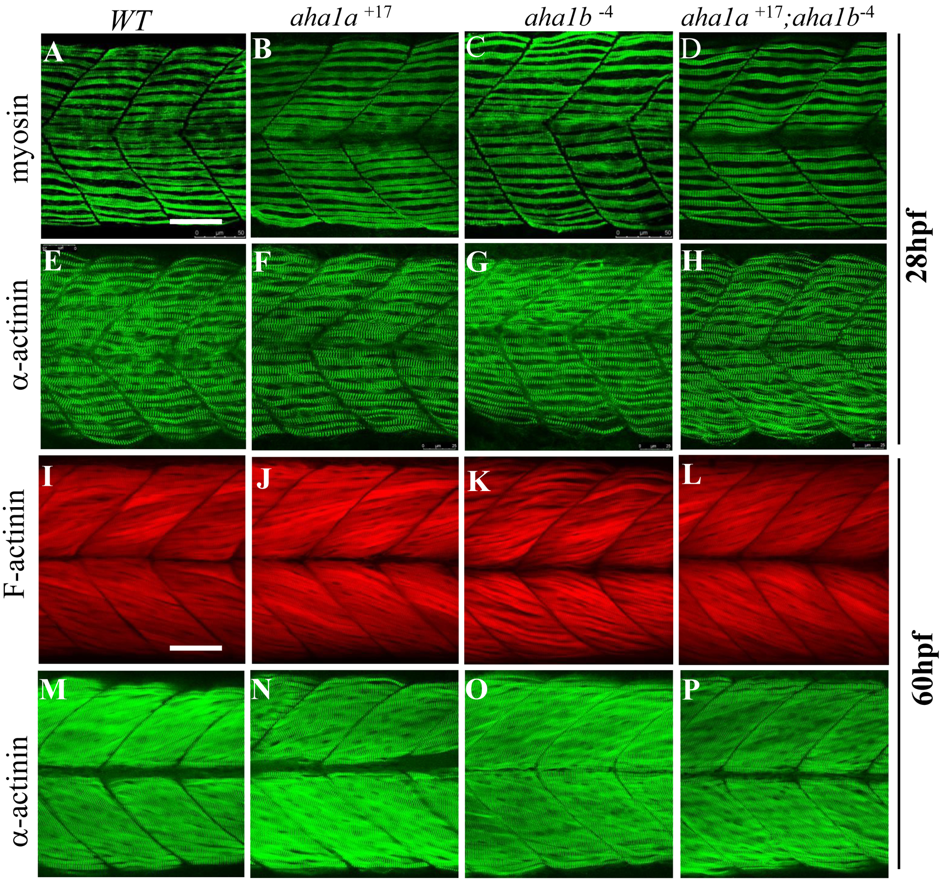
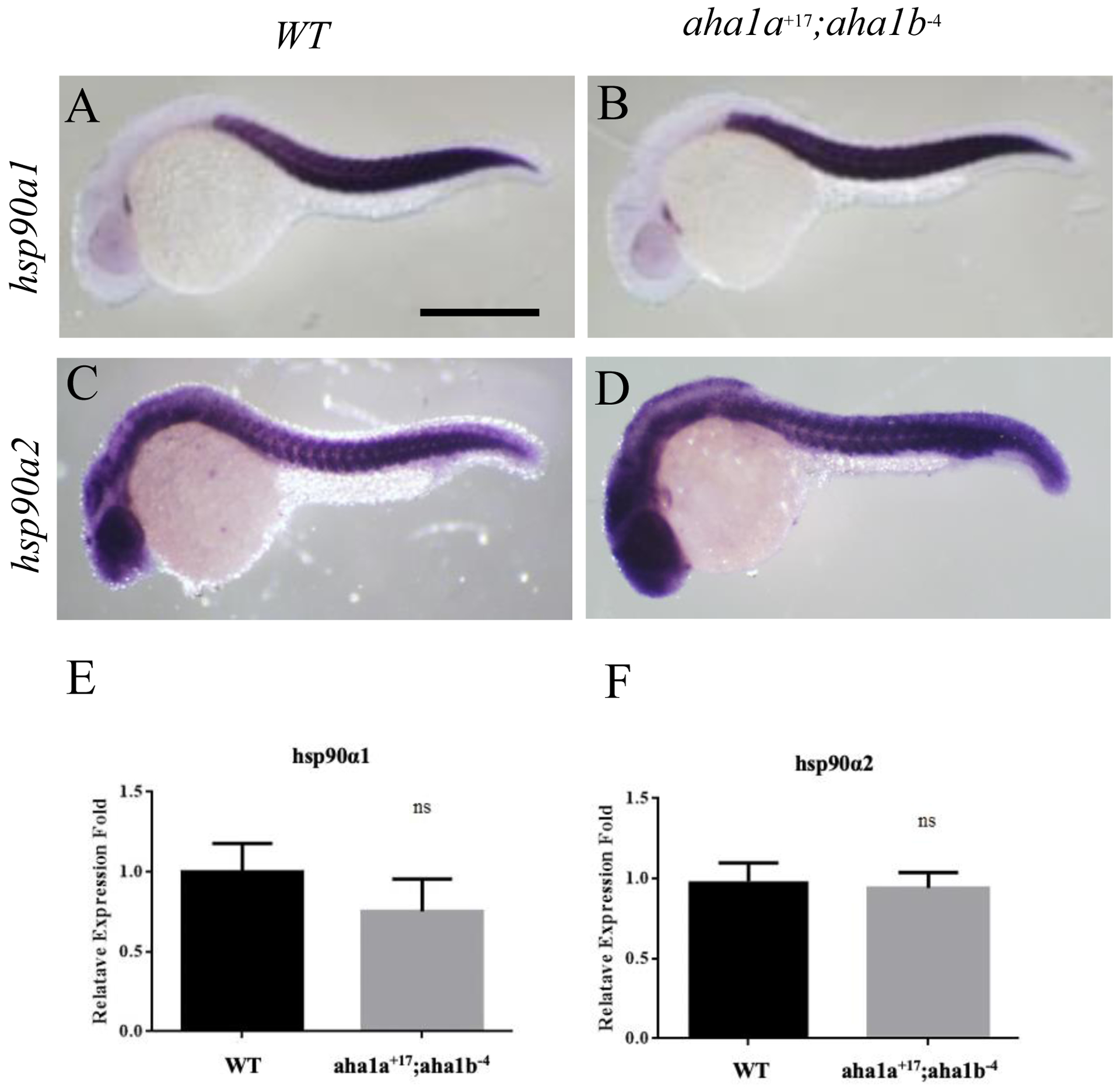
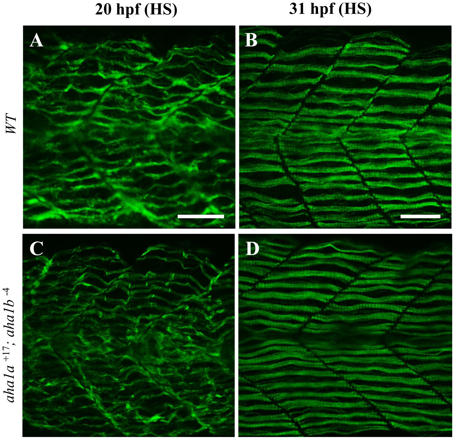
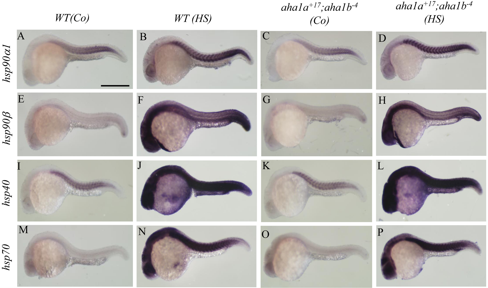
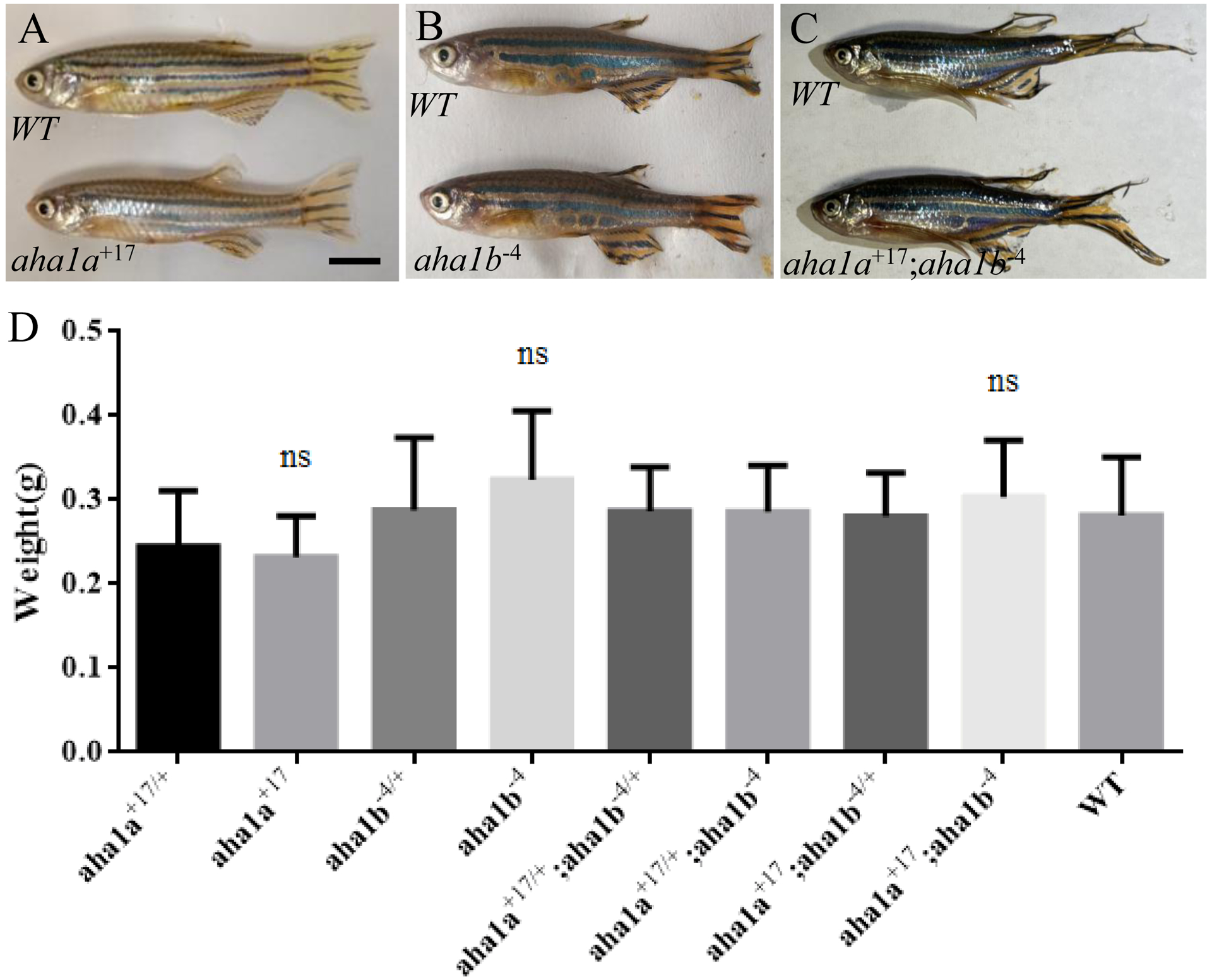
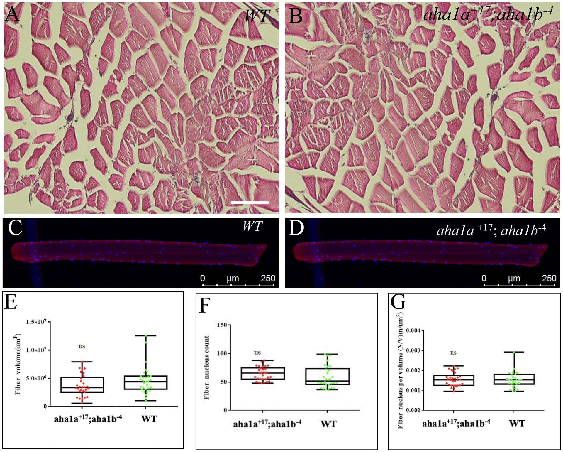
Activator of heat shock protein 90 (hsp90) ATPase (Aha1) is a Hsp90 co-chaperone required for Hsp90 ATPase activation. Aha1 is essential for yeast survival and muscle development in C. elegans under elevated temperature and hsp90-deficeiency induced stress conditions. The roles of Aha1 in vertebrates are poorly understood. Here, we characterized the expression and function of Aha1 in zebrafish. We showed that zebrafish genome contains two aha1 genes, aha1a and aha1b, that show distinct patterns of expression during development. Under the normal physiological conditions, aha1a is primarily expressed in skeletal muscle cells of zebrafish embryos, while aha1b is strongly expressed in the head region. aha1a and aha1b expression increased dramatically in response to heat shock induced stress. In addition, Aha1a-GFP fusion protein exhibited a dynamic translocation in muscle cells in response to heat shock. Moreover, upregulation of aha1 expression was also observed in hsp90a1 knockdown embryos that showed a muscle defect. Genetic studies demonstrated that knockout of aha1a, aha1b or both had no detectable effect on embryonic development, survival, and growth in zebrafish. The aha1a and aha1b mutant embryos showed normal muscle development and stress response in response to heat shock. Single or double aha1a and aha1b mutants could grow into normal reproductive adults with normal skeletal muscle structure and morphology compared with wild type control. Together, data from these studies indicate that Aha1a and Aha1b are involved in stress response. However, they are dispensable in zebrafish embryonic development, growth, and survival.

Keywords: Aha1(activator of Hsp90 ATPase); Heat shock protein; Heat shock response; Myosin chaperone.


Conflict of interest statement

Disclosures: The authors have no conflicts of interest to disclose.

Figures

Figure 1.
Figure 1.. Phylogenetic analysis of Aha-type co-chaperones among different species.
Phylogenetic tree was developed based on Aha-type protein sequences from yeast to mammals. The Aha-type cochaperones could be divided into four branches. The first branch is made up of the Ahsa1 (Aha1), Fish Aha1a and Aha1b subgroups. The other three branches are Ahsa2 (Aha2), invertebrate Aha1 and yeast Aha1 and Hch1, respectively.
Figure 2.
Figure 2.. The temporal and spatial patterns of aha1a and aha1b gene expression in zebrafish (Danio rerio) embryos and various adult tissues.
A. RT-PCR shows the temporal expression of aha1a and aha1b genes during embryonic development at various stages from newly fertilized eggs to six days postfertilization (6 dpf) zebrafish larvae. EF1α was included as an internal reference control. Two RT- RNA samples from 5d and 6d and water (N) were included as negative controls. B-K. In situ hybridization shows the expression of aha1a and aha1b in zebrafish embryos during early embryogenesis (10 – 48 hpf). Approximately 30–40 embryos were analyzed for each gene at each stage. The expression of aha1a was restricted to developing somite and skeletal muscles (B-F) and aha1b was expressed broadly in the whole embryos with stronger signals in the head regions (G-K) before 48 hpf. Scale bars =150 μm. L. aha1a and aha1b are ubiquitously expressed in adult tissues including skeletal muscle (Sm), heat (He), gill (Gi), gut (gu), eyes (Ey), liver (Li), testes (Te), ovaries (Ov), and brain (Br). Primers for RT-PCR were designed at the junctions of two adjacent exons to eliminate potential problems with genomic DNA contamination. Two RT- RNA samples from ovaries and brain and water (N) were included as negative controls.
Figure 3.
Figure 3.. The effects of heat shock on aha1a and aha1b mRNA expression in wildtype zebrafish embryos.
Wild type zebrafish embryos (n=50) were heat shocked for 1 hr at 37°C at 24 hpf or 48 hpf, respectively. The heat shocked embryos and respective untreated controls were fixed and used for expression analysis of aha1a and aha1b mRNA transcripts by whole mount in situ hybridization. The data show aha1a expression in controls at 24 hpf (A, E) and 48 hpf (C, G) and heat shocked treated groups at 24 hpf (B, F) and 48 hpf (D, H), respectively. A strong muscle-specific upregulation of aha1a expression was observed in heat shock treated embryos at 24 and 48 hpf. The data show aha1b expression in controls at 24 hpf (I, M) and 48 hpf (K, O) and heat shocked treated groups at 24 hpf (J, N) and 48 hpf (I, I), respectively. A dramatic global upregulation of aha1b expression was observed in heat shock treated embryos at 24 hpf (J, N). The upregulation was more pronounced at the head region at 48 hpf. A-D and I-L are side views. E-H and M-P are dorsal views. Scale bars = 100 μm.
Figure 4.
Figure 4.. Upregulation of aha1a and aha1b expression in defective muscle of hsp90a1 knockdown zebrafish embryos
Fertilized eggs were injected with control (Con-MO), hsp90α1-MO or hsp90α2-MO morpholinos at 1 or 2 cell stages. The aha1a and aha1b gene expression was analyzed by whole mount in situ hybridization and qRT-PCR in the injected embryos (n=120 for each MO) at 24 hpf. A-F, aha1a expression in Con-MO (A, D), hsp90α1-MO (B, E) and hsp90α2-MO (C, F) injected embryos, respectively. G-L, aha1b expression in Con-MO (G, J), hsp90α1-MO (H, K), and hsp90α2-MO (I, L) injected embryos, respectively. A-C, G-I: side views. D-F, J-L: dorsal views. Scale bar = 100 μm. M and N, qRT- qPCR analysis shows levels of aha1a (M) and aha1b (N) mRNA expression in hsp90α1, hsp90α2, and control embryos at 24 hfp.
Fig. 5
Fig. 5. Subcellular localization of Aha1a-GFP and Aha1b-GFP in myofibers of zebrafish embryos under normal and heat shock conditions.
DNA constructs expressing Aha1a-GFP or Aha1b-GFP fusion proteins were microinjected into zebrafish embryos. Each group of the injected embryos were randomly divided into two subgroups. One subgroup (~30 embryo) was subjected to heat shock (HS) treatment (37°C) for 1 hr at 48 hpf, another subgroup (~30 embryo) was used as untreated control. Subcellular localization of Aha1a-GFP and Aha1b-GFP was determined in myofibers of control and heat shock treated embryos together with M-line (Myomesin-3-RFP) and myosin (F59) markers. A-C, Confocal analysis shows Aha1a-GFP (A), Myomesin-3-RFP (B) and the merged (C) localization in myofibers of control embryos. D-F, Aha1b-GFP (D), Myomesin-3-RFP (E) and the merged (F) localization in myofibers of control embryos. G-I, Aha1a-GFP (G), myosin (F59, H) and the merged (I) localization in myofibers of heat shocked embryos. J-L, Aha1b-GFP (J), myosin (F59, K) and the merged (L) localization in myofibers of the heat shocked embryos. White arrows in (C) and (F) indicate sarcomeric Aha1a-GFP and Aha1b-GFP localization between the M-lines (Myomesin-3-RFP). Yellow arrows in (I) indicate the Aha1a-GFP colocalization with myosin (F59) in the A bands. Due to the mosaic pattern of gene expression via DNA injection, myofibers labeled with Myomesin and Myosin antibodies did not always express the aha1a-GFP or aha1b-GFP transgene. Scale bar = 6 μm.
Figure 6.
Figure 6.. Generation of aha1a and aha1b zebrafish mutants using CRISPR/Cas9.
Gene specific sgRNAs targeted to aha1a or aha1b were co-injected with Cas9 protein into fertilized embryos at 1–2 cells stages. Two mutant alleles were generated for each gene. A and B, the aha1a+17 and aha1a−20 mutant alleles were generated using aha1a-sgRNA-4 and aha1a-sgRNA- 6 targeted to aha1a gene exon 5 and 7, respectively. The aha1a+17 and aha1a−20 alleles carry a 17 bp insertion and 20 bp deletion, respectively. C, the aha1b−1 and aha1b−4 mutant alleles were generated using the same aha1b-sgRNA-3 targeted to exon 2 of aha1b gene. The aha1b−1 and aha1b−4 alleles carry a 1 bp and 4 bp deletion, respectively. All these indel mutations created reading frame shift in aha1a or aha1b, resulting in premature stop codons in their coding sequences.
Figure 7.
Figure 7.. Comparison of aha1a and aha1b mRNA expression in WT, aha1a and aha1b mutant embryos.
aha1a and aha1b mRNA expression was analyzed by whole mount in situ hybridization (A-F) and qRT-PCR (G, H) in WT, aha1a+17, aha1a−20, aha1b−4and aha1b−1mutant embryos (approximately 30 embryos for each group) at 24 hpf. A-C, aha1a expression in WT control (A) and aha1a+17 (B) aha1a−20 (C) mutant embryos. D-F, aha1b expression in WT control (D), aha1b−4(E) and aha1b−1(F) mutant embryos. All pictures are side views. Scale bar = 100 μm. G and H, qRT-PCR analysis of aha1a and aha1b mRNA expression in WT control and aha1a (G) and aha1b (H) mutant zebrafish embryos at 24 hpf. Expression of elongation factor 1-alpha (Ef-1 α) was used an internal reference gene. Error bars are mean ± SEM. ***P<0.001 were considered statistically significant. Nonsense- mediated mRNA decay was detected in aha1a+17, aha1b−4and aha1b−1mutant alleles.
Figure 8.
Figure 8.. Characterization of muscle cell differentiation and sarcomere organization in slow and fast muscles of aha1a and aha1b mutant embryos.
Myofibers in slow and fast muscles of WT, aha1a+17, aha1b−4single and aha1a+17; aha1b− 4 double mutant embryos (~80 fish embryos for each group) were analyzed by phalloidin and immunostaining that specifically label various sarcomere structures. A-D: Myosin organization in slow fibers revealed by F59 antibody staining of WT (A), aha1a+17(B), aha1b−4(C) and aha1a+17; aha1b−4(D) mutant embryos at 28 hpf. E-H: Z-line organization in slow fibers revealed by anti-α-actinin antibody staining of WT (E), aha1a+17(F), aha1b−4(G) and aha1a+17; aha1b−4(H) mutant embryos at 28 hpf. I-L: a-actin thin filament organization revealed by Phalloidin staining of WT (I), aha1a+17(J), aha1b−4(K) and aha1a+17; aha1b−4(L) mutant embryos at 60 hpf. M-P: Z-line organization in fast fibers shown by anti-α-actinin antibody staining in WT (M), aha1a+17(N), aha1b−4(O) and aha1a+17; aha1b−4(P) mutant embryos at 60 hpf. All pictures are side views of part of trunk muscles. Scale bar = 25 μm.
Figure 9.
Figure 9.. Knockout of aha1a and aha1b has no effect on hsp90α1 and hsp90α2 gene expression in fish embryos.
Expression of hsp90a1 and hsp90a2 was determined by whole mount in situ hybridization (A-D) and qRT-PCR (E, F) in WT control (A, C) and aha1a+17; aha1b−4double mutant (B, D) zebrafish embryos at 24hpf, respectively. WT embryos (n=51) and aha1a+17; aha1b−4double mutant embryos (n=50) were applied for in situ hybridization. Total RNA was extracted from zebrafish embryos (triplicates of 25 each) at 24 hpf in each group for qRT-PCR, respectively. All pictures here are side views. Scale bar = 100 μm.
Figure 10.
Figure 10.. The effect of heat shock on muscle development in Aha1a and Aha1b double mutant embryos.
WT (n=50) and aha1a+17; aha1b−4double mutant (n=50) embryos were subjected to heat shock (HS) at 20 hpf or 31 hpf under 37°C for 1h. Muscle development in the treated embryos was analyzed by immunostaining with F59 antibody that showed myosin thick filament organization in WT (A, B) and aha1a and aha1b double mutant (C, D) embryos at 20 and 31 hpf, respectively. No significant difference was detected between WT and aha1a+17; aha1b−4mutant embryos. All pictures are side views of part of trunk muscles. Scale bar = 25 μm.
Figure 11.
Figure 11.. Heat shock response in aha1a and aha1b double mutant embryos.
WT (n=120) and aha1a+17; aha1b−4double mutant (n=120) embryos were subjected to heat shock (HS) at 24 hpf under 37°C for 1h. mRNA expression of hsp90α1 (A-D), hsp90β (E-H), hsp40 (I-L) and hsp70 (M-P) were analyzed in untreated (A, E, I and M) and heat shock treated (B, F, J and N) WT control group as well as untreated (C, G, K and O) and heat shock treated (D, H, L and P) aha1a+17; aha1b−4 double mutant group. Comparable upregulation of hsp90α1, hsp90β, hsp40, and hsp70 was observed in WT and aha1a+17; aha1b−4 mutant embryos. All pictures here are side views. Scale bar = 100 μm.
Figure 12.
Figure 12.. Knockout of aha1a and aha1b has no effects on fish growth and survival.
A-C. Morphological comparison among WT, aha1a+17(A), aha1b−4(B) and aha1a+17; aha1b−4(C) mutant fish at 10-month-old. D. Body weight (g) comparison among fish generated from in-cross of aha1a+17/+, aha1b−4/+ or aha1a+17/+; aha1b−4/+ double heterozygous mutants at 10-month-old. Adult fish (n=162) generated above were used for body weight testing. Scale bar = 1.3 cm. Weight differences among different genotypes of zebrafish were analyzed using a Student’s t-test. No significant difference was detected in terms of body weight among these groups (P>0.05). Error bars are ± SEM.
Figure 13.
Figure 13.. The Aha1a and Aha1b double mutants displayed normal muscle growth.
Skeletal muscles were dissected from three WT and three aha1a+17; aha1b−4double mutant fish at 5- month-old. A and B, Skeletal muscle structures were analyzed by haematoxylin and eosin (HE) staining in cross sections of trunk muscles from WT (A) and aha1a+17; aha1b−4double mutant (B), respectively. Scale bar = 50 μm. C and D, single myofibers were dissected from skeletal muscles of WT (C) and aha1a+17; aha1b−4double mutant (D) fish at 5-month-old. The dissected single fibers (n=35, each) were stained with Hoechst 32258 and Phalloidin-TRITC. Average myofiber volume (E), nuclear count (F) and per nuclear volume (G) were calculated and compared between WT and aha1a+17; aha1b−4double mutant. No significant difference was detected between WT and aha1a+17; aha1b−4mutant fish. Error bars are Median± SEM. Differences between WT and mutants were analyzed using a Student’s t test

Similar articles

Cited by

References

    1. Armstrong H, Wolmarans A, Mercier R, Mai B & LaPointe P, 2012. The co-chaperone Hch1 regulates Hsp90 function differently than its homologue Aha1 and confers sensitivity to yeast to the Hsp90 inhibitor NVP-AUY922.PLoS One 7, e49322, doi: 10.1371/journal.pone.0049322. - DOI - PMC - PubMed
    1. Benjamin IJ, & McMillan DR,1998. Stress (heat shock) proteins: molecular chaperones in cardiovascular biology and disease. Circulation research, 83(2), 117–132. 10.1161/01.res.83.2.117 - DOI - PubMed
    1. Biebl MM, Buchner J, 2019. Structure, function, and regulation of the Hsp90 machinery. Cold Spring Harb. Perspect. Biol, 11, p. a034017. - PMC - PubMed
    1. Biebl MM, Riedl M, & Buchner J,2020. Hsp90 Co-chaperones Form Plastic Genetic Networks Adapted to Client Maturation. Cell reports, 32(8), 108063. 10.1016/j.celrep.2020.108063 - DOI - PubMed
    1. Bhattacharya K, Weidenauer L, Luengo TM et al.,2020. The Hsp70-Hsp90 co-chaperone Hop/Stip1 shifts the proteostatic balance from folding towards degradation. Nat Commun 11, 5975. 10.1038/s41467-020-19783-w - DOI - PMC - PubMed

MeSH terms

LinkOut - more resources