

Increased iron uptake in the bladder wall of racemose cysts of Taenia solium
- PMID: 35830923
- PMCID: PMC9869405
- DOI: 10.1016/j.molbiopara.2022.111496
Increased iron uptake in the bladder wall of racemose cysts of Taenia solium
Abstract
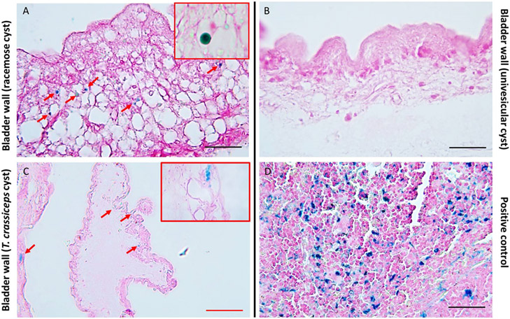
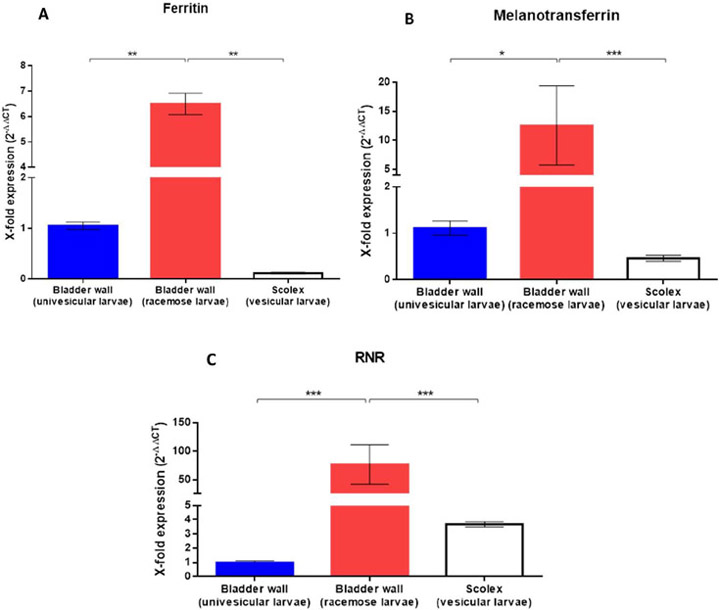
Racemose neurocysticercosis is an aggressive infection caused by the aberrant expansion and proliferation of the bladder wall of the Taenia solium cyst within the subarachnoid spaces of the human brain. The parasite develops and proliferates in a microenvironment with low concentrations of growth factors and micronutrients compared to serum. Iron is important for essential biological processes, but its requirement for racemose cyst viability and proliferation has not been studied. The presence of iron in the bladder wall of racemose and normal univesicular T. solium cysts was determined using Prussian blue staining. Iron deposits were readily detected in the bladder wall of racemose cysts but were not detectable in the bladder wall of univesicular cysts. Consistent with this finding, the genes for two iron-binding proteins (ferritin and melanotransferrin) and ribonucleotide reductase were markedly overexpressed in the racemose cyst compared to univesicular cysts. The presence of iron in the bladder wall of racemose cysts may be due to its increased metabolic rate due to proliferation.
Keywords: Cisticercosis; Ferritin; Iron metabolism; Neurocysticercosis; Ribonucleotide reductase; Subarachnoid cyst; Taenia solium.
Copyright © 2022 The Authors. Published by Elsevier B.V. All rights reserved.
Figures
References
-
- Pawlowski ZS. Taenia solium: Basic biology and Transmission. In: Singh G, Prabhakar S. Taenia solium Cysticercosis from basic to clinical science. NY, USA: CABI Publishing; 2002.
-
- Fleury A, Carrillo-Mezo R, Flisser A, Sciutto E, Corona T. Subarachnoid basal neurocisticercosis: focus on the most severe form of the disease. Expert. Rev. Anti. Infect. Ther 2011; 9(1): 123–133. - PubMed
Publication types
MeSH terms
Substances
Grants and funding
LinkOut - more resources
Full Text Sources
