. 2022 Aug 24;42(34):6566-6580.
doi: 10.1523/JNEUROSCI.0612-22.2022. Epub 2022 Jul 13.

Left-Right Locomotor Coordination in Human Neonates

Affiliations

Left-Right Locomotor Coordination in Human Neonates

Arthur H Dewolf et al. J Neurosci. .

Abstract

Terrestrial locomotion requires coordinated bilateral activation of limb muscles, with left-right alternation in walking or running, and synchronous activation in hopping or skipping. The neural mechanisms involved in interlimb coordination at birth are well known in different mammalian species, but less so in humans. Here, 46 neonates (of either sex) performed bilateral and unilateral stepping with one leg blocked in different positions. By recording EMG activities of lower-limb muscles, we observed episodes of left-right alternating or synchronous coordination. In most cases, the frequency of EMG oscillations during sequences of consecutive steps was approximately similar between the two sides, but in some cases it was considerably different, with episodes of 2:1 interlimb coordination and episodes of activity deletions on the blocked side. Hip position of the blocked limb significantly affected ipsilateral, but not contralateral, muscle activities. Thus, hip extension backward engaged hip flexor muscle, and hip flexion engaged hip extensors. Moreover, the sudden release of the blocked limb in the posterior position elicited the immediate initiation of the swing phase of the limb, with hip flexion and a burst of an ankle flexor muscle. Extensor muscles showed load responses at midstance. The variable interlimb coordination and its incomplete sensory modulation suggest that the neonatal locomotor networks do not operate in the same manner as in mature locomotion, also because of the limited cortical control at birth. These neonatal mechanisms share many properties with spinal mammalian preparations (i.e., independent pattern generators for each limb, and for flexor and extensor muscles, load, and hip position feedback).SIGNIFICANCE STATEMENT Bilateral coupling and reciprocal activation of flexor and extensor burst generators represent the fundamental mechanisms used by mammalian limbed locomotion. Considerable progress has been made in deciphering the early development of the spinal networks and left-right coordination in different mammals, but less is known about human newborns. We compared bilateral and unilateral stepping in human neonates, where cortical control is still underdeveloped. We found neonatal mechanisms that share many properties with spinal mammalian preparations (i.e., independent pattern generators for each limb, the independent generators for flexor and extensor muscles, load, and hip-position feedback. The variable interlimb coordination and its incomplete sensory modulation suggest that the human neonatal locomotor networks do not operate in the same manner as in mature locomotion.

Keywords: early development; human locomotion; interlimb coordination; neonatal stepping.


Figures

Figure 1.
Figure 1.
A, Illustration of one neonate performing unilateral stepping with one limb manually blocked in the middle position. B, Examples of raw EMGs and limb kinematics during bilateral and unilateral stepping with the limb blocked in the middle position in two different neonates. VMz, Vertical coordinate of the fifth metatarsal marker. Traces include the transition from bilateral to unilateral stepping or the contrary (vertical line marks transition). Notice that a brief segment of RF activity of the stepping limb of neonate b16 was corrupt and has been replaced by a zero line.
Figure 2.
Figure 2.
Kinematics of the stepping limb. A, Thigh, shank, and foot elevation angles of all steps of bilateral and unilateral stepping with the limb blocked in middle position in gray, ensemble-average traces (i.e., average across steps in all neonates) in black. B, Coefficient of correlation and time lag of the cross-correlations between each individual trace and the ensemble-average trace of bilateral stepping. C, Average stride and swing duration; interstride variability; range of motion of the thigh, shank, and foot angles; and the excursion of the fifth metatarso-phalangeal joint in horizontal (x), lateral (y), and vertical (z) direction. Asterisks denote significant differences.
Figure 3.
Figure 3.
Recorded bilateral EMG profiles during bilateral and unilateral stepping with the limb blocked in the middle position. A, Examples of raw EMGs of two consecutive steps in four different neonates during bilateral stepping. Note that alternations between left (light gray) and right (dark gray) limb muscles are mostly observed. B, Examples of raw EMGs of two consecutive steps in three different neonates during bilateral stepping. Both alternate and synchronous muscle activation of the stepping limb (light gray) and the blocked limb (blue) are observed. VMz, Vertical coordinate of the fifth metatarsal marker.
Figure 4.
Figure 4.
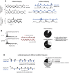
A, B, Interlimb coupling during bilateral (A) and unilateral (B) stepping with the blocked limb in middle position. A, B, First line, Polar histograms of center of activity for bilateral stepping versus normalized cycles discretized in 20 sectors; second and third lines, polar histograms of the phase lag between the center of activity of both limbs (ΔCoA) for bilateral stepping versus normalized cycles discretized in 20 sectors; black arrows, progression time, with angle that varies from 0° to 360° corresponding to a 0% and 100% cycle. Bar height denotes the percentage of cycles whose center of activity (or ΔCoA) is located in the corresponding sector. Red arrows, Resultant (circular mean) center of activity (Rayleigh test for nonuniform circular distributions, p < 0.05). The pie chart presents the percentage of steps with alternate or synchronous LG bilateral activation, or with no modulation of LG EMG activity (i.e., where the center of activity of one of the limbs was not identifiable; Rayleigh test, p > 0.05).
Figure 5.
Figure 5.
Decomposition of LG envelope time series into their Fourier series components during bilateral stepping (n = 9) and unilateral stepping (n = 13) with the leg blocked in the middle position. A, Examples of bilateral LG envelope during several consecutive steps with similar (top) or different left and right oscillation frequencies (bottom). B, Illustration of the fast Fourier transform analysis and the percentage of neonates with different and similar left and right oscillation frequencies during bilateral and unilateral stepping with leg blocked in the middle position. C, Two examples of the bilateral onset of LG bursts of activation during unilateral stepping with different limb oscillation frequencies. In most cases, there was one burst onset of the blocked limb during each step of the stepping limb (1:1 coordination). In some cases, an episode with two burst onsets of the blocked limb during one step of the stepping limb were observed (2:1 coordination). In other cases, an episode without LG activation of the blocked limb (0:1 coordination) occurred. The pie chart presents the quantification of the different episodes of coordination in unilateral stepping.
Figure 6.
Figure 6.
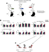
Effect of blocked limb position on stepping limb characteristics. A, Average (across steps in all neonates) stride and swing duration; range of motion of the thigh, shank, and foot angles; and orientation of the thigh, shank, and foot at touch-down and toe-off during unilateral stepping with the limb blocked in the anterior, middle, and posterior positions. B, Average FWHM, CoA, and mean EMG activity across one step of the four stepping limb muscles. Asterisks denote significant differences.
Figure 7.
Figure 7.
Effect of blocked limb position on blocked limb characteristics. A, Percentage of steps with alternate or synchronous LG bilateral activation, or with no modulation of LG EMG activity (i.e., where the center of activity of one of the limbs was not identifiable; Rayleigh test, p > 0.05). B, Average FWHM, CoA, and mean EMG activity of the four blocked limb muscles of the alternated (top) or synchronous steps (bottom). Asterisks denote significant differences. C, Schematic illustration of the effect of blocked limb position on muscle activation.
Figure 8.
Figure 8.
Top, Illustration of one neonate producing a dorsiflexion of the ankle after the release of the blocked limb in posterior position. Bottom, Examples of raw TA EMGs after the release of the blocked limb in posterior position in six different neonates.
Figure 9.
Figure 9.
Loading response of the stepping limb. Knee joint angle, RF and LG EMG envelope of all steps in which an extension of the knee was present during the first half of stance; black, the ensemble-average traces (average across steps in all neonates). The bar plot on the right presents the average knee extension (average across steps in all neonates) during the first 50% of stance.

References

    1. Andersson O, Grillner S (1981) Peripheral control of the cat's step cycle I. Phase dependent effects of ramp-movements of the hip during “fictive locomotion”. Acta Physiol Scand 113:89–101. 10.1111/j.1748-1716.1981.tb06867.x - DOI - PubMed
    1. Apgar V (1953) A proposal for a new method of evaluation of the newborn infant. Curr Res Anesth Analg 32:260–267. - PubMed
    1. Arber S (2012) Motor circuits in action: specification, connectivity, and function. Neuron 74:975–989. 10.1016/j.neuron.2012.05.011 - DOI - PubMed
    1. Batschelet E (1981) Circular statistics in biology. London, New York: Academic.
    1. Bernhardt NR, Memic F, Kullander K (2013) Genetic analysis of left-right coordination of locomotion. Front Biosci (Landmark Ed) 18:21–35. 10.2741/4085 - DOI - PubMed

Publication types

LinkOut - more resources