. 2022 Jul 12;13(7):602.
doi: 10.1038/s41419-022-05049-4.

Targeting sphingosine kinase 1/2 by a novel dual inhibitor SKI-349 suppresses non-small cell lung cancer cell growth

Affiliations

Targeting sphingosine kinase 1/2 by a novel dual inhibitor SKI-349 suppresses non-small cell lung cancer cell growth

Yuhang Xue et al. Cell Death Dis. .

Abstract

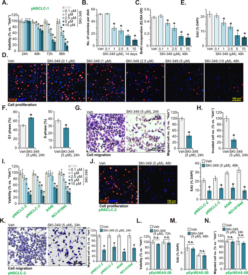
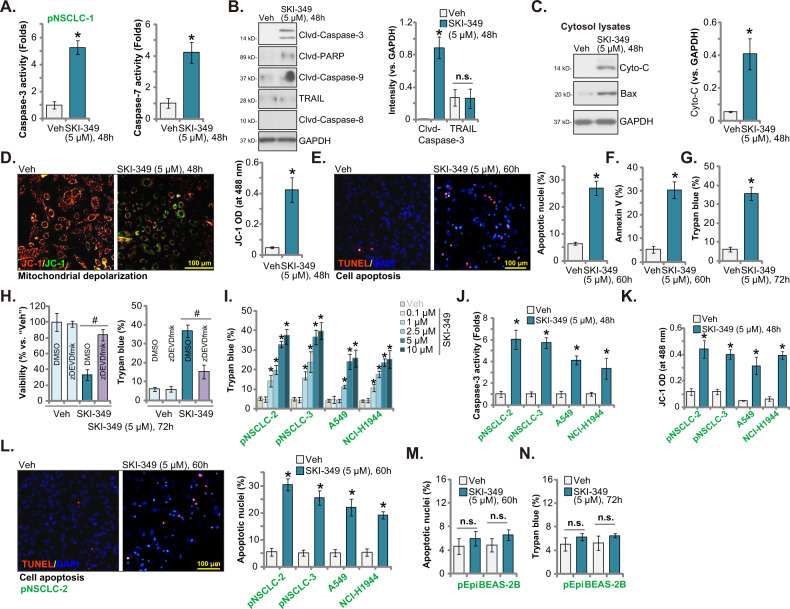
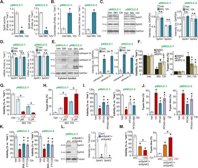
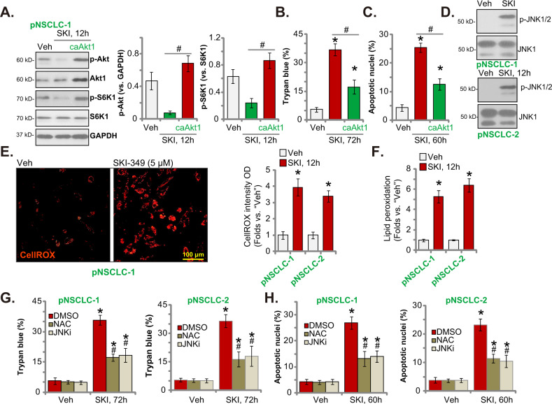
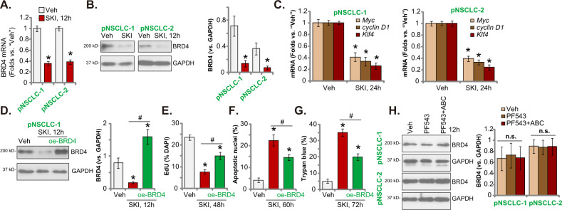
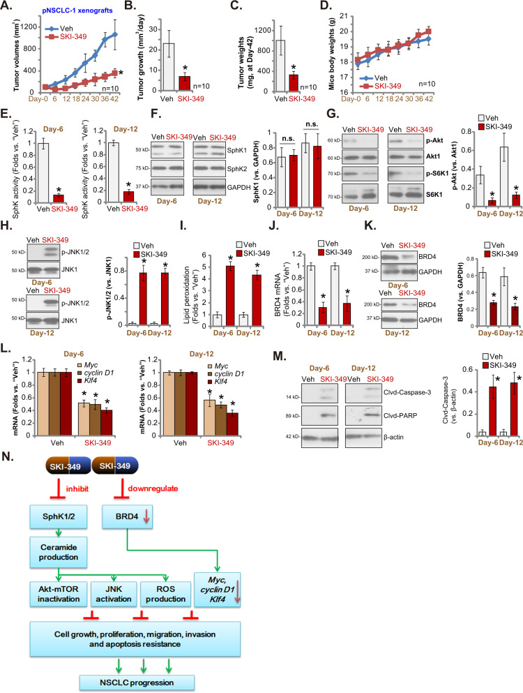
Sphingosine kinase 1 (SphK1) and sphingosine kinase (SphK2) are both important therapeutic targets of non-small cell lung cancer (NSCLC). SKI-349 is a novel, highly efficient and small molecular SphK1/2 dual inhibitor. Here in primary human NSCLC cells and immortalized cell lines, SKI-349 potently inhibited cell proliferation, cell cycle progression, migration and viability. The dual inhibitor induced mitochondrial depolarization and apoptosis activation in NSCLC cells, but it was non-cytotoxic to human lung epithelial cells. SKI-349 inhibited SphK activity and induced ceramide accumulation in primary NSCLC cells, without affecting SphK1/2 expression. SKI-349-induced NSCLC cell death was attenuated by sphingosine-1-phosphate and by the SphK activator K6PC-5, but was potentiated by the short-chain ceramide C6. Moreover, SKI-349 induced Akt-mTOR inactivation, JNK activation, and oxidative injury in primary NSCLC cells. In addition, SKI-349 decreased bromodomain-containing protein 4 (BRD4) expression and downregulated BRD4-dependent genes (Myc, cyclin D1 and Klf4) in primary NSCLC cells. At last, SKI-349 (10 mg/kg) administration inhibited NSCLC xenograft growth in nude mice. Akt-mTOR inhibition, JNK activation, oxidative injury and BRD4 downregulation were detected in SKI-349-treated NSCLC xenograft tissues. Taken together, targeting SphK1/2 by SKI-349 potently inhibits NSCLC cell growth in vitro and in vivo.


Conflict of interest statement

The authors declare no competing interests.

Figures

Fig. 1
Fig. 1. SKI-349 potently inhibits NSCLC cell progression in vitro.
The patient-derived primary NSCLC cells, pNSCLC-1, were treated with SKI-349 at the applied concentration or the vehicle control (“Veh”, 0.1% DMSO), and cells were further cultivated for designated time; Cell viability (by measuring CCK-8 OD, A), colony number (B) and cell proliferation (by testing BrdU incorporation and EdU-positive nuclei percentage, CE) as well as cell cycle distributions (F), in vitro cell migration and invasion (G, H) were examined by the designated assays. The patient-derived primary NSCLC cells (pNSCLC-2 and pNSCLC-3), the immortalized cell lines (A549 and NCI-H1944), the BEAS-2B lung epithelial cells or the primary human lung epithelial cells (pEpi) were treated with SKI-349 (5 μM, except for I) or the vehicle control (“Veh”, 0.1% DMSO), and cells were further cultivated for designated time; Cell viability (I and L), proliferation (by measuring the EdU-positively-stained nuclei percentage, J and M) and in vitro cell migration (“Transwell” assays, K and N) were tested. Data were presented as mean ± SD (n = 5). *P < 0.05 versus “Veh” treatment. “n.s.” stands for non-statistical difference (P > 0.05) (LN). Experiments in this figure were repeated five times, and similar results were obtained. Scale bar = 100 μm.
Fig. 2
Fig. 2. SKI-349 provokes apoptosis in NSCLC cells.
The patient-derived primary human NSCLC cells, pNSCLC-1, were treated with SKI-349 (5 μM) or the vehicle control (“Veh”, 0.1% DMSO), and cells were further cultivated for designated time; The relative caspase-3 and caspase-7 activities (A), expression of the apoptosis-associated proteins (B, C) and depolarization of mitochondria (by measuring JC-1 green monomer intensity, (D)) were tested. Cell apoptosis was tested by measuring TUNEL-positively stained nuclei percentage (E) and Annexin-V positively gated cell percentage (F). Cell death was tested by measuring the ratio of Trypan blue positively-stained cells (G). pNSCLC-1 cells were pretreated with z-DEVD-fmk (45 μM, 30 min pretreatment) or vehicle control (0.1% DMSO), followed by SKI-349 (5 μM) stimulation and cultivated for additional 72 h, cell viability (by measuring CCK-8 OD) and death (by measuring Trypan blue positively-stained cells) were tested (H). The patient-derived primary NSCLC cells (pNSCLC-2 and pNSCLC-3), the immortalized cell lines (A549 and NCI-H1944), the BEAS-2B lung epithelial cells or the primary human lung epithelial cells (pEpi) were treated with SKI-349 (5 μM, expect for (I)) or the vehicle control (“Veh”, 0.1% DMSO), and cells were cultivated for designated time; cell death (I and N), relative caspase-3 activity (J), depolarization of mitochondria (K), cell apoptosis (L, M) were tested using the described methods. Data were presented as mean ± SD (n = 5). *P < 0.05 versus “Veh” treatment. #P < 0.05 (H). “n.s.” stands for non-statistical difference (P > 0.05, B, M, N). Experiments in this figure were repeated five times, and similar results were obtained. Scale bar = 100 μm.
Fig. 3
Fig. 3. SKI-349 inactivates SphK in NSCLC cells.
The patient-derived primary NSCLC cells (pNSCLC-1 and pNSCLC-2) were treated with SKI-349 (“SKI”, 5 μM) or the vehicle control (“Veh”, 0.1% DMSO), cells were further cultivated for 12 h; The relative SphK activity (A), cellular ceramide contents (B), and expression of listed proteins were tested (CE). pNSCLC-1 primary cells were pretreated for 45 min with sphingosine-1-phosphate (S1P, 15 μM), K6PC-5 (15 μM) or 0.1% DMSO, followed by SKI-349 (“SKI”, 5 μM) stimulation and cells were further cultivated for 72 h; Cell viability (by measuring CCK-8 OD) and death (by measuring Trypan blue positively-stained cell ratio) were tested (F). pNSCLC-1 primary cells were treated with SKI-349 (1 or 5 μM), together with or without the short-chain C6 ceramide (10 μg/mL), and cells were further cultivated for 72 h, and cell viability (G) and cell death (H) were tested. pNSCLC-1 and pNSCLC-2 cells were treated with SKI-349 (“SKI”, 5 μM), PF-543 (25 μM), PF-543 (25 μM) plus ABC294640 (25 μM, “PF543 + ABC”), BML-258 (10 μM) or FTY720 (10 μM), or the vehicle control (“Veh”, 0.1% DMSO) for 72 h; Cell viability and death were tested (IK). Expression of listed proteins in stable pNSCLC-1 cells expressing the scramble control shRNA (“shC”) or SphK1/2 shRNA lentiviral particles (“shSphK1 + shSphK2”) was shown (L); The “shSphK1 + shSphK2” pNSCLC-1 cells were further treated with or without SKI-349 (“SKI”, 5 μM) for 72 h, and cell viability (CCK-8 OD) and death (Trypan blue ratio) were tested (M). Data were presented as mean ± SD (n = 5). *P < 0.05 versus “Veh” treatment or shC treatment. #P < 0.05 versus DMSO pretreatment (F). #P < 0.05 (G, H and M). #P < 0.05 versus SKI-349 treatment (IK). “n.s.” stands for non-statistical difference (P > 0.05). Experiments in this figure were repeated five times, and similar results were obtained.
Fig. 4
Fig. 4. SKI-349 induces Akt-mTOR inactivation, JNK activation, ROS production and oxidative injury in primary NSCLC cells.
The patient-derived pNSCLC-1 primary cells, with or without the constitutively-active S473D mutant Akt1 (caAkt1), were treated with SKI-349 (“SKI”, 5 μM), control cells were treated with the vehicle control (“Veh”, 0.1% DMSO) and cells were cultivated for designated time, expression of listed proteins was shown (A); Cell death (by measuring Trypan blue positively-stained cell percentage, (B)) and apoptosis (by measuring TUNEL-positively stained nuclei percentage, C) were tested. pNSCLC-1 and pNSCLC-2 cells were treated with SKI-349 (“SKI”, 5 μM) or the vehicle control (“Veh”, 0.1% DMSO), and cells were further cultivated for designated time; Expression of listed proteins (D), ROS contents (by measuring CellROX intensity (E)) and lipid peroxidation (by measuring TBAR activity, (F)) were tested. pNSCLC-1 and pNSCLC-2 cells were pretreated for 45 min with the antioxidant NAC (n-acetyl-L-cysteine 500 μM), the JNK inhibitor SP600125 (JNKi, 10 μM) or 0.1% DMSO, followed by SKI-349 (“SKI”, 5 μM) stimulation and cells were further cultivated for 72 h; Cell death (G) and apoptosis (H) were tested. Data were presented as mean ± SD (n = 5). *P < 0.05 versus “Veh” treatment. #P < 0.05 (AC). #P < 0.05 versus “DMSO group (G, H). Experiments in this figure were repeated five times, and similar results were obtained. Scale bar = 100 μm (E).
Fig. 5
Fig. 5. SKI-349 silences BRD4 cascade in primary NSCLC cells.
The patient-derived pNSCLC-1 and pNSCLC-2 cells were treated with SKI-349 (5 μM) or the vehicle control (“Veh”, 0.1% DMSO), cells were further cultivated for designated time; Expression of listed genes and proteins were shown (AC). pNSCLC-1 cells, with or without the lentiviral BRD4-expressing construct (“oe-BRD4”), were treated with SKI-349 (5 μM), control cells were treated with the vehicle control (“Veh”, 0.1% DMSO) and cells were cultivated for designated time, expression of listed proteins was shown (D); Cell proliferation (by measuring EdU-positively stained nuclei percentage, (E)), death (by measuring Trypan blue-positive cell percentage, (F)) and apoptosis (by measuring TUNEL-positively stained nuclei percentage, (G)) were tested. pNSCLC-1 cells were treated with PF-543 (25 μM), PF-543 (25 μM) plus ABC294640 (25 μM, “PF543 + ABC”), or the vehicle control (“Veh”, 0.1% DMSO) for 12 h, expression of listed proteins was shown (H). Data were presented as mean ± SD (n = 5). *P < 0.05 versus “Veh” treatment. #P < 0.05 group (D–G). “n.s.” stands for non-statistical difference (P > 0.05). Experiments in this figure were repeated five times, and similar results were obtained.
Fig. 6
Fig. 6. SKI-349 administration inhibits NSCLC xenograft in nude mice.
The mice bearing pNSCLC-1 xenografts were subject to the applied SKI-349 administration or vehicle control treatment (“Veh”), with 10 mice per group (n = 10). The tumor volumes (A) and the mice body weights (D) were measured every 6 days (“Day-0” to “Day-42”); The estimated daily tumor growth (B) and weights of pNSCLC-1 xenografts at “Day-42” (C) were measured. At “Day-6” and “Day-12”, one pNSCLC-1 tumor per group were isolated, and the relative SphK activity was tested (E). Expression of listed genes and proteins in the described tumor tissues were tested (F, G, H, J, K, L and M). The relative lipid peroxidation intensity (TBAR activity, (I)) was examined as well. The proposed signaling pathway of the study (N). Data were presented as mean ± SD. *P < 0.05 versus “Veh” treatment. “n.s.” stands for non-statistical difference (P > 0.05).

Similar articles

Cited by

References

    1. Siegel RL, Miller KD, Fuchs HE, Jemal A. Cancer statistics, 2021. CA Cancer J Clin. 2021;71:7–33. doi: 10.3322/caac.21654. - DOI - PubMed
    1. Siegel RL, Miller KD, Jemal A. Cancer statistics, 2020. CA Cancer J Clin. 2020;70:7–30. doi: 10.3322/caac.21590. - DOI - PubMed
    1. Thai AA, Solomon BJ, Sequist LV, Gainor JF, Heist RS. Lung cancer. Lancet. 2021;398:535–54. doi: 10.1016/S0140-6736(21)00312-3. - DOI - PubMed
    1. Hiley CT, Le Quesne J, Santis G, Sharpe R, de Castro DG, Middleton G, et al. Challenges in molecular testing in non-small-cell lung cancer patients with advanced disease. Lancet. 2016;388:1002–11. doi: 10.1016/S0140-6736(16)31340-X. - DOI - PubMed
    1. Zhang Y, Wang Y, Wan Z, Liu S, Cao Y, Zeng Z. Sphingosine kinase 1 and cancer: a systematic review and meta-analysis. PLoS One. 2014;9:e90362. doi: 10.1371/journal.pone.0090362. - DOI - PMC - PubMed

Publication types

MeSH terms