. 2022 Jul 13;8(1):319.
doi: 10.1038/s41420-022-01106-1.

Grb2 interacts with necrosome components and is involved in rasfonin-induced necroptosis

Affiliations

Grb2 interacts with necrosome components and is involved in rasfonin-induced necroptosis

Bolin Hou et al. Cell Death Discov. .

Abstract

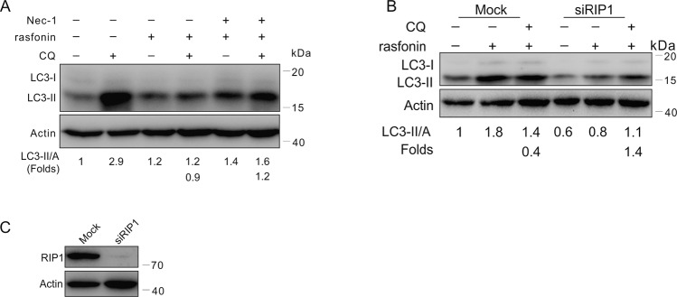
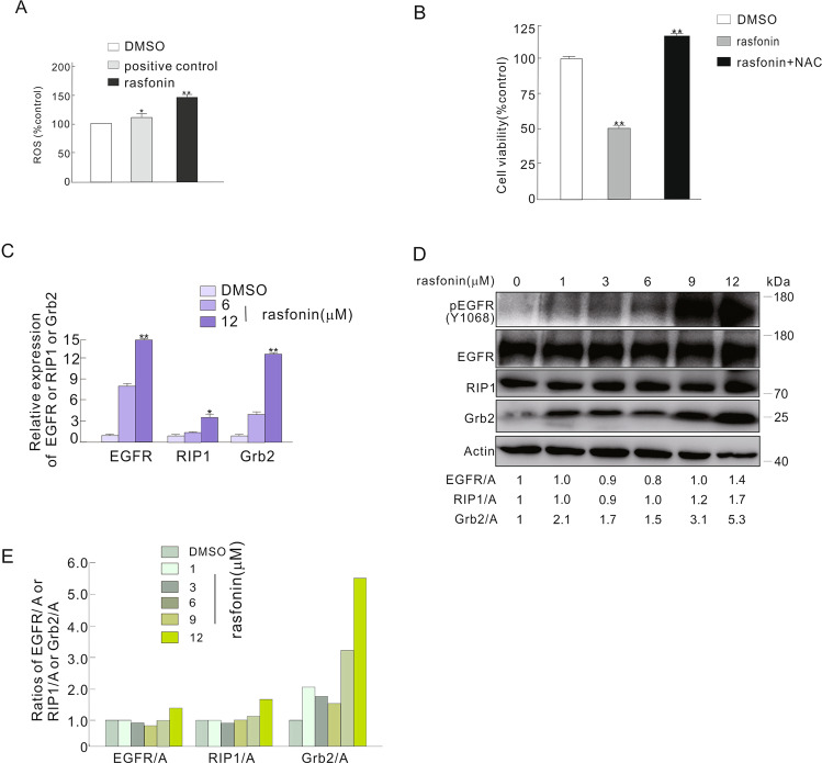
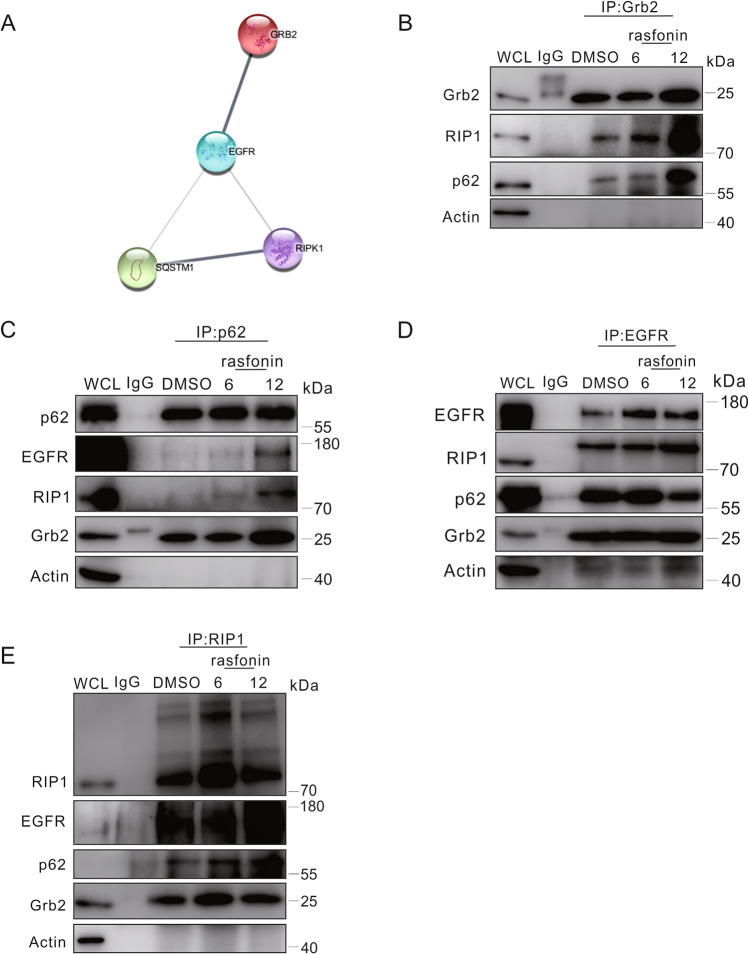
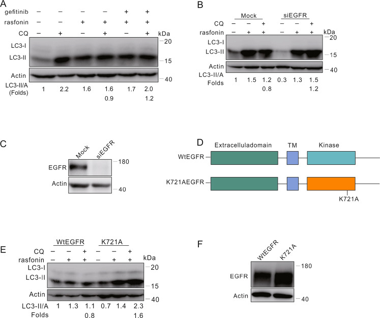
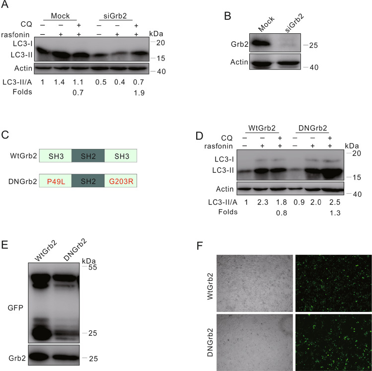
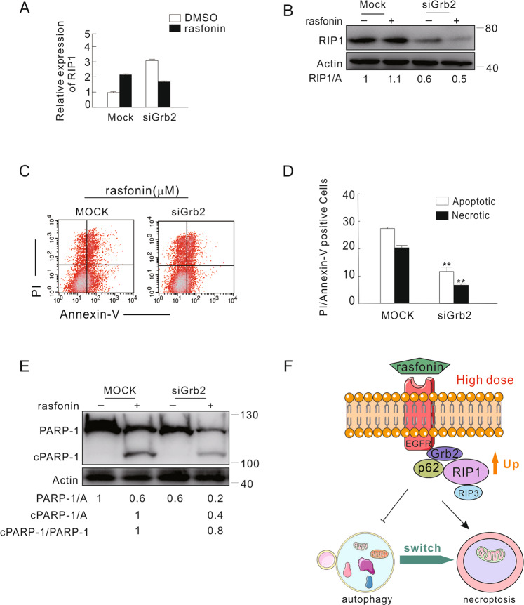
The underlying mechanism by which growth factor receptor-bound protein 2 (Grb2) regulates necroptosis remains unexplored. In the present study, we found that rasfonin, a fungal natural product and an activator of necroptosis, enhanced Grb2 binding to receptor-interacting serine/threonine kinase 1 (RIP1), which plays a critical role in regulating programmed necrosis. Moreover, we observed that SQSTM/p62 (p62), a protein that can form necrosomes with RIP1, increased its interaction with Grb2 upon rasfonin challenge. Although it has been used as an activator of autophagy in our previous study, here we found that a high dose of rasfonin was able to inhibit autophagic process. Inhibition of RIP1 either chemically or genetically reversed the inhibition of rasfonin on autophagy, whereas knockdown of Grb2 markedly reduced rasfonin-induced necrosis. Additionally, we found that the compound failed to upregulate the expression of RIP1 in Grb2-deprived cells. In summary, our data revealed that Grb2 actively participated in rasfonin-induced necroptosis by interacting with the components of necrosome and mediating their expression.


Conflict of interest statement

The authors declare no competing interests.

Figures

Fig. 1
Fig. 1. High concentration of rasfonin inhibited autophagy and induced necroptosis.
A ACHN cells were treated with rasfonin (0–12) μM for 4 h, the cells were lysed and subjected to immunoblotting with the antibodies indicated. Actin was used as a loading control. B, C ACHN cells were split onto coverslips, cultured overnight, following rasfonin (6 or 12 μM) for 6 h. the cells were fixed with 4% paraformaldehyde, immunostained with anti-LC3 antibody and visualized by fluorescence microscopy The number of the punctate LC3 in each cell was counted, and at least 50 cells were included for each group. Data representing the mean ± S.D. were shown in graph. D ACHN cells were treated with rasfonin (12 μM) in the presence or absence of necrostatin 1 (Nec-1, 30 μM) for 12 h; cell viability was analyzed by MTS assay as described in Materials and Methods. E, F ACHN cells were treated with rasfonin (12 μM) alone or together with Nec-1 (30 μM) for 6 h, the induced apoptosis and necrosis were determined by flow cytometry. Apoptotic: AV-positive and PI-negative; necrotic: PI-positive. The data are presented as mean ± S.D. from three independent experiments. The double asterisk denotes the group is statistically different from the control groups (P < 0.01). G Electron microscopy was performed in ACHN cells following treatment of rasfonin (12 μM) for 6 h as described in “Materials and methods”. Arrow: vesiculation of cytoplasmic organelle; Double Arrows: rupture of the plasma membrane. Similar experiments were repeated at least three times.
Fig. 2
Fig. 2. Rasfonin failed to inhibit autophagy in RIP1-deprived cells.
A ACHN cells were treated with rasfonin (12 μM) with or without Nec-1 (30 μM) for 4 h, and then cell lysates were prepared and analyzed by immunoblotting using the indicated antibodies. B, C After transfection with the control (Mock) or RIP1 siRNA (siRIP1) for 48 h, ACHN cells were treated with rasfonin (12 μM) for 4 h. Cell lysates were analyzed by immunoblotting with the indicated antibodies. Actin was used as a loading control. Similar experiments were repeated at least three times.
Fig. 3
Fig. 3. Rasfonin increased the expression of EGFR, Grb2 and RIP1.
A The level of reactive oxygen species (ROS) was detected as described in Materials and Methods following ACHN cells were treated with rasfonin (12 μM) or positive control for 12 h. B ACHN cells were treated with rasfonin (12 μM) in the presence or absence of N-acetyl-L-cysteine (NAC, 0.5 mM) for 12 h; cell viability was analyzed by MTS assay as described in Materials and Methods. C ACHN cells were challenged with rasfonin (6 and 12 μM) for 4 h and then, the total RNA was extracted, reversed, and detected by real-time PCR. D, E ACHN cells were treated with rasfonin (0–12 μM) for 4 h, the cells were lysed and subjected to immunoblotting with the antibodies indicated. Actin was used as a loading control. Data are presented as mean ± SD and are representatives of three independent experiments. Each performed in triplicate, and the data was analyzed by t-test. Single asterisk denotes that the group is statistically different from the control groups (p < 0.05), whereas double asterisk means (p < 0.01). The ratios between EGFR, RIP1 or Grb2 and Actin (A) were shown below the blots (D). Similar experiments were repeated at least three times.
Fig. 4
Fig. 4. Rasfonin enhanced the interaction of Grb2 with components of necrosomes.
A The STRING network analysis of possible interactions among EGFR, Grb2, RIP1 and p62. B–E ACHN cells were exposed to rasfonin (6 or 12 μM) for 6 h, and equal amounts of cell lysates were immunoprecipitated with the mouse monoclonal antibodies of Grb2, p62, EGFR, RIP1 or IgG. Immunoprecipitates were then immunoblotted for the indicated polyclonal (Grb2, p62 and EGFR) or monoclonal (RIP1) antibodies. Similar experiments were repeated twice.
Fig. 5
Fig. 5. Knockdown of EGFR reversed the inhibitory effect of rasfonin on autophagy.
A ACHN cells were treated with rasfonin (12 μM) with or without gefitinib (5 μM) for 4 h. B After transfection with the control (Mock) or EGFR siRNA (siEGFR) for 48 h, ACHN cells were treated with rasfonin (12 μM) for 4 h. Cell lysates in (A, B) were analyzed by immunoblotting with the indicated antibodies. C The efficiency of siRNA interference was determined with sample in (B). D Schematic representation of the WT EGFR and K721A EGFR. E, F HEK293T cells were transfected transiently with the WT EGFR or K721A EGFR plasmids for 36 h, and treated with rasfonin (12 μM) for 4 h. Cell lysates were analyzed by immunoblotting with the indicated antibody. Actin was used as a loading control. Similar experiments were repeated at least three times.
Fig. 6
Fig. 6. Rasfonin failed to inhibit autophagy in Grb2-depleted cells.
A Following transfection with the control (Mock) or Grb2 siRNA (siGrb2) for 48 h, ACHN cells were treated with rasfonin (12 μM) for 4 h. Cell lysates were analyzed by immunoblotting with the indicated antibodies. B The efficiency of siRNA interference was determined with sample in (A). C Schematic representation of the WT Grb2 and mutant DN Grb2. D, E HEK293T cells were transfected transiently with the WT Grb2 or DN Grb2 plasmids for 36 h, and treated with rasfonin (12 μM) for 4 h. Cell lysates were analyzed by immunoblotting with the indicated antibody. Actin was used as a loading control. F The transfection efficiency was detected and acquired by using fluorescence microscope. Similar experiments were repeated at least three times.
Fig. 7
Fig. 7. Grb2 mediated the expression of RIP1 and was involved in rasfonin-induced necroptosis.
A–E After transfection with the control (Mock) or Grb2 siRNA (siGrb2) for 48 h, ACHN cells were exposed to 12 μM rasfonin for 6 h, and the total RNA was extracted, reversed, and detected by real-time PCR (A); cell lysates were analyzed by immunoblotting with the indicated antibodies, Actin was used as a loading control (B); the apoptosis and necrosis induced by rasfonin were determined by flow cytometry (C and D). Apoptotic: AV-positive and PI-negative; necrotic: PI-positive. The data are presented as mean ± S.D. from three independent experiments. The double asterisk denotes the group is statistically different from the control groups (P < 0.01). E Cell lysates in (B) were analyzed by immunoblotting with the indicated antibodies, and ratios between cleaved PARP-1 (cPARP-1) and PARP-1, or between cPARP-1 and Actin (A), or between PARP-1 and Actin (A) were shown below the blots. Similar experiments were repeated at least three times. F Schematic mechanism of EGFR/Grb2 in rasfonin-dependent autophagy and necroptosis.

Similar articles

Cited by

References

    1. Fath I, Schweighoffer F, Rey I, Multon MC, Boiziau J, Duchesne M, et al. Cloning of a Grb2 isoform with apoptotic properties. Science. 1994;264:971–4. doi: 10.1126/science.8178156. - DOI - PubMed
    1. Wang Z. ErbB receptors and cancer. Methods Mol Biol. 2017;1652:3–35. doi: 10.1007/978-1-4939-7219-7_1. - DOI - PubMed
    1. Giubellino A, Burke TR, Jr., Bottaro DP. Grb2 signaling in cell motility and cancer. Expert Opin Ther Targets. 2008;12:1021–33. doi: 10.1517/14728222.12.8.1021. - DOI - PMC - PubMed
    1. Ijaz M, Wang FS, Shahbaz M, Jiang WJ, Fathy AH, Nesa EU. The role of Grb2 in cancer and peptides as Grb2 antagonists. Protein Pept Lett. 2017;24:1084–95. - PubMed
    1. Hou B, Xu S, Xu Y, Gao Q, Zhang C, Liu L, et al. Grb2 binds to PTEN and regulates its nuclear translocation to maintain the genomic stability in DNA damage response. Cell Death Dis. 2019;10:546. doi: 10.1038/s41419-019-1762-3. - DOI - PMC - PubMed