. 2022 Jul 13;13(1):4065.
doi: 10.1038/s41467-022-31815-1.

A SOX17-PDGFB signaling axis regulates aortic root development

Affiliations

A SOX17-PDGFB signaling axis regulates aortic root development

Pengfei Lu et al. Nat Commun. .

Abstract

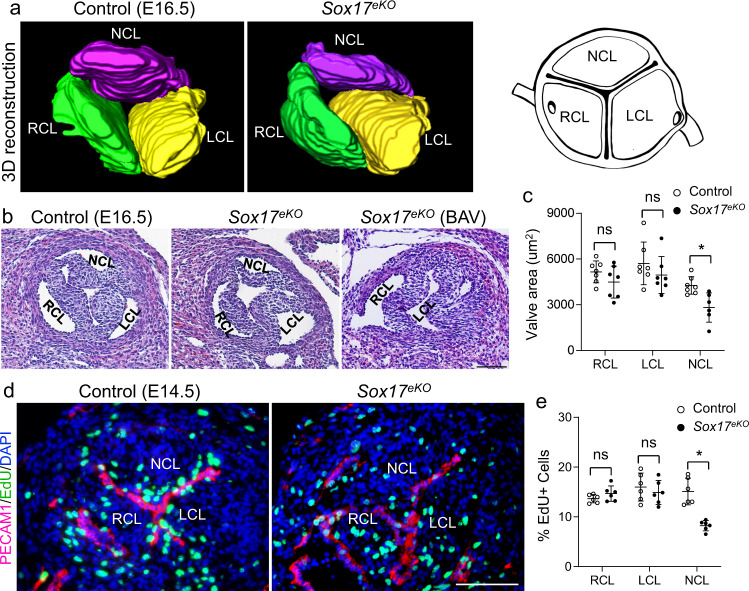
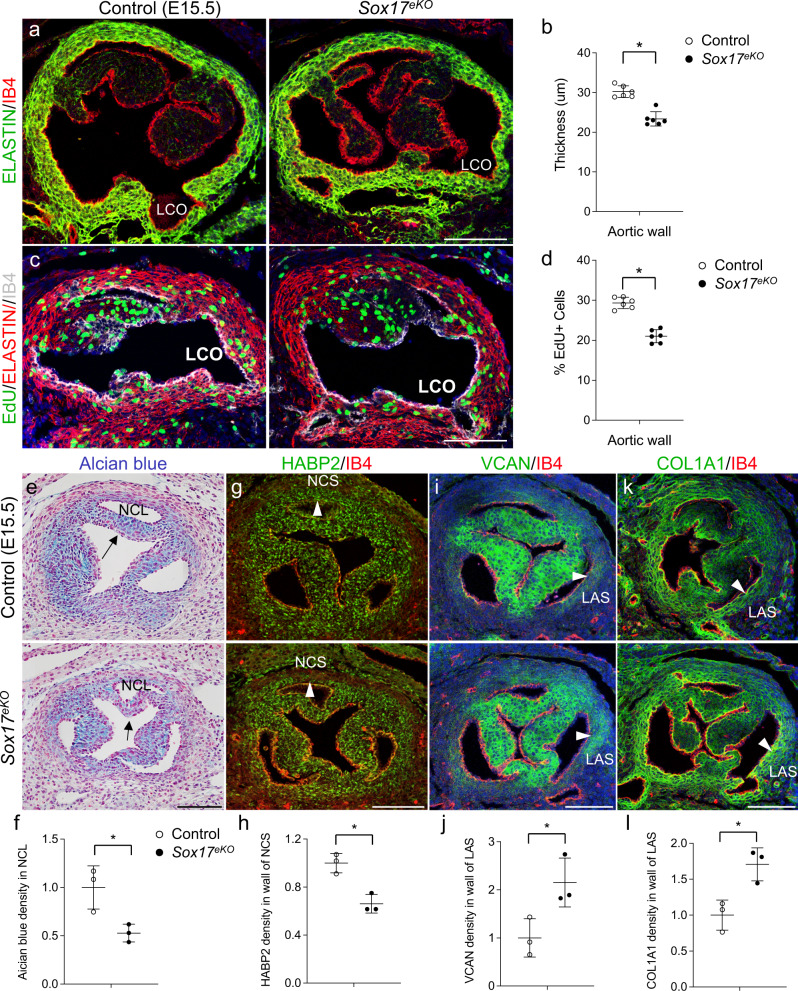
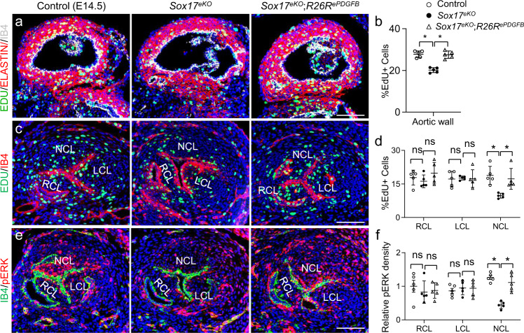
Developmental etiologies causing complex congenital aortic root abnormalities are unknown. Here we show that deletion of Sox17 in aortic root endothelium in mice causes underdeveloped aortic root leading to a bicuspid aortic valve due to the absence of non-coronary leaflet and mispositioned left coronary ostium. The respective defects are associated with reduced proliferation of non-coronary leaflet mesenchyme and aortic root smooth muscle derived from the second heart field cardiomyocytes. Mechanistically, SOX17 occupies a Pdgfb transcriptional enhancer to promote its transcription and Sox17 deletion inhibits the endothelial Pdgfb transcription and PDGFB growth signaling to the non-coronary leaflet mesenchyme. Restoration of PDGFB in aortic root endothelium rescues the non-coronary leaflet and left coronary ostium defects in Sox17 nulls. These data support a SOX17-PDGFB axis underlying aortic root development that is critical for aortic valve and coronary ostium patterning, thereby informing a potential shared disease mechanism for concurrent anomalous aortic valve and coronary arteries.


Conflict of interest statement

The authors declare no competing interests.

Figures

Fig. 1
Fig. 1. Sox17 expression in the aortic root endothelium is necessary for heart development and embryonic survival.
a Schematic view of aortic root (AR) shows the anatomic relationship of aortic valve, aortic sinus and coronary ostium. LCL/RCL/NCL: left, right and non-coronary leaflet; LCO/RCO: left and right coronary ostium, LCA/RCA: left and right coronary artery. b Representative immunofluorescence (IF) (n = 3/group) shows specific and developmentally regulated SOX17 expression (red) in the aortic root endothelium of E11.5 and E14.5 hearts. c IF (n = 5/group) shows diminished SOX17 expression (red) in the aortic root endothelium of E12.5 Sox17eKO hearts. AV: aortic valve. d IF (n = 5/group) shows reduced SOX17 expression (red) in the aortic root endothelium in E15.5 Sox17eKO hearts. Note that the SOX17 expression in the coronary artery endothelium is not affected by the valve specific deletion using the transgenic Nfatc1-enhancer Cre which expresses specifically in the aortic root endothelium including the valve endothelium. e H&E staining (n = 5/group) of frontal (top panels) and transverse sections (bottom panels) show thin ventricular wall in all 8 E16.5 Sox17eKO embryos examined. LV/RV: left and right ventricle. f Echocardiography (n = 3/group) shows decreased aortic velocity in E16.5 Sox17eKO hearts. Scale bar: 100 µm in bd, 200 µm in e.
Fig. 2
Fig. 2. Deletion of Sox17 in the aortic root endothelium causes aortic valve defects.
a 3D reconstruction of continuous H&E stained sections of aortic root shows reduced NCL volume in the E16.5 Sox17eKO heart (n = 5/group). A schematic view of aortic valve leaflets is on the right. b Representative H&E stained transverse sections show underdeveloped NCL and BAV without NCL in the E16.5 Sox17eKO heart. c Quantitative analysis shows significantly decreased area of NCL in E16.5 Sox17eKO hearts. (n = 7 for control, n = 6 for Sox17eKO, mean ± SD, unpaired two-tailed t-test, p = 0.0063 for NCL, *p < 0.05). BAV was excluded from the quantitative analysis. d Representative images from EdU (green) assays on sections of E14.5 hearts show reduced cell proliferation in NCL. e Quantitative analysis of EdU assays. (n = 6/group, mean ± SD, unpaired two-tailed t-test, p = 0.0002 for NCL, *p < 0.05). Source data are provided as a Source Data file. Scale bars: 100 µm.
Fig. 3
Fig. 3. Sox17 deletion in the aortic root endothelium impairs aortic root growth and maturation.
a IF for ELASTIN (green) shows thin aortic root wall in E15.5 Sox17eKO hearts. b Quantitative analysis shows a significant reduction in the wall thickness. c EdU (green) assay shows reduced proliferation of VSMCs in the aortic root wall marked by red ELSTAIN staining in Sox17eKO hearts. IB4 (white) stains the endothelium. d Quantitative analysis shows a significant reduction in VSMC proliferation. (For (ad) Figures, n = 6/group, mean ± SD, unpaired two-tailed t-test, p = 0.00003 for (b), p = 0.000003 for (d), *p < 0.05). e, f Representative Alcian blue staining images and quantitative analysis of E15.5 control and Sox17eKO hearts show reduced proteoglycans in NCL (arrow) of the Sox17eKO hearts. g, h Representative IF images and quantitative analysis of E15.5 control and Sox17eKO hearts indicate decreased HABP2 (green) in the wall of NCS (arrowhead). i, j Representative IF images and quantitative analysis of E15.5 control and Sox17eKO hearts show increased VCAN (green) in the wall of LAS (arrowhead). k, l Representative IF images and quantitative analysis of E15.5 control and Sox17eKO hearts showing upregulated COL1A1 (green) in the wall of LAS (arrowhead). In el, n = 3/group, mean ± SD, unpaired two-tailed t-test, p = 0.027 for (f), p = 0.006 for (h), p = 0.036 for (j), p = 0.017 for (l), *p < 0.05. Source data are provided as a Source Data file. Scale bars: 100 µm.
Fig. 4
Fig. 4. Deletion of Sox17 results in high-takeoff left coronary ostium.
a Schematic view shows the coronary ostium position within the aortic root. Top plane indicates the sino-tubular junction between the aortic sinus and the ascending aorta. Bottom plane indicates the ventricular-aortic junction between the left ventricle and the aortic root. Dashed line a’ indicates the coronary ostia (origin of each main coronary stem in the respective aortic sinus). Dashed line b’ indicates the base of aortic valve leaflets. b, c IF images (n = 5/group) for the GFP reporter (green, indicating the lineage of Nfatc1enCre-marked aortic root endothelial cells) and for SOX17 staining (red) for aortic root and coronary arterial endothelium. Note a sharp tissue boundary (b, double arrows) between the GFP-positive/SOX17-deleted aortic root endothelium and the GFP-negative/SOX17-expressing coronary artery endothelium, which marks the coronary entries or ostia. The main coronary arteries are negative for GFP but positive for SOX17, indicating that the deletion does not affect the coronary artery endothelium. d, e Representative H&E stained continuous sections E16.5 control and Sox17eKO hearts from the base of aortic valve leaflets to the coronary ostia shows that LCO in E16.5 Sox17eKO hearts is shifted up and posteriorly toward the left coronary sinus. Arrowheads indicate the main coronary arteries and ostia. f Quantitative analysis of (d, e) confirms the high-takeoff LCO. (n = 7 for control, n = 5 for Sox17eKO, mean ± SD, unpaired two-tailed t-test, p = 0.015, *p < 0.05). g, h Representative smMHC IF images (n = 5/group) show the high-takeoff LCO (single arrow) in E16.5 Sox17eKO hearts and upshifted ventricular (smMHC-negative)-aortic (smMHC-positive) junction (double arrows). Source data are provided as a Source Data file. Scale bars: 100 µm.
Fig. 5
Fig. 5. Misplaced LCO causes reduced coronary flow, leading to lethal anomalous coronary arteries.
a Representative whole heart smMHC IF images (n = 5/group) of E16.5 control and Sox17eKO hearts show narrowed LCA (arrow) and collateral vessels extended from RCA to LCA (arrowheads) in the mutant hearts. b Representative images of whole heart PECAM1 IF (n = 3/group) of E16.5 control and Sox17eKO embryos show narrowed LCA (arrows) in Sox17eKO hearts. c, d Representative IF images (n=5/group) of E16.5 control and Sox17eKO hearts shows reduced endothelial expression of KLF4 (red, arrow) in LCO (c) and LCA (d) of Sox17eKO hearts. e Quantitative analysis of (c, d) confirms reduced aortic-coronary flow at LCO. (n = 5/group, mean ± SD, unpaired two-tailed t-test, p = 0.001 for LCO, p = 0.0014 for LCA, *p < 0.05). f Computational simulation shows reduced wall sheer stress (WSS) from 82 to 55 cgs (centimetre–gram–second system of units) at LCO in E16.5 Sox17eKO hearts. g, h Representative hypoxia probe staining images (n = 4/group) of E13.5 or E15.5 control and Sox17eKO hearts shows severe hypoxia developed in the left ventricle (LV) of E15.5 Sox17eKO hearts (h), which also affects the right ventricle (RV) after the establishment of coronary circulation around E14.5, but not E13.5 Sox17eKO hearts before functional coronary circulation starts (g). Source data are provided as a Source Data file. Scale bars: 500 µm in a, b, g, h; 50 µm in c, d.
Fig. 6
Fig. 6. SOX17 regulates PDGFB signaling in the NCL mesenchyme.
a Representative RNAscope ISH images (n = 3/group) of E13.5 control and Sox17eKO hearts show diminished Pdgfb expression (red) in the aortic valve endothelium in Sox17eKO hearts. Noted that only the large dots are considered the real signals. b, c IF images of E15.5 control and Sox17eKO hearts show significantly reduced phosphorylated ERK (pERK) density (red) in the NCL mesenchyme of Sox17eKO hearts. (n = 4/group, mean ± SD, unpaired two-tailed t-test, p = 0.011, *p < 0.05). d, e IF images of E14.5 control and Sox17eKO hearts show significantly reduced PDGFRB density (red) in the aortic valve region, especially in NCL, of the Sox17eKO hearts. (n = 5/group, mean ± SD, unpaired two-tailed t-test, p = 0.007 for RCL, p = 0.006 for LCL, p = 0.0001 for NCL, *p < 0.05). Source data are provided as a Source Data file. Scale bars: 100 µm.
Fig. 7
Fig. 7. SOX17 regulates Pdgfb expression by directly binding to Pdgfb distal enhancer.
a UCSC Genome browser tracks show two Pdgfb transcriptional enhancers containing the consensus SOX17 binding motifs, one located at the ~10 Kb upstream of the first exon of Pdgfb (enhancer 1) and the other between exon 2 and 3 (enhancer 2) identified for E14.5 and adult mouse hearts based on the mouse ENCODE project. b ChIP-qPCR shows the binding of SOX17 at enhancer 1. (n = 3/group, mean ± SD, unpaired two-tailed t-test, p = 0.03 for enhancer 1, *p < 0.05). c Reporter gene assays show that enhancer 1 activates the promoter of reporter gene and the mutagenesis analysis indicates that the activation is dependent on the intact SOX17 binding motifs. (n = 4/group, mean ± SD, unpaired two-tailed t-test, p = 0.000003 for enhancer 1-WT, p = 0.002 for enhancer 1-Del, *p < 0.05). Source data are provided as a Source Data file.
Fig. 8
Fig. 8. PDGFB re-expression rescues aortic root defects.
a Representative RNAscope images (n = 3/group) of E13.5 control and Sox17eKO;R26RePDGFB hearts showing the human PDGFB expression (red) in aortic endothelium in the Sox17eKO;R26RePDGFB heart. b, c H&E staining (n=5/group) showing normal ventricular wall and NCL, respectively, in all 5 E16.5 Sox17eKO;R26RePDGFB embryos examined. LV/RV: left and right ventricle. d Quantitative analysis shows comparable NCL area in E16.5 Sox17eKO;R26RePDGFB and control hearts. n = 5/group, mean ± SD, unpaired two-tailed t-test, ns, no significance. e Representative H&E stained continuous sections E16.5 control and Sox17eKO;R26RePDGFB hearts from the base of aortic valve leaflets to the coronary ostia showing that LCO is correctly positioned in E16.5 Sox17eKO;R26RePDGFB hearts compared to the controls. Arrowheads indicate the main coronary arteries and ostia. f Quantitative analysis showing no difference in the LCO position related to aortic valve base between Sox17eKO;R26RePDGFB hearts and controls. n = 5/group, mean ± SD, unpaired two-tailed t-test, ns, no significance. g, h H&E staining and quantitative analysis show the normal aortic wall thickness in E16.5 Sox17eKO;R26RePDGFB hearts. n = 5/group, mean ± SD, unpaired two-tailed t-test, ns, no significance. i Representative hypoxia probe staining images (n = 5/group) showing the similar staining pattern and intensity between E16.5 control and Sox17eKO;R26P hearts. Source data are provided as a Source Data file. Scale bars: 100 µm in a, c, e, g; 200 µm in b, i.
Fig. 9
Fig. 9. PDGFB re-expression rescues reduced cell proliferation of VSMCs and NCL and decreased pERK activity resulted from the Sox17 deletion.
a, b EdU (green) assay and quantitative analysis showing the reduced proliferation of VSMCs in the aortic root wall marked by ELSTAIN (red) staining in the Sox17eKO hearts was rescued in the Sox17eKO;R26RePDGFB heart. IB4 (white) stains the endothelium. n = 5/group, mean ± SD, one-way ANOVA Tukey’s test, p = 0.00002 for Sox17eKO, p = 0.00006 for Sox17eKO;R26RePDGFB, *p < 0.05). c, d EdU (green) assay and quantitative analysis showing the reduced proliferation of NCL in the Sox17eKO hearts was rescued in the Sox17eKO;R26RePDGFB hearts. n = 5/group, mean ± SD, one-way ANOVA Tukey’s test, p = 0.001 for NCL of Sox17eKO, p = 0.008 for NCL of Sox17eKO;R26RePDGFB, *p < 0.05. e, f IF images and quantitative analysis showing the reduced phosphorylated ERK (pERK) density (red) in the NCL mesenchyme of the Sox17eKO hearts was restored in the Sox17eKO;R26RePDGFB heart. n = 5/group, mean ± SD, one-way ANOVA Tukey’s test, p = 0.000004 for NCL of Sox17eKO, p = 0.0006 for NCL of Sox17eKO;R26RePDGFB, *p < 0.05. Source data are provided as a Source Data file. Scale bars: 100 µm.
Fig. 10
Fig. 10. Summary of molecular, cellular and tissue structural changes in the mouse model of BAV syndrome caused by the Sox17 inactivation in aortic root endothelium.
A working model showing the SOX17 deficiency results in downregulated Pdgfb transcription in aortic root endothelium and reduced endothelial to mesenchymal PDGFB signaling that lead to cell proliferation and maturation defects in the developing aortic root. These defects cause complex aortic root malformations including BAV, hypoplastic aortic wall, as well as high takeoff left coronary ostium that lead to lethal coronary artery anomalies.

References

    1. Siu SC, Silversides CK. Bicuspid aortic valve disease. J. Am. Coll. Cardiol. 2010;55:2789–2800. doi: 10.1016/j.jacc.2009.12.068. - DOI - PubMed
    1. Villa AD, et al. Coronary artery anomalies overview: the normal and the abnormal. World J. Radio. 2016;8:537–555. doi: 10.4329/wjr.v8.i6.537. - DOI - PMC - PubMed
    1. Rashid A, Saucedo JF, Hennebry TA. Association of single coronary artery and congenital bicuspid aortic valve with review of literature. J. Inter. Cardiol. 2005;18:389–391. doi: 10.1111/j.1540-8183.2005.00067.x. - DOI - PubMed
    1. Fedak PW, et al. Clinical and pathophysiological implications of a bicuspid aortic valve. Circulation. 2002;106:900–904. doi: 10.1161/01.CIR.0000027905.26586.E8. - DOI - PubMed
    1. Meindl C, et al. Coronary Artery Ectasia Are Frequently Observed in Patients With Bicuspid Aortic Valves With and Without Dilatation of the Ascending Aorta. Circ. Cardiovasc. Interv. 2016;9:e004092. doi: 10.1161/CIRCINTERVENTIONS.116.004092. - DOI - PubMed

Publication types