. 2022 Jul 13;12(1):11918.
doi: 10.1038/s41598-022-15756-9.

Efficacy of auranofin as an inhibitor of desmoid progression

Affiliations

Efficacy of auranofin as an inhibitor of desmoid progression

Kan Ito et al. Sci Rep. .

Abstract

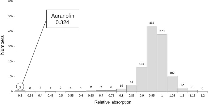
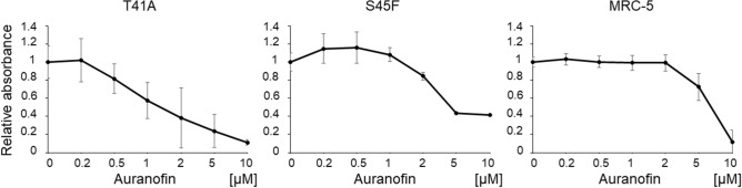
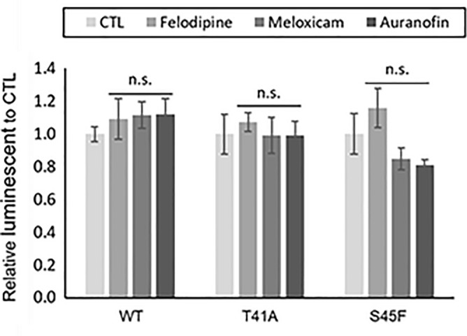
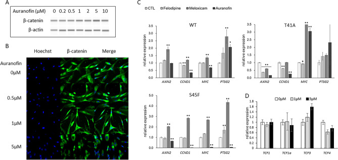
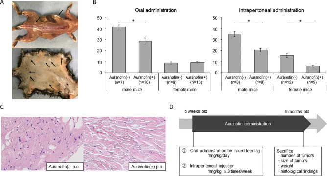
Anticancer drugs and molecular targeted therapies are used for refractory desmoid-type fibromatosis (DF), but occasionally cause severe side effects. The purpose of this study was to identify an effective drug with fewer side effects against DF by drug repositioning, and evaluate its efficacy. FDA-approved drugs that inhibit the proliferation of DF cells harboring S45F mutations of CTNNB1 were screened. An identified drug was subjected to the investigation of apoptotic effects on DF cells with analysis of Caspase 3/7 activity. Expression of β-catenin was evaluated with western blot analysis, and immunofluorescence staining. Effects of the identified drug on in vivo DF were analyzed using Apc1638N mice. Auranofin was identified as a drug that effectively inhibits the proliferation of DF cells. Auranofin did not affect Caspase 3/7 activity compared to control. The expression level of β-catenin protein was not changed regardless of auranofin concentration. Auranofin effectively inhibited the development of tumorous tissues by both oral and intraperitoneal administration, particularly in male mice. Auranofin, an anti-rheumatic drug, was identified to have repositioning effects on DF. Since auranofin has been used for many years as an FDA-approved drug, it could be a promising drug with fewer side effects for DF.


Conflict of interest statement

The authors declare no competing interests.

Figures

Figure 1
Figure 1
Screening for FDA-approved drugs. The S45F-mutated cells were cultured for 24 h in a 96 well culture plate of 5 × 103 cells/well in the presence of 10 μM 1200 FDA-approved compounds. The cell proliferation was measured using the MTS assay, and the absorbance (at 490 nm) was measured using a microplate reader. The number of drugs is plotted for each growth inhibition rate of desmoid cells. If the relative absorption is less than 1, the growth inhibition will be stronger. Auranofin was extracted as an effective and safe drug, which had a proliferation rate of 0.324.
Figure 2
Figure 2
Effects of auranofin on DF cell proliferation. Inhibitory effects of auranofin on cell proliferation were evaluated with MTS assay. The DF cells were seeded on a 96-well plate (5 × 103 cells/well) for 12 h. Thereafter, the effects of auranofin at each concentration (0.2 to 10 µM) on cell proliferation was measured using the MTS assay kit after 24 h.
Figure 3
Figure 3
Apoptosis assay for DF cells under drug administration. The DF cells (WT, T41A, and S45F) were seeded on a 96-well plate (5 × 103 cells/well) for 12 h. Thereafter, the effects of auranofin, felodipine, and meloxicam on the activity of Caspase 3/7 was measured using a Caspase assay kit). CTL: control, n.s.: not significant.
Figure 4
Figure 4
Effects of auranofin on expression of β-catenin and downstream genes of Wnt/β-catenin signaling pathway. (A) Effects of auranofin (0–10 µM) on the expression of β-catenin protein. The T41A-mutated cells were seeded on a 6-well plate (1 × 105 cells per well) for 12 h followed by auranofin (0–10 µM) treatment for 24 h. These cells were subjected to western blotting using Simple Western for β-catenin and β-actin. (B) Fluorescent immunostaining for β-catenin. The T41A-mutated cells were seeded onto chamber slides (1.0 × 105 cells/ml) for 12 h followed by auranofin treatment (0–5 µM) for 24 h. Cells were fluorescently visualized using mouse anti-β-catenin antibody. (C) Expression levels of mRNA, AXIN2, CCND1, MYC, PTGS2. The DF cells (WT, T41A and S45F: 1 × 104 cells per well) were seeded in a 96-well plate for 12 h. Subsequently the cells were cultured in a medium containing felodipine, meloxicam, and auranofin (5 μM) for 6 h, and subjected to RT-PCR. CTL: control. (D) Effects of auranofin (0–5 µM) on the mRNA expression of four TCFs. The T41A cells (1 × 104 cells per well) were seeded in a 96-well plate for 12 h. Subsequently the cells were cultured in a medium containing auranofin (0–5 µM) for 6 h, and subjected to RT-PCR. *P < 0.05, **P < 0.01.
Figure 5
Figure 5
Schedule of auranofin administration for Apc1638N mice and effects of auranofin in in vivo DF development model mice. (A) Representative findings of tumor development in a Apc1638N mouse (6 months of age) (black arrows: tumorous tissues). Small white tumorous tissues of a few millimeters in size occurred subcutaneously and on the fascia. (B) The number of tumors that developed in Apc1638N (6 months of age) with or without treatment of auranofin by oral or intraperitoneal administration. Oral dose for mice was set at 1.0 mg/kg/day and the intraperitoneal dose was set as 1 mg/kg three times a week. The average number of tumors developed with oral administration of auranofin (10 male mice and 13 female mice) and control (7 male mice and 8 female mice), ant that with intraperitoneal administration of auranofin (8 male mice and 9 female mice) and control (8 male mice and 12 female mice) were plotted. *P < 0.01. (C) Pathological findings of the tumors developed in mice with or without treatment of auranofin by oral administration (6 months of age) (H&E, × 200). p.o.: per OS. (D) Experimental procedure in vivo. Auranofin was administered from 5 weeks to 6 months of age. Two separate experiments were performed based on the administration route, oral and intraperitoneal. Mice were sacrificed at 6 months of age, and the number of tumors, tumor size, tumor weight were calculated. Tumorous tissues were also subjected to histological examination.

Similar articles

Cited by

References

    1. Nishida Y, et al. Transition of treatment for patients with extra-abdominal desmoid tumors: Nagoya university modality. Cancers (Basel) 2012;4:88–99. doi: 10.3390/cancers4010088. - DOI - PMC - PubMed
    1. Alman B, et al. The management of desmoid tumours: A joint global consensus-based guideline approach for adult and paediatric patients. Eur. J. Cancer. 2020;127:96–107. doi: 10.1016/j.ejca.2019.11.013. - DOI - PubMed
    1. Penel N, et al. Surgical versus non-surgical approach in primary desmoid-type fibromatosis patients: A nationwide prospective cohort from the French Sarcoma Group. Eur. J. Cancer. 2017;83:125–131. doi: 10.1016/j.ejca.2017.06.017. - DOI - PubMed
    1. Nishida Y, et al. Successful treatment with meloxicam, a cyclooxygenase-2 inhibitor, of patients with extra-abdominal desmoid tumors: A pilot study. J. Clin. Oncol. 2010;28:e107–e109. doi: 10.1200/JCO.2009.25.5950. - DOI - PubMed
    1. Hansmann A, Adolph C, Vogel T, Unger A, Moeslein G. High-dose tamoxifen and sulindac as first-line treatment for desmoid tumors. Cancer. 2004;100:612–620. doi: 10.1002/cncr.11937. - DOI - PubMed

Publication types