. 2022 Jul 13;12(1):11935.
doi: 10.1038/s41598-022-13670-8.

Ultrafast-UV laser integrating cavity device for inactivation of SARS-CoV-2 and other viruses

Affiliations

Ultrafast-UV laser integrating cavity device for inactivation of SARS-CoV-2 and other viruses

Sharad Ambardar et al. Sci Rep. .

Abstract

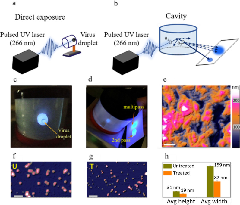
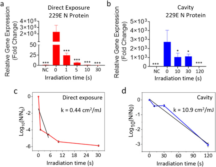
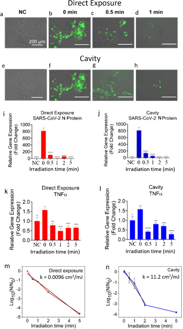
Ultraviolet (UV) irradiation-based methods used for viral inactivation have provided an important avenue targeting severe acute respiratory-syndrome coronavirus-2 (SARS-CoV-2) virus. A major problem with state-of-the-art UV inactivation technology is that it is based on UV lamps, which have limited efficiency, require high power, large doses, and long irradiation times. These drawbacks limit the use of UV lamps in air filtering systems and other applications. To address these limitations, herein we report on the fabrication of a device comprising a pulsed nanosecond 266 nm UV laser coupled to an integrating cavity (LIC) composed of a UV reflective material, polytetrafluoroethylene. Previous UV lamp inactivation cavities were based on polished walls with specular reflections, but the diffuse reflective UV ICs were not thoroughly explored for virus inactivation. Our results show that LIC device can inactivate several respiratory viruses including SARS-CoV-2, at ~ 1 ms effective irradiation time, with > 2 orders of magnitude higher efficiency compared to UV lamps. The demonstrated 3 orders of magnitude cavity enhancement relative to direct exposure is crucial for the development of efficient real-time UV air and water purification systems. To the best of our knowledge this is the first demonstration of LIC application for broad viral inactivation with high efficiency.


Conflict of interest statement

The authors declare no competing interests.

Figures

Figure 1
Figure 1
Virus inactivation using ultrafast UVC laser integrating cavity. (a) Schematic of the direct exposure of pulsed UVC laser on a droplet of virus solution in a vial. (b) Schematic of the LICD exposure of UVC laser irradiation on a droplet of virus placed inside the cavity at the location of aperture a2. (c) Photograph of the cavity with a virus droplet inside a vial. (d) Photograph of the cavity filled with UVC laser light and the reflection of fluorescence from the 2nd pass and multipass scattering as two bright spots on a white card, placed at the a2 aperture. (e) AFM height image of PTFE sheet used as a diffuse reflective LICD cavity wall. (f, g) AFM height images of the untreated (U) and UVC laser treated (T) HCoV-229E virions. Scale bar is 0.3 µm. (h) Average height and width of the untreated and treated HCoV-229E virions.
Figure 2
Figure 2
HCoV-229E virus was exposed to direct pulsed UVC laser (a) and cavity (b) for the indicated times. Calu-3 cells were treated 24 h after seeding with the indicated groups of 229E (3 MOI). At 72 h post-infection RNA was extracted and qPCR performed. Average fold change ± SEM, compared to the negative control (NC), is shown (N = 3). A 1-Way ANOVA and Dunnett’s post hoc test was used to determine significance compared to 0 s. HCoV-229E survival as a function of UV irradiation time via direct exposure (c) and cavity (d). *p < 0.05, **p < 0.01, ***P < 0.001, ****P < 0.0001.
Figure 3
Figure 3
SARS-CoV-2 virus was exposed to indicated irradiation times of direct or cavity UVC laser light. Calu-3 cells were infected 24 h after seeding with the indicated groups of CoV-2 (0.1 MOI). (ah) Images were taken 48 h post-infection using the EVOS microscope (Thermo Fisher). 200X. Scale bar = 200 µm. (ij) SARS-CoV-2 N protein expression in Calu-3 cells. At 72 h post-infection RNA was extracted and qPCR performed. Average fold change ± SEM, compared to the negative control (NC), is shown (N = 3). A 1-Way ANOVA and Dunnett’s post hoc test was used to determine significance compared to 0 s. (kl) Inflammatory marker expression in Calu-3 cells after SARS-CoV-2 infection. For the negative control, CoV-2 was exposed to UVC light under a handheld wand for 2 min. At 48 h post-infection RNA was extracted and qPCR performed. Average fold change ± SEM, compared to the negative control (NC), is shown (N = 3). A 1-Way ANOVA and Dunnett’s post hoc test was used to determine significance compared to 0 s. SARS-CoV-2 survival as a function of irradiation time via direct exposure (m) and cavity (n). *p < 0.05, **p < 0.01, ***P < 0.001, ****P < 0.0001.
Figure 4
Figure 4
Reinfection of Calu-3 cells with culture supernatant from SARS-CoV-2 infected Calu-3 cells that were exposed to direct or cavity UVC laser light (Fig. 3). (ad) Images were taken 48 h post-infection using EVOS microscope (Thermo Fisher). 200X. Scale bar = 200 µm. (ef) SARS-CoV-2 N protein expression in Calu-3 cells. At 48 h post-infection RNA was extracted and qPCR performed. Average fold change ± SEM, compared to the negative control (NC), is shown (N = 3). A 1-Way ANOVA and Dunnett’s post hoc test was used to determine significance compared to 0 s. *p < 0.05, **p < 0.01, ***P < 0.001, ****P < 0.0001.

References

    1. McGill A, et al. SARS-CoV-2 Immuno-pathogenesis and potential for diverse vaccines and therapies: opportunities and challenges. Infect. Dis. Rep. 2021;13:102–125. doi: 10.3390/idr13010013. - DOI - PMC - PubMed
    1. Tiwari S, et al. Antibacterial and antiviral high-performance nano-systems to mitigate new SARS-CoV-2 variants of concerns. Curr. Opin. Biomed. Eng. 2021;21:100363. doi: 10.1016/j.cobme.2021.100363. - DOI - PMC - PubMed
    1. Kaushik AK, Dhau JS. Photoelectrochemical oxidation assisted air purifiers; perspective as potential tools to control indoor SARS-CoV-2 exposure. Appl. Surf. Sci. Adv. 2022;9:100236. doi: 10.1016/j.apsadv.2022.100236. - DOI
    1. Sabino CP, et al. Light-based technologies for management of COVID-19 pandemic crisis. J. Photochem. Photobiol. B Biol. 2020;212:111999. doi: 10.1016/j.jphotobiol.2020.111999. - DOI - PMC - PubMed
    1. Darnell ME, Subbarao K, Feinstone SM, Taylor DR. Inactivation of the coronavirus that induces severe acute respiratory syndrome, SARS-CoV. J. Virol. Methods. 2004;121:85–91. doi: 10.1016/j.jviromet.2004.06.006. - DOI - PMC - PubMed

Publication types