. 2022 Jul;607(7920):816-822.
doi: 10.1038/s41586-022-04952-2. Epub 2022 Jul 13.

Mechanisms and inhibition of Porcupine-mediated Wnt acylation

Affiliations

Mechanisms and inhibition of Porcupine-mediated Wnt acylation

Yang Liu et al. Nature. 2022 Jul.

Abstract

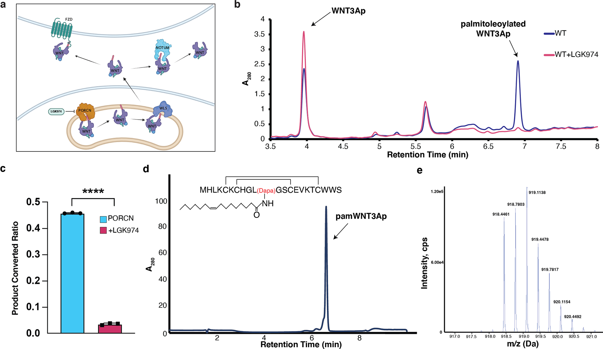
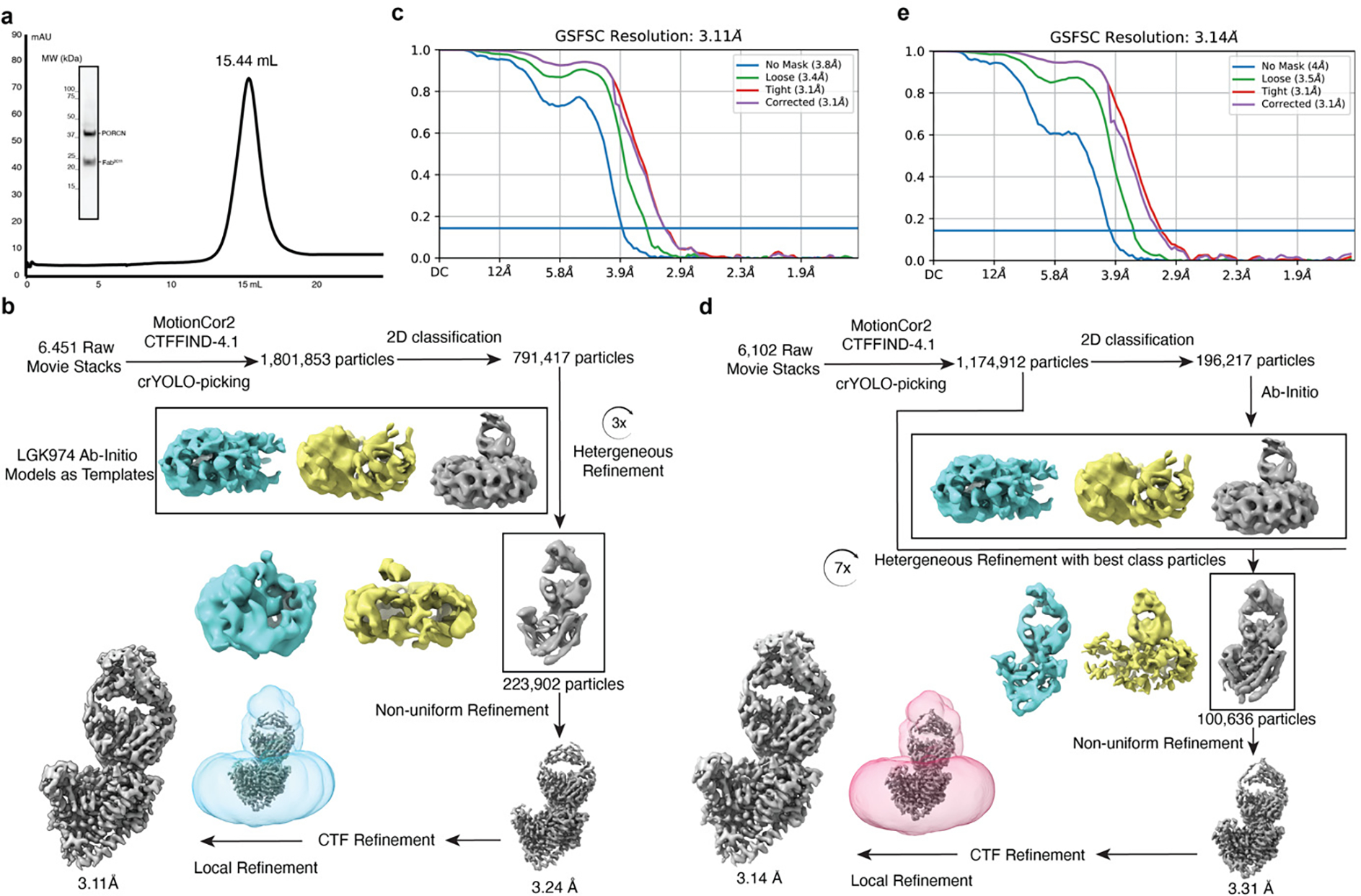
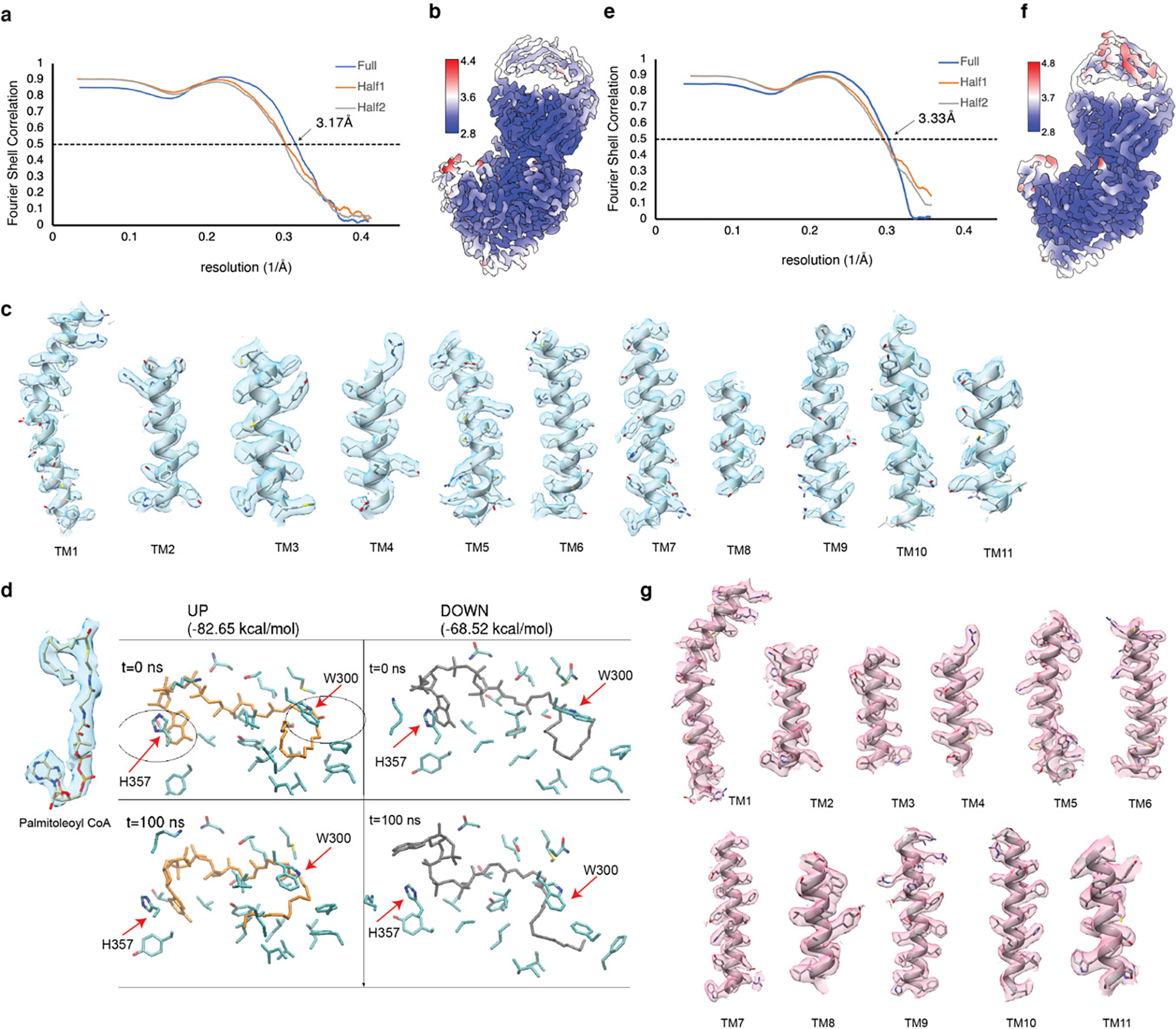
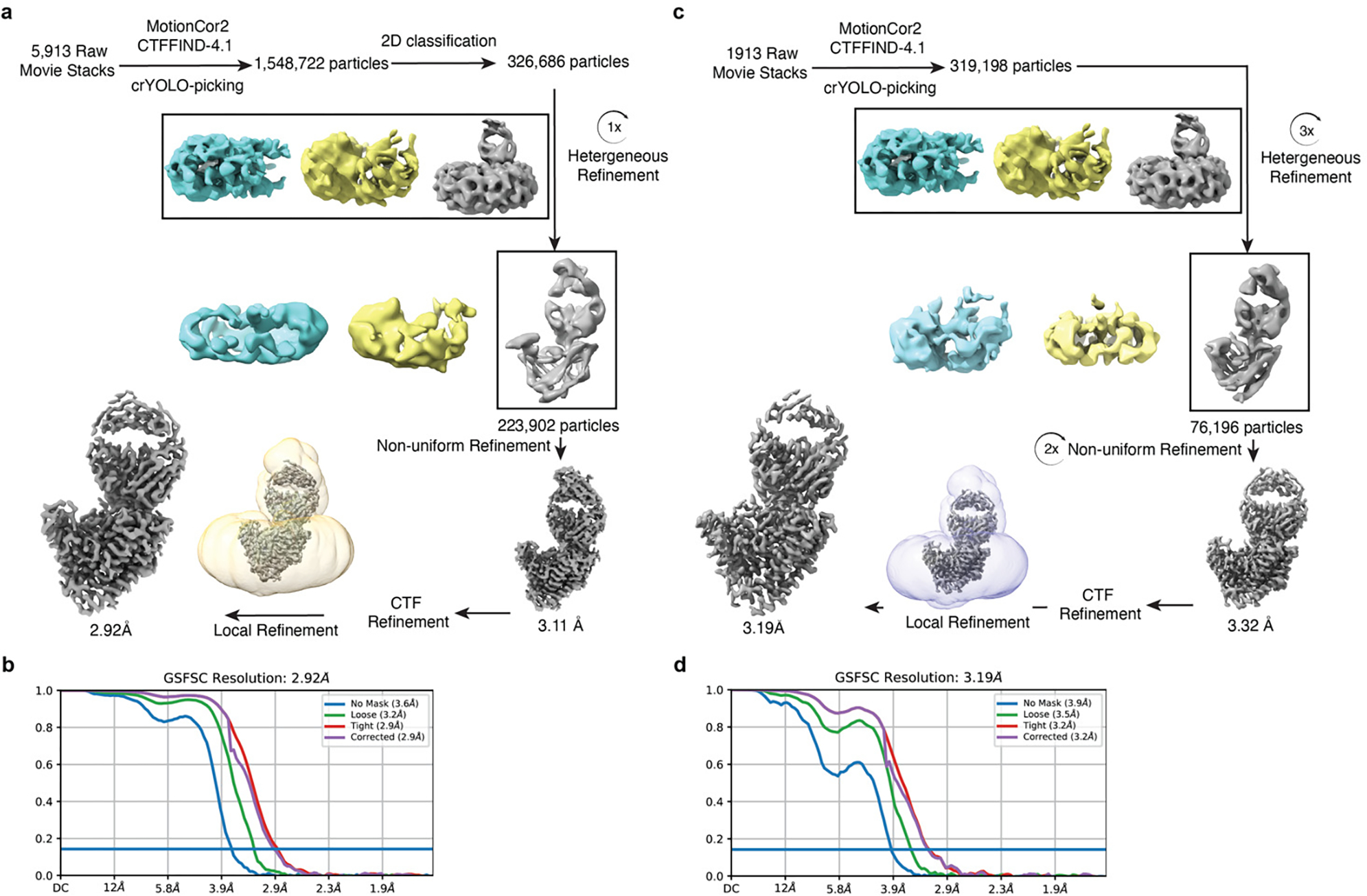
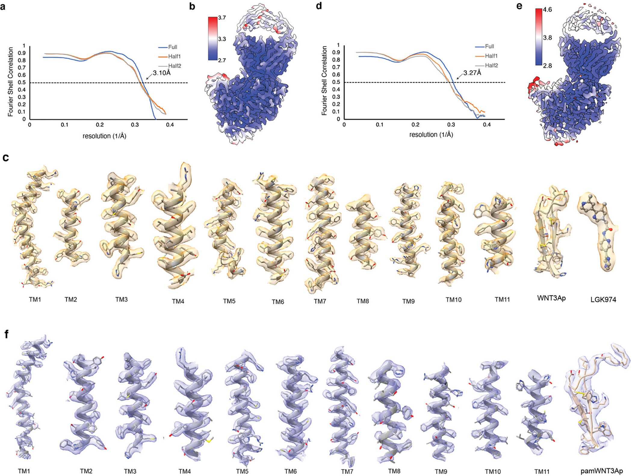
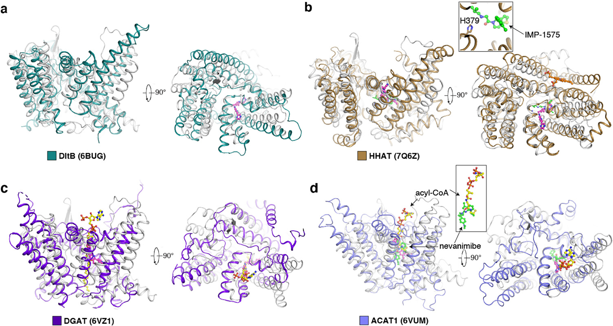
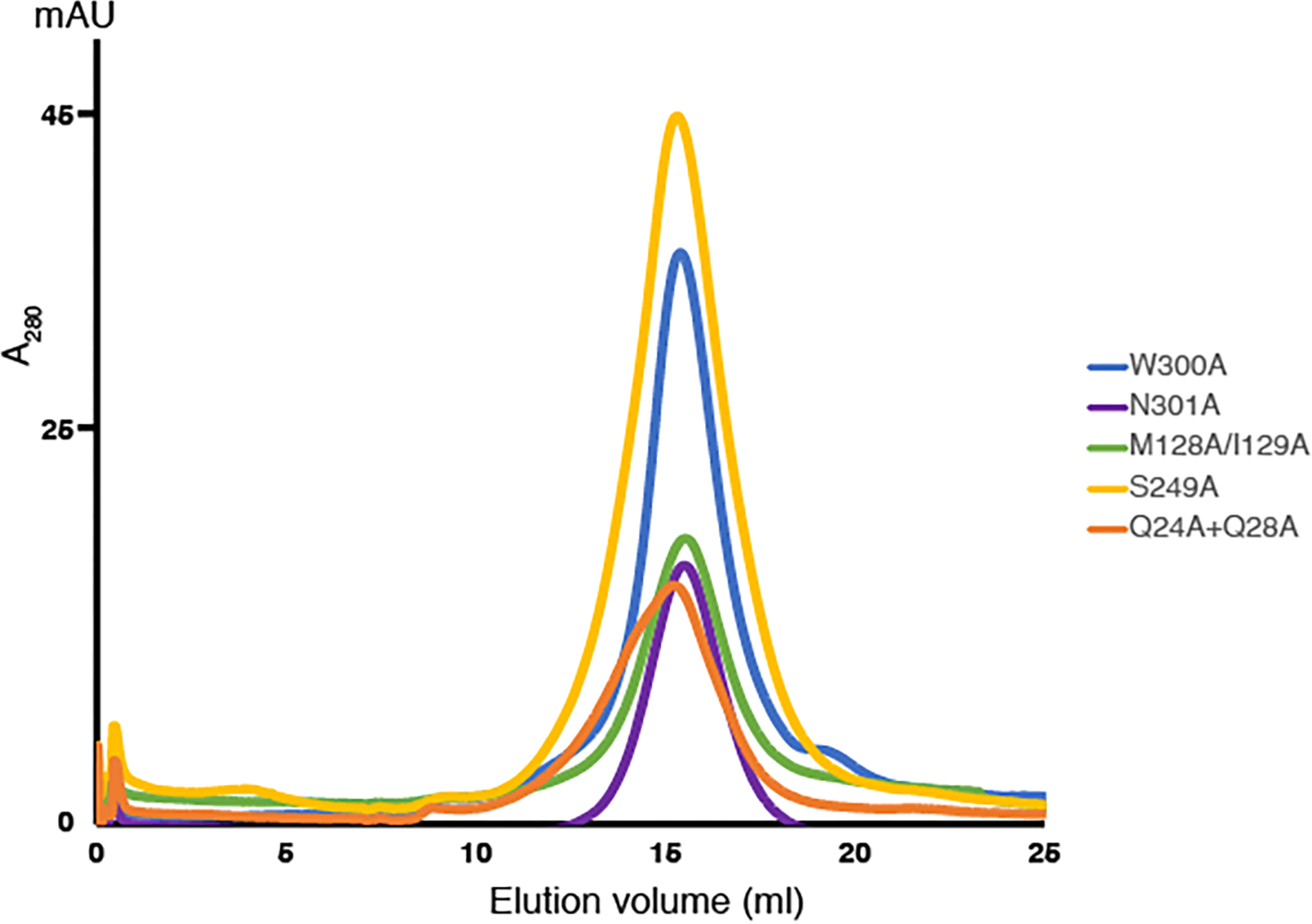
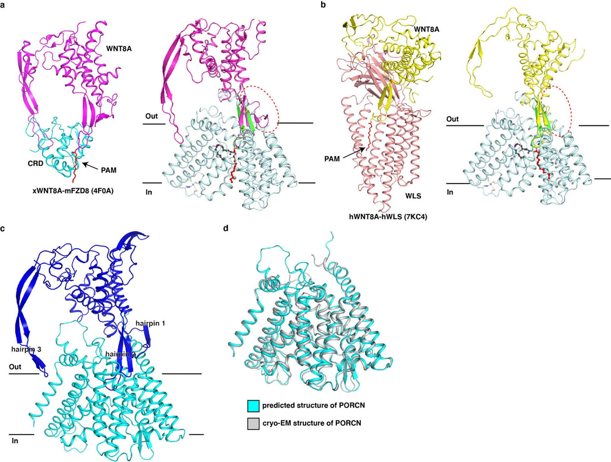
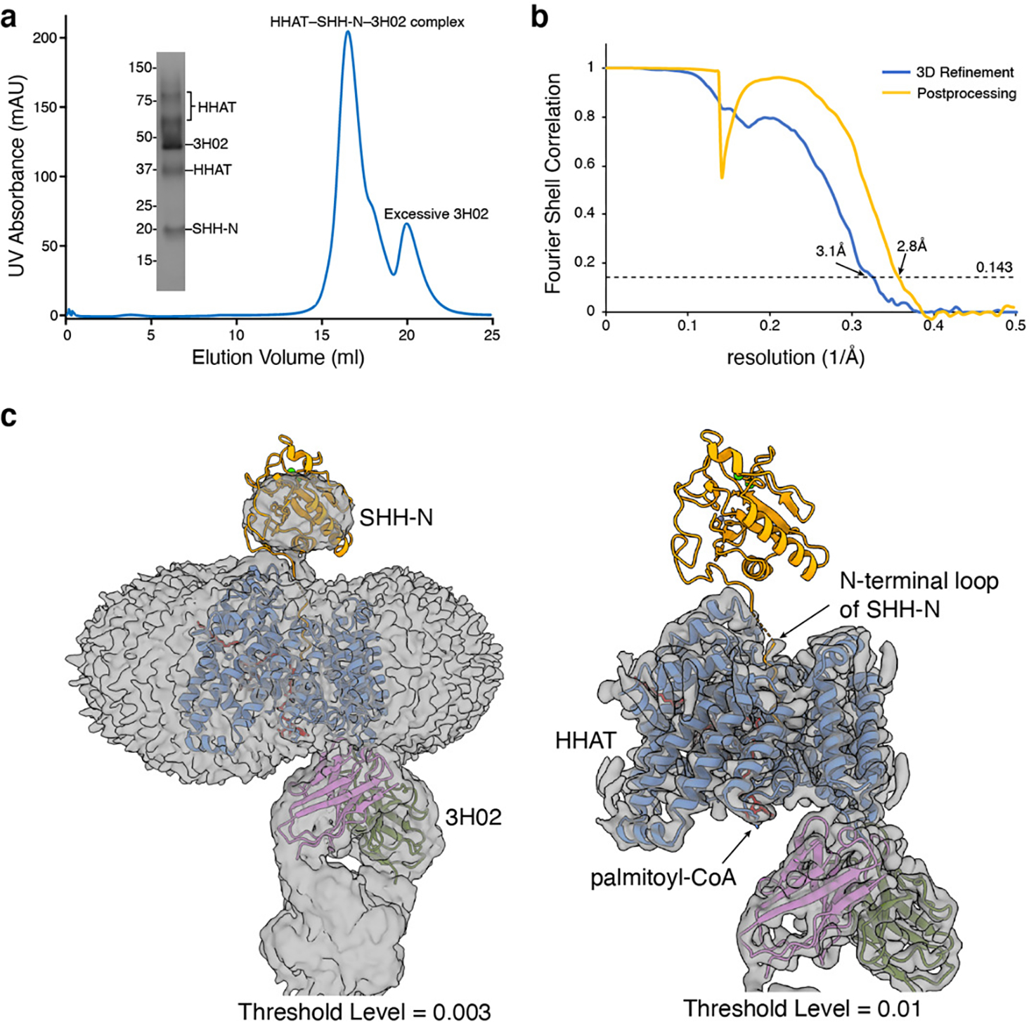
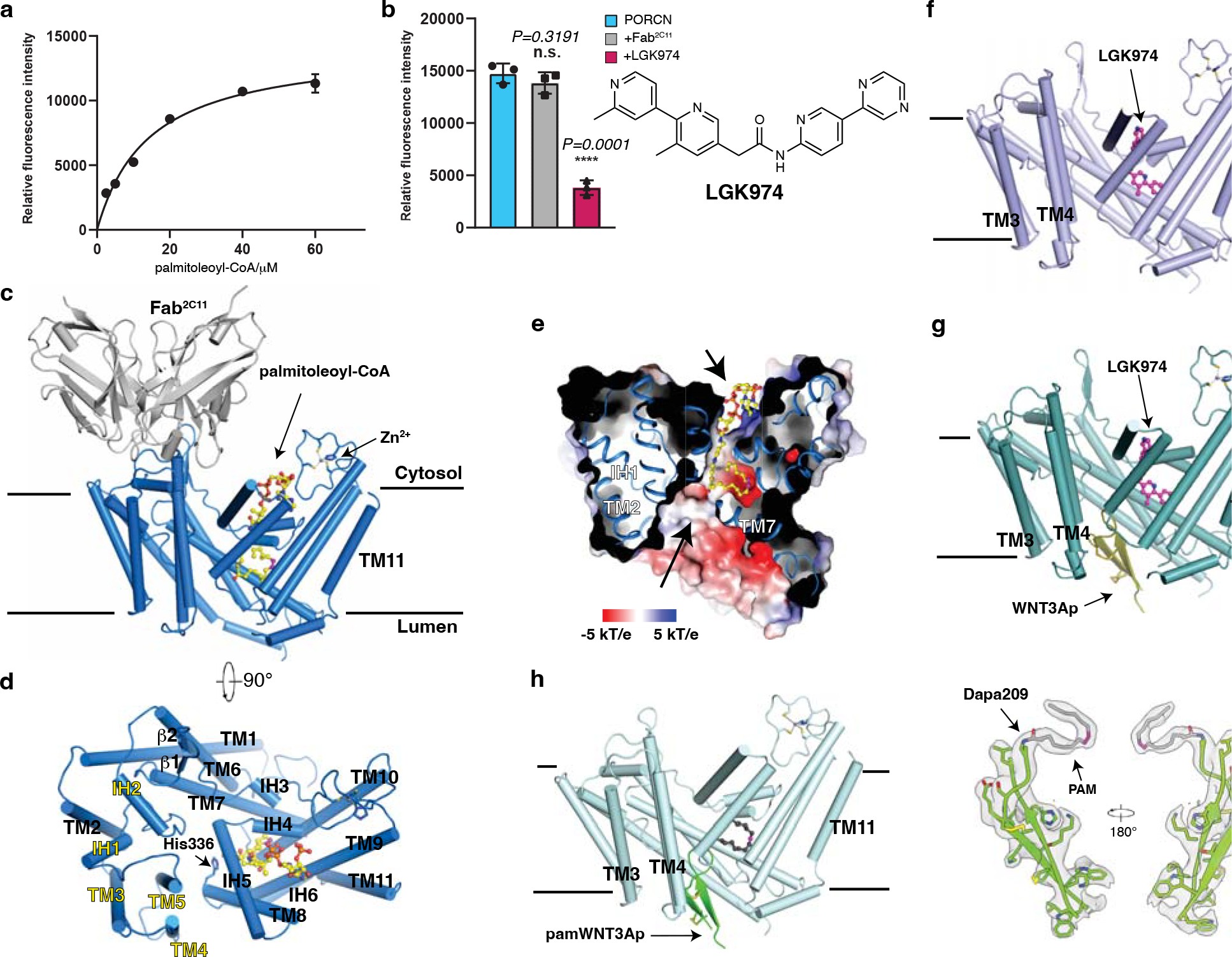
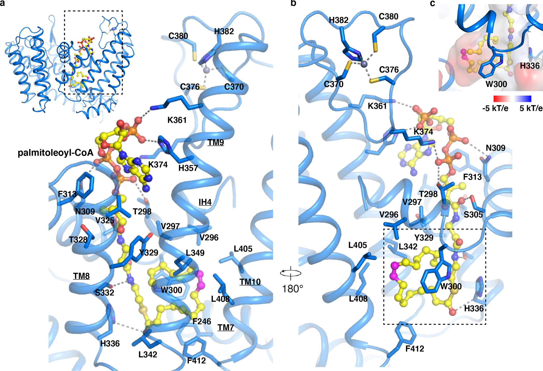
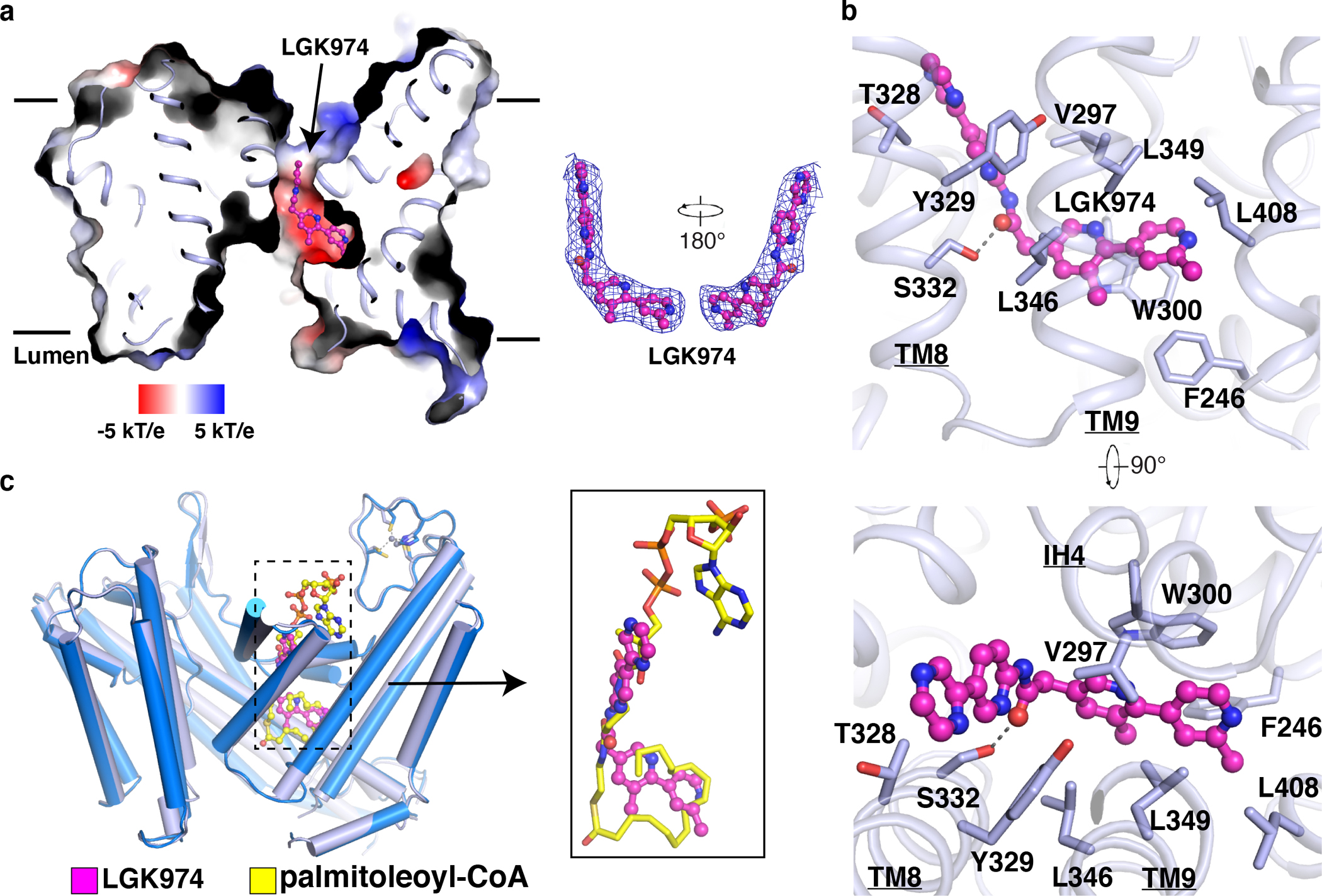
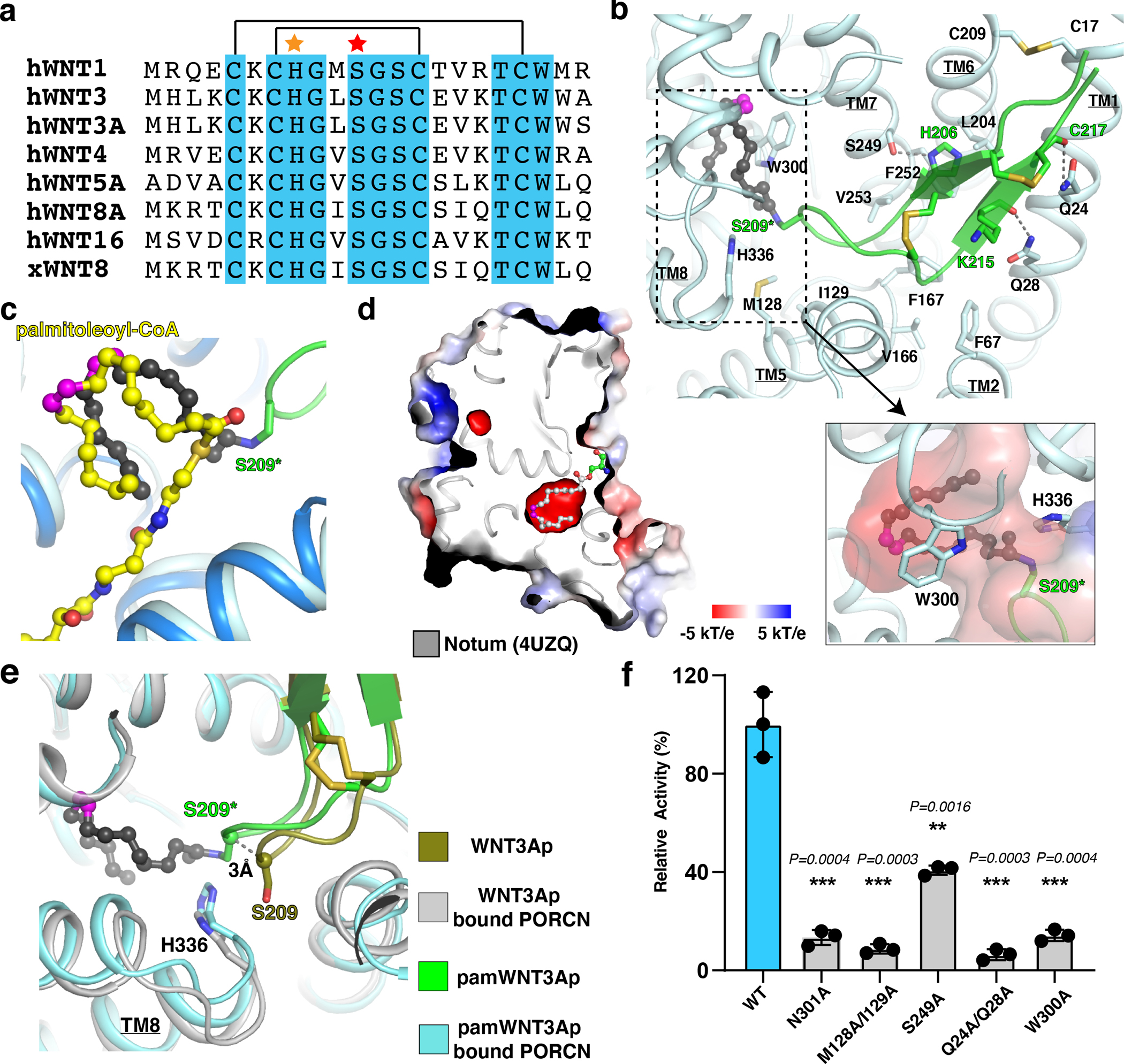
Wnt signalling is essential for regulation of embryonic development and adult tissue homeostasis1-3, and aberrant Wnt signalling is frequently associated with cancers4. Wnt signalling requires palmitoleoylation on a hairpin 2 motif by the endoplasmic reticulum-resident membrane-bound O-acyltransferase Porcupine5-7 (PORCN). This modification is indispensable for Wnt binding to its receptor Frizzled, which triggers signalling8,9. Here we report four cryo-electron microscopy structures of human PORCN: the complex with the palmitoleoyl-coenzyme A (palmitoleoyl-CoA) substrate; the complex with the PORCN inhibitor LGK974, an anti-cancer drug currently in clinical trials10; the complex with LGK974 and WNT3A hairpin 2 (WNT3Ap); and the complex with a synthetic palmitoleoylated WNT3Ap analogue. The structures reveal that hairpin 2 of WNT3A, which is well conserved in all Wnt ligands, inserts into PORCN from the lumenal side, and the palmitoleoyl-CoA accesses the enzyme from the cytosolic side. The catalytic histidine triggers the transfer of the unsaturated palmitoleoyl group to the target serine on the Wnt hairpin 2, facilitated by the proximity of the two substrates. The inhibitor-bound structure shows that LGK974 occupies the palmitoleoyl-CoA binding site to prevent the reaction. Thus, this work provides a mechanism for Wnt acylation and advances the development of PORCN inhibitors for cancer treatment.


Conflict of interest statement

Competing interests: The authors declare no competing interests.

Figures

Extended Data Fig. 1
Extended Data Fig. 1. LC-MS analysis of PORCN-mediated WNT3Ap acylation and MS analysis of pamWNT3Ap.
a, The secretion pathway of WNT ligand. In the endoplasmic reticulum (yellow), WNT (purple) is modified by PORCN (orange). Transport of the modified WNT is mediated by WLS (blue). WNTs are secreted to extracellular space via vesicles. After binding to FZD (green), WNTs trigger the signal transduction. Notum (light blue) acts as a deacylase, removing the lipid of WNT to abolish the signal. The structure of palmitoleated WNT3A hairpin 2 is shown. b, Representative LC chromatograms of PORCN-mediated WNT3Ap acylation with (red) and without (blue) LGK974. ESI-MS: m/z calculated for [M+3H]3+ 840.041, found 840.039, and m/z calculated for [palmitoleated WNT3Ap+3H]3+ 918.779, found 918.778. c, Quantitative analysis of the product ratio after the reaction. Data are mean ± s.d. (n=3 biologically independent experiments). ****P ≤ 0.0001, two-tailed unpaired t-test using GraphPad Prism 8. d, LC analysis of pamWNT3Ap. The samples (panels b and d) were applied to a HPLC column (DiscoveryBIO wide pore C5, 4.6×150mm, 5 μm) using a gradient in which percentage of solvent B increases from 20% to 80% in 6 minutes at 1.5 mL/min. e, Mass spectrometry analysis of [pamWNT3Ap+3H]3+. ESI-MS: m/z calculated for [M+3H]3+ (C124H196N33O28S5) 918.451, found 918.446.
Extended Data Fig. 2
Extended Data Fig. 2. Protein Purification and data processing of palmitoleoyl-CoA-bound and LGK974-bound PORCN.
a, Representative Superose 6 increase 10/300 GL gel-filtration chromatogram of PORCN complex with Fab2C11. The peak fraction is shown on SDS-PAGE with molecular markers. Each protein is indicated. For gel source data, see Supplementary Fig. 1a. b, The data processing of palmitoleoyl-CoA-bound PORCN. The cryo-EM 3D classes as well as the mask used for the refinement are shown. The final cryo-EM map after cryoSPARC refinement was sharpened with a B-factor value of −170 Å2. c, Fourier shell correlation (FSC) curve of palmitoleoyl-CoA-bound PORCN map as a function of resolution using cryoSPARC output. d, The data processing of LGK974-bound PORCN. The cryo-EM 3D classes as well as the mask used for the refinement are shown. The final cryo-EM map after cryoSPARC refinement was sharpened with a B-factor value of −130 Å2. e, Fourier shell correlation (FSC) curve of LGK974-bound PORCN map as a function of resolution using cryoSPARC output.
Extended Data Fig. 3
Extended Data Fig. 3. cryo-EM map of structural elements of palmitoleoyl-CoA-bound and LGK974-bound PORCN.
a, The Fourier shell correlation (FSC) curves calculated between the refined structure model and the half map used for refinement (yellow), the other half map (gray) and the full map (blue) of palmitoleoyl-CoA-bound PORCN. b, Density map colored by local resolution estimation using cryoSPARC. c, The major helices of PORCN. d, MD simulation suggests that palmitoleoyl-CoA binds to PORCN in the curled-up conformation. After 100 ns simulations, the interaction between residues W300, H357 and the curled-down palmitoleoyl-CoA disrupts (the right bottom panel). The palmitoleoyl-CoA in either conformation with the cryo-EM map is shown. e, The Fourier shell correlation (FSC) curves calculated between the refined structure model and the half map used for refinement (yellow), the other half map (gray) and the full map (blue) of palmitoleoylated WNT3A-bound PORCN. f, Density map colored by local resolution estimation using cryoSPARC. g, The major helices of PORCN.
Extended Data Fig. 4
Extended Data Fig. 4. Data processing of WNT3Ap/LGK974-bound PORCN and pamWNT3Ap-bound PORCN.
a, The data processing of WNT3Ap/LGK974-bound PORCN. The cryo-EM 3D classes as well as the mask used for the refinement are shown. The final cryo-EM map after cryoSPARC refinement was sharpened with a B-factor value of −147 Å2. b, Fourier shell correlation (FSC) curve of WNT3Ap/LGK974-bound PORCN map as a function of resolution using cryoSPARC output. c, The data processing of pamWNT3Ap-bound PORCN. The cryo-EM 3D classes as well as the mask used for the refinement are shown. The final cryo-EM map after cryoSPARC refinement was sharpened with a B-factor value of −140 Å2. d, Fourier shell correlation (FSC) curve of pamWNT3Ap-bound PORCN map as a function of resolution using cryoSPARC output.
Extended Data Fig. 5
Extended Data Fig. 5. cryo-EM map of structural elements of WNT3Ap/LGK974-bound PORCN and pamWNT3Ap-bound PORCN.
a, The Fourier shell correlation (FSC) curves calculated between the refined structure model and the half map used for refinement (yellow), the other half map (gray) and the full map (blue) of WNT3Ap/LGK974-bound PORCN. b, Density map colored by local resolution estimation using cryoSPARC. c, The major helices of PORCN, WNT3Ap and LGK974. d, The Fourier shell correlation (FSC) curves calculated between the refined structure model and the half map used for refinement (yellow), the other half map (gray) and the full map (blue) of pamWNT3Ap-bound PORCN. e, Density map colored by local resolution estimation using cryoSPARC. f, The major helices of PORCN and pamWNT3Ap.
Extended Data Fig. 6
Extended Data Fig. 6. The structure of DHHC20 and the structural comparison of PORCN (gray) with the other MBOAT proteins.
a, The structure of DHHC20. The zinc ions are shown as gray balls. b, The structural comparison with DltB. c, The structural comparison with HHAT. IMP-1575, an HHAT inhibitor, is shown as green sticks. The catalytic H379 of HHAT is shown as sticks. d, The structural comparison with DGAT1. e, The structural comparison with ACAT1. The LGK974 is shown as magenta sticks. Nevanimibe, an ACAT1 inhibitor, is shown as green sticks. The acyl-CoA in the DGAT and ACAT1 is shown as yellow sticks.
Extended Data Fig. 7
Extended Data Fig. 7. Purification of human PORCN variants for activity assays.
Representative Superose 6 increase 10/300 GL gel-filtration chromatogram of PORCN variants in buffer containing 20 mM HEPES, pH 7.5, 150 mM NaCl, and 0.06% Digitonin.
Extended Data Fig. 8
Extended Data Fig. 8. Superimposing the WNT8A-FZD and WNT8A-WLS complexes into the structure of pamWNT3Ap-bound PORCN and the model of WNT3A-bound PORCN.
a, Superimposing the hairpin 2 of the structure of WNT8A-FZD complex into PORCN-pamWNT3Ap complex. The clash between WNT8A ligand and PORCN is indicated by a dashed circle. b, Superimposing the hairpin 2 of the structure of WNT8A-WLS complex into PORCN-pamWNT3Ap complex. The clash between WNT8A ligand and PORCN is indicated by a dashed circle. PAM, palmitoleic acid (red sticks). c, The predicted structure of WNT3A-bound PORCN. Three hairpins of WNT3A have been indicated. d, Structural comparison of the predicted PORCN structure to the cryo-EM structure (PDB:7URA).
Extended Data Fig. 9
Extended Data Fig. 9. The structure of HHAT-SHH-N complex.
a, Representative Superose 6 increase 10/300 GL gel-filtration chromatogram of HHAT-SHH-N complex with Fab3H02. The peak fraction is shown on SDS-PAGE with molecular markers. Each protein is indicated. For gel source data, see Supplementary Fig. 1b. b, Fourier shell correlation (FSC) curve as a function of resolution using RELION-3 output. c, Cryo-EM map of the complex after 3D refinement reveals the mean body of SHH-N protein at the threshold level of 0.003 but not 0.01.
Fig. 1
Fig. 1. Functional characterization and overall structures of human PORCN.
a, Concentration curve of the WNT3Ap acylation by PORCN with palmitoleoyl-CoA (Km=14.29 μM, fitted and calculated using GraphPad Prism 8). Data are mean ± s.d. (n=3 biologically independent experiments). b, LGK974 but not Fab2C11 inhibits the activity of PORCN in vitro. The chemical structure of LGK974 is shown on the right. Data are mean ± s.d. (n=3 biologically independent experiments). ****P ≤ 0.0001, two-tailed unpaired t-test using GraphPad Prism 8. c and d, Overall structure showing palmitoleoyl-CoA-bound PORCN viewed from the side of the membrane (c) or from the cytosol (d). The structural elements that bind to Fab2C11 are highlighted in yellow. e, Electrostatic surface representation of catalytic cavity viewed from the side of the membrane. The routes of substrates access are indicated by arrows. f, Overall structure showing LGK974-bound PORCN. g, Overall structure showing WNT3Ap-bound PORCN in the presence of LGK974. h, Overall structure showing pamWNT3Ap-bound PORCN. The cryo-EM map of pamWNT3Ap is shown. The palmitoleoyl-CoA is shown as sticks in yellow and LGK974 is shown as sticks in magenta. The cis double bond at C9 position of palmitoleoyl chain is colored in magenta. PAM, palmitoleic acid.
Fig. 2
Fig. 2. Overall structure of PORCN with palmitoleoyl-CoA.
a and b, Overall view of the palmitoleoyl-CoA and zinc ion with the bound residues in PORCN. Dashed lines indicate the polar interactions. The cis double bond at C9 position of palmitoleoyl-CoA is colored in magenta. c, Electrostatic surface representation of palmitoleate bound cavity (dashed box in panel b).
Fig. 3
Fig. 3. Overall structure of LGK974-bound PORCN.
a, Electrostatic surface representation of LGK974-bound PORCN. LGK974 is shown as sticks in magenta. The cryo-EM map of LGK974 is shown at 5σ level. b, Interaction of LGK974 with cavity residues. c, The comparison of LGK974-bound structure with palmitoleoyl-CoA bound structure. The palmitoleoyl-CoA is shown as sticks in yellow and LGK974 is shown as sticks in magenta.
Fig. 4
Fig. 4. Overall structures of PORCN with WNT3Ap and palmitoleoylated WNT3Ap.
a, Sequence alignment of the hairpin 2 from the Wnt ligands. The conserved residues among Wnt ligands are highlighted in cyan. The disulfide bonds are indicated by black lines. The His206 is indicated by an orange star. The acylation serine of Wnt ligands is marked by a red star. b, Overall view of the pamWNT3Ap with the bound residues in PORCN. S209 in pamWNT3Ap is replaced by (L)-2,3-diaminoproprionic acid (S209*). PAM, palmitoleic acid (dark gray sticks). Electrostatic surface representation of palmitoleate bound cavity (dashed box) is shown. c, Comparison of palmitoleate moiety in the palmitoleoyl-CoA bound PORCN (yellow sticks) and pamWNT3Ap bound PORCN structures (black sticks). d, Electrostatic surface representation of Notum. The palmitoleate is shown as gray sticks. The cis double bond at C9 position of palmitoleoyl-CoA is colored in magenta. e, Structural comparison of unmodified WNT3Ap and pamWNT3Ap. f, Functional validation of residues Q24/Q28, M128/I129, S249, W300, and N301. Data are mean ± s.d. (n=3 biologically independent experiments). **P ≤ 0.01, ***P ≤ 0.001, two-tailed unpaired t-test using GraphPad Prism 8.
Fig. 5
Fig. 5. The structural comparison with HHAT.
a, Membrane view of the structure of PORCN (blue) compared to that of acyl-CoA bound HHAT (gray). The acyl-CoA in the archway and heme of HHAT are indicated. The cis double bond at C9 position of palmitoleoyl-CoA is colored in magenta. A close-up view of the heme (salmon sticks) binding site in HHAT compared to that in PORCN. The dashed line indicates the interaction between HHAT residues and heme. b, Electrostatic surface representation of catalytic cavity of HHAT. The acyl-CoA in the cavity is shown in sticks. PLM, palmitic acid (gray sticks). c, Lumenal view of the structure of palmitoleoyl-CoA bound PORCN compared to that of acyl-CoA bound HHAT. The conformational difference of PORCN-TM10 is indicated by the red arrow. Residue Leu405 of PORCN is colored in red. TM11 of PORCN and the acyl-CoA in the archway are indicated. Palmitoleoyl-CoA is shown in yellow sticks. d, Lumenal view of the structure of pamWNT3Ap bound PORCN (light cyan) compared to that of HH product bound HHAT. The conformational difference between HHAT-TM2 (gold) and PORCN-TM1 is indicated by the red arrow. e, Docking palmitoleoyl-CoA into WNT3Ap/LGK974 bound PORCN (dark cyan). f, A proposed “one-step” working model.

References

    1. Nusse R & Clevers H Wnt/beta-Catenin Signaling, Disease, and Emerging Therapeutic Modalities. Cell 169, 985–999, doi:10.1016/j.cell.2017.05.016 (2017). - DOI - PubMed
    1. Taipale J & Beachy PA The Hedgehog and Wnt signalling pathways in cancer. Nature 411, 349–354, doi:10.1038/35077219 (2001). - DOI - PubMed
    1. MacDonald BT, Tamai K & He X Wnt/beta-catenin signaling: components, mechanisms, and diseases. Developmental cell 17, 9–26, doi:10.1016/j.devcel.2009.06.016 (2009). - DOI - PMC - PubMed
    1. Zhan T, Rindtorff N & Boutros M Wnt signaling in cancer. Oncogene 36, 1461–1473, doi:10.1038/onc.2016.304 (2017). - DOI - PMC - PubMed
    1. Willert K et al. Wnt proteins are lipid-modified and can act as stem cell growth factors. Nature 423, 448–452, doi:10.1038/nature01611 (2003). - DOI - PubMed

MeSH terms