. 2022 Aug;608(7924):819-825.
doi: 10.1038/s41586-022-04930-8. Epub 2022 Jul 13.

Reconstitution of a telomeric replicon organized by CST

Affiliations

Reconstitution of a telomeric replicon organized by CST

Arthur J Zaug et al. Nature. 2022 Aug.

Abstract

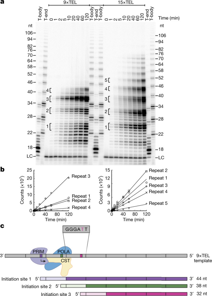
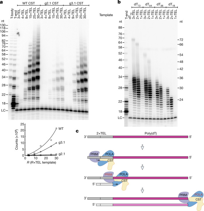
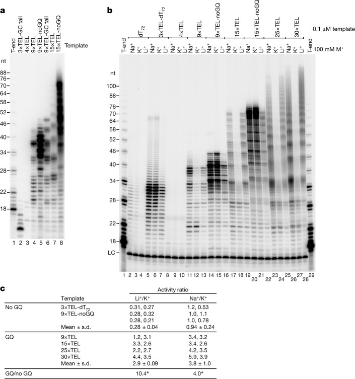
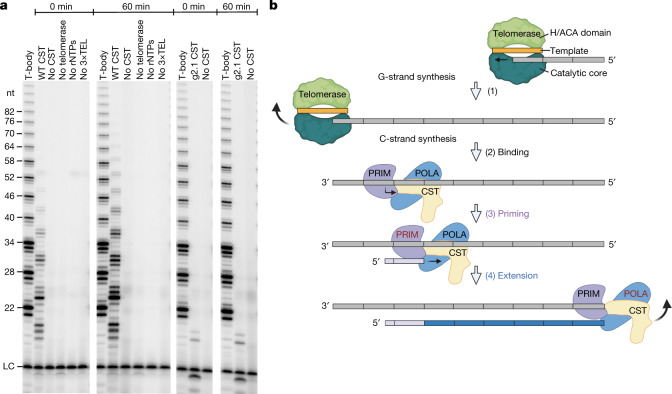
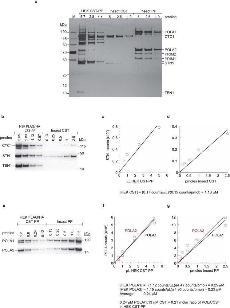
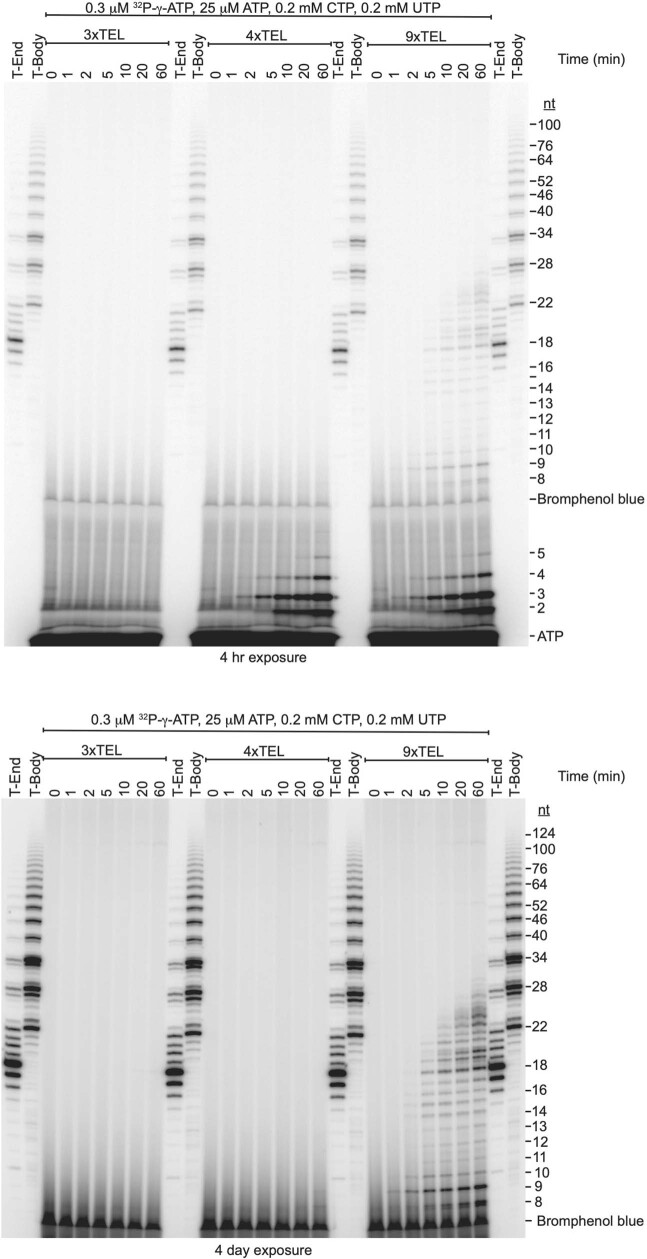
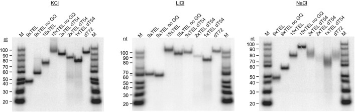
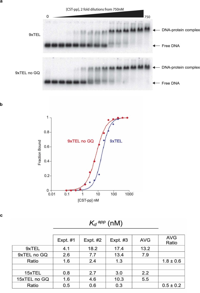
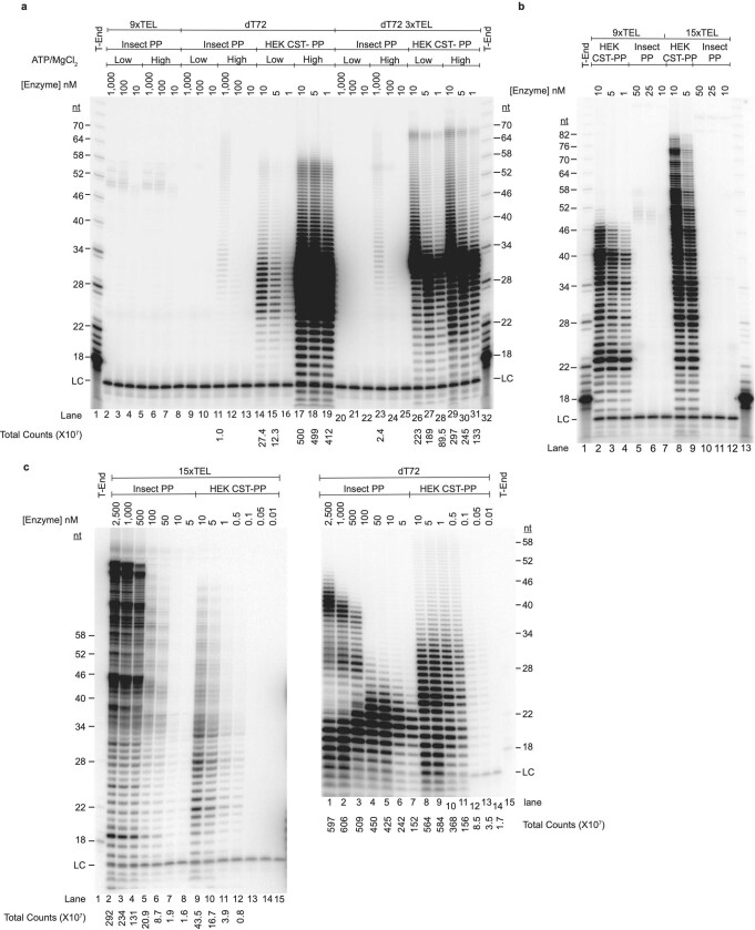
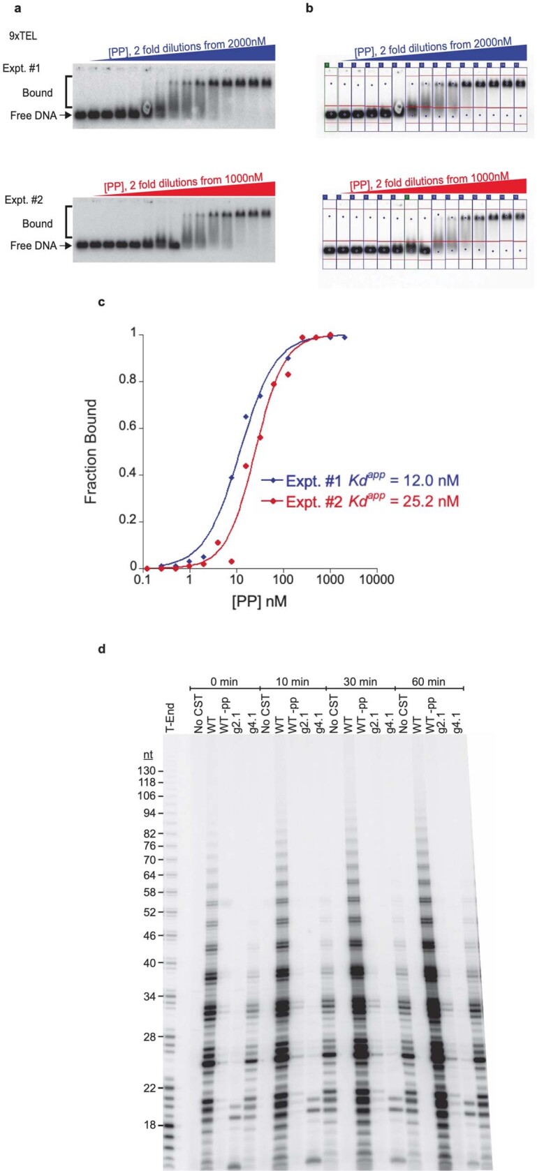
Telomeres, the natural ends of linear chromosomes, comprise repeat-sequence DNA and associated proteins1. Replication of telomeres allows continued proliferation of human stem cells and immortality of cancer cells2. This replication requires telomerase3 extension of the single-stranded DNA (ssDNA) of the telomeric G-strand ((TTAGGG)n); the synthesis of the complementary C-strand ((CCCTAA)n) is much less well characterized. The CST (CTC1-STN1-TEN1) protein complex, a DNA polymerase α-primase accessory factor4,5, is known to be required for telomere replication in vivo6-9, and the molecular analysis presented here reveals key features of its mechanism. We find that human CST uses its ssDNA-binding activity to specify the origins for telomeric C-strand synthesis by bound Polα-primase. CST-organized DNA polymerization can copy a telomeric DNA template that folds into G-quadruplex structures, but the challenges presented by this template probably contribute to telomere replication problems observed in vivo. Combining telomerase, a short telomeric ssDNA primer and CST-Polα-primase gives complete telomeric DNA replication, resulting in the same sort of ssDNA 3' overhang found naturally on human telomeres. We conclude that the CST complex not only terminates telomerase extension10,11 and recruits Polα-primase to telomeric ssDNA4,12,13 but also orchestrates C-strand synthesis. Because replication of the telomere has features distinct from replication of the rest of the genome, targeting telomere-replication components including CST holds promise for cancer therapeutics.


Conflict of interest statement

T.R.C. is a scientific advisor for Storm Therapeutics and Eikon Therapeutics. The other authors declare no competing interests.

Figures

Fig. 1
Fig. 1. CST–Polα–primase uses telomeric repeats as origins of priming and replication.
a, Time courses of C-strand synthesis on telomeric DNA templates, with products labelled with [α-32P]dCTP. Size markers are telomerase reaction products with end-labelled primer (T-end, 5′-phosphate) and body-labelled products (T-body, 5′-hydroxyl) as described in Methods. LC, labelled oligonucleotide loading control. b, Quantification of C-strand synthesis products in the experiment directly above. c, Model explaining the observed 6-nt ladders of C-strand reaction products. CST–Polα–primase binds the template (grey) at different sets of telomeric repeats, such that runoff synthesis gives products in 6-nt increments. The binding site shown at the top (initiation site 1) gives the purple C-strand product, while initiation at sites 2 and 3 gives the green and red products, respectively. In each case, the RNA primer is indicated by a lighter shade and telomeric repeats are indicated by vertical bars. The location of CST relative to Polα–primase and the template DNA is taken from the recent cryo-EM structure. For gel source data, see Supplementary Fig. 1. Source Data
Fig. 2
Fig. 2. C-strand synthesis requires DNA binding by CST–Polα–primase, and a CST–Polα–primase-binding site greatly enhances replication of a poly(dT) template.
a, C-strand synthesis for 1 h with DNA templates (100 nM) comprising increasing numbers of telomeric repeats catalysed by WT and two different DNA-binding mutants of CST–Polα–primase (20 nM). The apparent products in the third lane (3×TEL template) are spillover from the adjacent marker lane; repeat experiments confirmed that no product is formed. LC, loading control. Below, quantification of the experiment in a. Total incorporation was normalized to the loading control as a function of the number of telomeric repeats in the template. b, Replication products of CST–Polα–primase on a poly(dT) template, dT72, and on templates with various numbers of telomeric repeats added to the 3′ end of the poly(dT) sequence. c, Model for the initiation of CST–Polα–primase at the telomeric repeats and extension along the template. Whether CST remains bound to Polα–primase during extension is unknown. The length of the rose-coloured C-strand is limited by the template length (shown here) or, for longer templates, by the intrinsic processivity of the enzyme. For gel source data, see Supplementary Fig. 1. Source Data
Fig. 3
Fig. 3. C-strand synthesis overcomes GQ structures in the template DNA.
a, Mutating the telomeric repeat sequence (noGQ) greatly increases C-strand synthesis (compare lane 4 with lane 5 and lane 7 with lane 8). The GC tail templates have 10-nt GC sequences added on their 5′ ends to test whether the C-strand synthesis proceeds to the end of the template (compare lanes 4 and 6). Although 3×TEL is inactive, adding the 10-nt tail to 3×TEL provides a template for C-strand synthesis (lane 2). b, C-strand synthesis when GQ structures are destabilized (LiCl) or stabilized (KCl). NaCl is an intermediate condition. CST–Polα–primase at 25 nM; DNA templates at 100 nM. c, Activity with 100 mM LiCl relative to that in 100 mM KCl or activity with 100 mM NaCl relative to that with 100 mM KCl; quantification of two experiments including the one in b. *P < 0.0001, two-tailed t-test; n = 6 independent experiments for the noGQ templates and n = 8 independent experiments for the GQ templates. For gel source data, see Supplementary Fig. 1.
Fig. 4
Fig. 4. Reconstitution of complete telomere end replication.
a, Telomerase products are unlabelled (except in the T-body marker lanes), and C-strands are labelled with [α-32P]dCTP. Time between initiation of the telomerase reaction and addition of 57 nM CST–Polα–primase (WT CST) is indicated at top. Control lanes are reactions identical to WT CST except containing no CST, no telomerase, no ribonucleotides (rNTPs) or no 3×TEL primer for telomerase. In the two right-hand sets of lanes, 57 nM g2.1 CST, a DNA-binding-defective mutant of CST, is substituted for WT CST. The uncropped gel is shown in Supplementary Fig. 1, and an experiment with intermediate time points is shown in Extended Data Fig. 10d. b, Model for reconstituted telomere replication. (1) Telomerase (RNA template in orange) binds to the 3×TEL DNA primer (grey) and extends it with telomeric repeats (grey rectangles). (2) Telomerase dissociates, and CST–Polα–primase binds to telomeric repeats and begins RNA primer synthesis with ATP. (3) Primase synthesizes RNA primer (~8 nt). (4) Template–primer pair is handed off to Polα, which catalyses C-strand DNA synthesis (blue bar). The continued presence of CST during extension is unknown.
Extended Data Fig. 1
Extended Data Fig. 1. Purified CST–Polα-primase and stoichiometry of the subunits.
a, Silver-stained SDS-PAGE of one of the HEK cell CST–Polα-primase preparations used in this study (HEK CST-PP), recombinant human CST overexpressed in insect cells (Insect CST), and recombinant human Polα-primase overexpressed in insect cells (Insect PP). M, marker proteins. The HEK cell and insect cell STN1 proteins have slightly different mobilities due to a Myc tag on the former. b, Western blot with antibodies to STN1 and to epitope tags on the HEK cell CTC1 and TEN1 subunits (thus no signal for CTC1 or TEN1 in the Insect CST). The HEK cell-expressed CTC1 consistently runs as a doublet. Amount of protein loaded (pmoles) is based on direct determination of protein concentration for Insect CST but is calculated from the STN1 western blots for HEK CST-PP. HEK FLAG/HA CST-PP indicates double immunopurification via FLAG and HA tags. c and d, Quantification of STN1 from the western blot gives (c) the phosphorimager counts of STN1 per µL for the HEK cell preparation and (d) the counts of STN1 per pmol for purified insect cell CST; the calculation below the graphs gives the concentration of STN1 in the HEK cell CST–Polα-primase preparation. e, Western blot with antibodies to the two POLA subunits. The endogenous POLA1 from HEK cells consistently runs as a doublet, likely due to processing between lys 123 and lys 124 producing a stable 165 kDa species; in contrast, the POLA1 in the Polα-primase overexpressed in insect cells is mostly a single species with a minor smaller species. Amount of protein loaded (pmoles) is based on direct determination of protein concentration for Insect PP but is calculated from the POLA western blots for HEK CST-PP. f, g, Quantification of POLA1 and POLA2 from the western blot gives (f) the counts of POLA1 and POLA2 per µL of the HEK cell preparation and (g) the counts per pmol for the purified insect cell Polα-primase; the calculation below the graphs gives the concentration of POLA in the HEK cell CST–Polα-primase preparation. Dividing the concentration of POLA from f and g by the concentration of CST from c and d gives the stoichiometry of 0.21 POLA per CST in the HEK cell preparation. These results were consistent between two biological replicates of the entire set of experiments. For gel source data, see Supplementary Fig. 1 Source Data
Extended Data Fig. 2
Extended Data Fig. 2. Validation of the C-strand synthesis reaction on telomeric DNA templates.
a, Product formation requires DNA template, CST–Polα-primase, and ribonucleotides (rNTPs). +pp: CST–Polα-primase. -pp: CST with Polα-primase removed by 300 mM NaCl elution. DNA templates are None, or 100 nM 3xTEL, 9xTEL and 15xTEL. Products labeled with α-32P-dCTP. Nucleotide (nt) sizes are based on telomerase reaction with a 5’-end labelled primer (T-End). b, Longer DNA templates give larger ladders of C-strand products. Templates consist of the number of telomeric repeats indicated. 3xTEL and 4xTEL do not support DNA synthesis under these conditions. Products labeled with α-32P-dCTP. T-Body is a marker lane of body-labelled telomerase products, which run about one nt slower than the T-End products; the T-End products have a 5’-phosphate which increases their electrophoretic mobility. c, Adding a 10-nt tail to the 5’ end of a 9xTEL template confirms that at least some products runoff the end of the template, and adding an antisense oligonucleotide to pair with the 10-nt tail shows that C-strand synthesis stops at double-stranded DNA. 'GC tail' is 5’-CGCCGCCGCC, followed by (TTAGGG)9, and the 'anti' oligo is 5’-CTAAGGCGGCGGCG, designed to pair with the GC tail and the first four nt of the adjacent telomeric repeat. Products labelled with α-32P-dATP. Reactions performed at two MgCl2 concentrations as indicated. Reactions +dGTP allowed DNA synthesis to use the GC tail as template (brackets), while the sets of lanes without dGTP still gave some synthesis because of misincorporation. Addition of the anti oligo (lanes 5, 9, 14, 18) inhibited use of the GC tail as template; as a control (lanes 3, 7, 12, 16), the anti oligo had no effect with the 9xTEL template, which contains no complementary sequence. LC, loading control. d, POT1-TPP1N bound to telomeric DNA templates does not prevent CST–Polα-primase action but inhibits partially at high molar excess of POT1-TPP1N/template. Four DNA templates at 50 nM preincubated for 30 min at 30 °C with indicated concentrations of POT1-TPP1N prior to 1 h CST–Polα-primase reaction at 30 °C. Comparing 9xTEL-GC tail without and with the antisense oligonucleotide (anti) that binds the GC tail again confirms that C-strand synthesis stops when it encounters dsDNA. LC, loading control. e, POT1-TPP1N binding to trace amounts of radiolabelled 9xTEL and 15xTEL DNA as determined by electrophoretic mobility shift assay (EMSA). Binding for 30 min at 30 °C in C-strand synthesis buffer. Kdapp values ± SD (n = 3 independent binding curves). For gel source data, see Supplementary Fig. 1 Source Data
Extended Data Fig. 3
Extended Data Fig. 3. Processivity of CST–Polα-primase on poly(dT) templates.
a, Reactions as in Fig. 2b but with increasing concentrations of the indicated templates. Products labelled with α-32P-dATP. LC, loading control. Arrows, rare extension to the ends of these templates. Note that the templates containing 3xTEL give much more product than poly(dT), but the length of the products (which at high [template] is limited by processivity) is similar for all templates. b, The intensity of each extension product (that is, the counts in each band of a lane in a) was divided by the number of As in that product and the median length was calculated. Diamonds, 3xTELdT72. Squares, 3xTELdT54. Circles, dT72. The observation that the median extension is largely independent of the template DNA concentration means that extension is limited by the processivity of the enzyme on that template. For gel source data, see Supplementary Fig. 1 Source Data
Extended Data Fig. 4
Extended Data Fig. 4. Processivity of CST–Polα-primase on 9xTEL and 15xTEL templates; increasing DNA template concentration distinguishes processive C-strand synthesis from distributive re-priming.
a, Reaction of 25 nM CST–Polα-primase with increasing concentrations of 9xTEL or 15xTEL DNA template. b, Quantification of groups of extension products ('repeats') from a. Numbering of repeats is the same as in Fig. 1a. Increase of counts of repeat 1 products with increasing template concentration is interpreted as distributive re-priming, whereas the fraction of repeat 4 or 5 products that persists at high template concentration (1 µM template is 40-fold excess over concentration of CST–Polα-primase) is interpreted as processive extension. Repeat 4 is synthesized by approximately 70% re-priming and 30% processive synthesis. Products longer than repeat 4 are synthesized more by re-priming. Products shorter than repeat 4 persist at high [template], so they are due to processive extension from multiple initiation sites to the end of the template. For gel source data, see Supplementary Fig. 1 Source Data
Extended Data Fig. 5
Extended Data Fig. 5. RNA primers initiate with ATP and are approximately 8 nt long.
a, Reactions with γ-32P-ATP (trace concentration) as the only labelled nucleotide, with increasing concentrations of unlabelled ATP as indicated. Label incorporation decreases at 100 and 200 µM rATP, because above Km, dilution of the radiolabelled rATP is no longer compensated by increased reaction velocity. b, Hydrolysis of the RNA primer with NaOH or RNase A shortens the dC-labelled C-strand. WT, wild-type CST–Polα-primase, T-End, 5’-end labeled telomerase reaction products as size markers. RNase A, reaction products from lane WT were boiled and then treated with RNase A. NaOH, reaction products from lane WT were treated with NaOH. The 8-nt reduction in dC-labeled product size upon NaOH treatment indicates that the C-strands initiate with pppAACCCUAA/dCdCdC…, where the RNA primer is underlined and the slash indicates the 3’-most site of hydrolysis. RNase A, which cleaves after pyrimidines, would then cleave as follows: pppAACCU/AAdCdCdC…. This would result in the RNase A products being two nt longer than the NaOH products, as observed. The lines connecting the bands on the gel indicate the reduction in product size upon NaOH or RNase A treatment. For gel source data, see Supplementary Fig. 1.
Extended Data Fig. 6
Extended Data Fig. 6. RNA primers synthesized by CST–Polα-primase on three different DNA templates, directly visualized by omitting deoxynucleotides from the reaction.
Left half, production of short RNA oligonucleotides initiating with pppA on the 4xTEL and 9xTEL DNA templates. Their mobilities are consistent with 2, 3 and 4 nt, as indicated, although these sizes have not been independently confirmed. These products may represent abortive initiation by primase. Right half, the bottom of the gel containing the unincorporated 32P-ATP was cut off to allow a longer exposure of the larger products. The 9xTEL template allowed synthesis of RNA primers of length 8 and 9 nt; size estimates have ± 1 nt uncertainty because they are based on the telomerase size markers, which have a different 5’ end and different base composition. There are also some longer products formed (14 – 27 nt), presumably indicating some ability of Polα to incorporate ribonucleotides when there are no dNTPs in the reaction. The 4xTEL template, which supported synthesis of the putative abortive initiation products, was too short to allow synthesis of the 8–9 nt primers. For gel source data, see Supplementary Fig. 1.
Extended Data Fig. 7
Extended Data Fig. 7. Testing if DNA templates fold into G-quadruplex structures.
Template DNAs were 5’-end labelled with 32P, incubated at 30 °C in buffer containing 100 mM of the indicated salt, and then subjected to electrophoresis in a polyacrylamide gel containing 100 mM of the same salt in both the gel and the running buffer. Gels were run in a warm room at 30 °C, the same temperature as the DNA replication reactions. In LiCl, oligonucleotides ran largely according to their length; there are small differences due to base composition (that is, the 72-nt dT72 has an electrophoretic mobility similar to that of the 90-nt 15xTEL and 15xTEL-noGQ oligos). In KCl and NaCl, two of the oligos – 9xTEL and 15xTEL – showed anomalously fast mobility due to G-quadruplex formation. M, DNA length standards purchased from IDT and then 5’-end labelled. For gel source data, see Supplementary Fig. 1.
Extended Data Fig. 8
Extended Data Fig. 8. CST–Polα-primase binds similarly to GQ-forming and non-GQ-forming template DNAs.
a, Typical EMSA measuring binding of labelled template DNA by CST–Polα-primase. b, Quantification of fraction bound = DNA-protein complex/(free DNA + DNA-protein complex) as a function of CST–Polα-primase concentration. Data points were fit to a binding equation where the Hill coefficient was allowed to vary to give best fit. c, Summary of apparent Kd values obtained from 12 independent experiments similar to those shown in a and b. The better binding of 9xTEL-noGQ relative to 9xTEL changed with the 15xTEL series, where 15xTEL bound better than 15xTEL-noGQ. This difference was not anticipated and is not understood, but it was reproducible; in any case, the differences in binding affinity between GQ and noGQ templates are small, not exceeding two-fold. For gel source data, see Supplementary Fig. 1 Source Data
Extended Data Fig. 9
Extended Data Fig. 9. CST–Polα-primase purified from HEK cells is much more active on telomeric DNA templates than recombinant human Polα-primase purified from insect cells.
a, Under two reaction conditions (low, 0.2 mM rATP and 1 mM MgCl2; high, 1.0 mM rATP and 2 mM MgCl2), CST–Polα-primase purified from HEK cells (HEK CST-PP) is more than 10,000x more reactive than human Polα-primase purified from insect cells (Insect PP). For example, compare 1000 nM Insect PP with 1 nM HEK CST-PP under the same reaction conditions: lanes 11 and 19, lanes 20 and 28, lanes 23 and 31. Phosphorimager counts are given for those lanes that had a signal above background. b, The extremely large difference in activity between CST-PP and PP also pertains to the 9xTEL and 15xTEL templates using the 'high' reaction conditions of a (1.0 mM rATP and 2 mM MgCl2). In both a and b, template DNA was 100 nM. c, Reactions under the conditions of ref. , with 50-fold higher DNA template concentrations (5 µM). For the 15xTEL template, the difference in activity was greater than 10-fold; compare the two 10 nM reactions, lanes 7 and 9; or 100 nM Insect PP with 10 nM HEK CST-PP, lanes 5 and 9; or 50 nM Insect PP with 5 nM HEK CST-PP, lanes 6 and 10. However, the processivity of the two enzymes was similar; compare lanes 5 and 6 with lanes 9 and 10. (The high-concentration Insect PP points in lanes 2–4 show re-priming, not high processivity.) For the dT72 template, there was very little difference in activity under these conditions; compare the two 10 nM reactions, lanes 6 and 8, or the two 5 nM reactions, lanes 7 and 9. Note that the HEK CST-PP is somewhat more processive than the Insect PP; compare dT72 lanes 8-11 with lanes 4-7. (The high-concentration Insect PP points (lanes 1-3) show re-priming, not high processivity.) For this entire figure, note that the HEK CST-PP concentrations are based on the concentration of CST, so the concentration of PP in these preparations is 5-fold lower (see Extended Data Fig. 1). Thus, in terms of PP, the large activity advantage of CST-PP indicated above is actually 5-fold larger. Each gel contains a telomerase ladder (T-End) as markers. Each lane contains the same loading control (LC), a labelled 16 nt DNA. For gel source data, see Supplementary Fig. 1.
Extended Data Fig. 10
Extended Data Fig. 10. The lower activity of recombinant Polα-primase cannot be explained entirely by weaker binding to the ssDNA template and reconstitution of telomere end-replication.
a, Two EMSA experiments measuring binding of Polα-primase purified from insect cells (Insect PP in Extended Data Fig. 1) with labelled 9xTEL DNA. b, Because these DNA-PP complexes dissociate during electrophoresis, the definition we used for free and bound DNA is shown here by the red horizontal lines. The bound species are taken to include both the fully bound complex and the smear of counts between bound and free DNA. c, Quantification of fraction bound as a function of Polα-primase concentration, performed as in Extended Data Fig. 8b. Although the apparent Kd values determined here for Insect cell PP are similar to those determined in Extended Data Fig. 8 for HEK cell CST-PP, recall that CST-PP is only 21% PP. Thus, in terms of PP binding, the CST-PP has 5-fold higher affinity. d, Reconstitution of complete telomere end-replication. C-strands were labelled with α-32P-dCTP. Time between initiation of telomerase reaction and addition of CST–Polα-primase is indicated at top. CST–Polα-primase is present at 75 nM. WT-pp is CST largely depleted of Polα-primase by conducting IP purification at high (300 mM) salt concentration. g2.1 is a DNA-binding defective mutant of CST, and g4.1 is a control mutant of CST that retains DNA binding but is present at only one-third the concentration as WT CST owing to lower yield during purification. For gel source data, see Supplementary Fig. 1 Source Data

Similar articles

Cited by

References

    1. Lim CJ, Cech TR. Shaping human telomeres: from shelterin and CST complexes to telomeric chromatin organization. Nat. Rev. Mol. Cell Biol. 2021;22:283–298. - PMC - PubMed
    1. Kim NW, et al. Specific association of human telomerase activity with immortal cells and cancer. Science. 1994;266:2011–2015. - PubMed
    1. Greider CW, Blackburn EH. Identification of a specific telomere terminal transferase activity in Tetrahymena extracts. Cell. 1985;43:405–413. - PubMed
    1. Goulian M, Heard CJ. The mechanism of action of an accessory protein for DNA polymerase α/primase. J. Biol. Chem. 1990;265:13231–13239. - PubMed
    1. Casteel DE, et al. A DNA polymerase-α·primase cofactor with homology to replication protein A-32 regulates DNA replication in mammalian cells. J. Biol. Chem. 2009;284:5807–5818. - PMC - PubMed

LinkOut - more resources