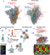
Engineering viral genomics and nano-liposomes in microfluidic platforms for patient-specific analysis of SARS-CoV-2 variants
- PMID: 35832078
- PMCID: PMC9254234
- DOI: 10.7150/thno.72339
Engineering viral genomics and nano-liposomes in microfluidic platforms for patient-specific analysis of SARS-CoV-2 variants
Abstract
New variants of severe acute respiratory syndrome coronavirus 2 (SARS-CoV-2) are continuing to spread globally, contributing to the persistence of the COVID-19 pandemic. Increasing resources have been focused on developing vaccines and therapeutics that target the Spike glycoprotein of SARS-CoV-2. Recent advances in microfluidics have the potential to recapitulate viral infection in the organ-specific platforms, known as organ-on-a-chip (OoC), in which binding of SARS-CoV-2 Spike protein to the angiotensin-converting enzyme 2 (ACE2) of the host cells occurs. As the COVID-19 pandemic lingers, there remains an unmet need to screen emerging mutations, to predict viral transmissibility and pathogenicity, and to assess the strength of neutralizing antibodies following vaccination or reinfection. Conventional detection of SARS-CoV-2 variants relies on two-dimensional (2-D) cell culture methods, whereas simulating the micro-environment requires three-dimensional (3-D) systems. To this end, analyzing SARS-CoV-2-mediated pathogenicity via microfluidic platforms minimizes the experimental cost, duration, and optimization needed for animal studies, and obviates the ethical concerns associated with the use of primates. In this context, this review highlights the state-of-the-art strategy to engineer the nano-liposomes that can be conjugated with SARS-CoV-2 Spike mutations or genomic sequences in the microfluidic platforms; thereby, allowing for screening the rising SARS-CoV-2 variants and predicting COVID-19-associated coagulation. Furthermore, introducing viral genomics to the patient-specific blood accelerates the discovery of therapeutic targets in the face of evolving viral variants, including B1.1.7 (Alpha), B.1.351 (Beta), B.1.617.2 (Delta), c.37 (Lambda), and B.1.1.529 (Omicron). Thus, engineering nano-liposomes to encapsulate SARS-CoV-2 viral genomic sequences enables rapid detection of SARS-CoV-2 variants in the long COVID-19 era.
Keywords: COVID-19; Organ on-a-chip; microfluidics; mutations, variants of concerns; nano-liposomes; viral genomics.
© The author(s).
Conflict of interest statement
Competing Interests: The authors have declared that no competing interest exists.
Figures


Similar articles
-
Competitive SARS-CoV-2 Serology Reveals Most Antibodies Targeting the Spike Receptor-Binding Domain Compete for ACE2 Binding.mSphere. 2020 Sep 16;5(5):e00802-20. doi: 10.1128/mSphere.00802-20. mSphere. 2020. PMID: 32938700 Free PMC article.
-
SARS-CoV-2 pandemic and research gaps: Understanding SARS-CoV-2 interaction with the ACE2 receptor and implications for therapy.Theranostics. 2020 Jun 12;10(16):7448-7464. doi: 10.7150/thno.48076. eCollection 2020. Theranostics. 2020. PMID: 32642005 Free PMC article. Review.
-
The SARS-CoV-2 Spike Glycoprotein as a Drug and Vaccine Target: Structural Insights into Its Complexes with ACE2 and Antibodies.Cells. 2020 Oct 22;9(11):2343. doi: 10.3390/cells9112343. Cells. 2020. PMID: 33105869 Free PMC article. Review.
-
Optimized Pseudotyping Conditions for the SARS-COV-2 Spike Glycoprotein.J Virol. 2020 Oct 14;94(21):e01062-20. doi: 10.1128/JVI.01062-20. Print 2020 Oct 14. J Virol. 2020. PMID: 32788194 Free PMC article.
-
Perspectives on development of vaccines against severe acute respiratory syndrome coronavirus 2 (SARS-CoV-2).Hum Vaccin Immunother. 2020 Oct 2;16(10):2366-2369. doi: 10.1080/21645515.2020.1787064. Epub 2020 Sep 22. Hum Vaccin Immunother. 2020. PMID: 32961082 Free PMC article. Review.
Cited by
-
Activation of cancer immunotherapy by nanomedicine.Front Pharmacol. 2022 Dec 22;13:1041073. doi: 10.3389/fphar.2022.1041073. eCollection 2022. Front Pharmacol. 2022. PMID: 36618938 Free PMC article. Review.
-
Microfluidic Organ-Chips and Stem Cell Models in the Fight Against COVID-19.Circ Res. 2023 May 12;132(10):1405-1424. doi: 10.1161/CIRCRESAHA.122.321877. Epub 2023 May 11. Circ Res. 2023. PMID: 37167356 Free PMC article. Review.
-
Recent updates on liposomal formulations for detection, prevention and treatment of coronavirus disease (COVID-19).Int J Pharm. 2023 Jan 5;630:122421. doi: 10.1016/j.ijpharm.2022.122421. Epub 2022 Nov 19. Int J Pharm. 2023. PMID: 36410670 Free PMC article. Review.
-
Opportunities and Challenges for Inhalable Nanomedicine Formulations in Respiratory Diseases: A Review.Int J Nanomedicine. 2024 Feb 17;19:1509-1538. doi: 10.2147/IJN.S446919. eCollection 2024. Int J Nanomedicine. 2024. PMID: 38384321 Free PMC article. Review.
-
Disruptive 3D in vitro models for respiratory disease investigation: A state-of-the-art approach focused on SARS-CoV-2 infection.Biomater Biosyst. 2023 Jul 16;11:100082. doi: 10.1016/j.bbiosy.2023.100082. eCollection 2023 Sep. Biomater Biosyst. 2023. PMID: 37534107 Free PMC article.
References
-
- Liu J, Liu Y, Xia H, Zou J, Weaver SC, Swanson KA. et al. BNT162b2-elicited neutralization of B.1.617 and other SARS-CoV-2 variants. Nature. 2021;596(7871):273–5. - PubMed
-
- Kupferschmidt K. New mutations raise specter of 'immune escape'. Science. 2021;371(6527):329–30. Epub 2021/01/23. - PubMed
-
- Countries CC-dri. Coronavirus (COVID-19) death rate in countries.
Publication types
MeSH terms
Substances
Supplementary concepts
Grants and funding
LinkOut - more resources
Full Text Sources
Medical
Research Materials
Miscellaneous

