Morphological Changes of Frontal Areas in Male Individuals With HIV: A Deformation-Based Morphometry Analysis
- PMID: 35832184
- PMCID: PMC9271794
- DOI: 10.3389/fneur.2022.909437
Morphological Changes of Frontal Areas in Male Individuals With HIV: A Deformation-Based Morphometry Analysis
Abstract
Objective: Previous studies on HIV-infected (HIV+) individuals have revealed brain structural alterations underlying HIV-associated neurocognitive disorders. Most studies have adopted the widely used voxel-based morphological analysis of T1-weighted images or tracked-based analysis of diffusion tensor images. In this study, we investigated the HIV-related morphological changes using the deformation-based morphometry (DBM) analysis of T1-weighted images, which is another useful tool with high regional sensitivity.
Materials and methods: A total of 157 HIV+ (34.7 ± 8.5 years old) and 110 age-matched HIV-uninfected (HIV-) (33.7 ± 10.1 years old) men were recruited. All participants underwent neurocognitive assessments and brain scans, including high-resolution structural imaging and resting-state functional imaging. Structural alterations in HIV+ individuals were analyzed using DBM. Functional brain networks connected to the deformed regions were further investigated in a seed-based connectivity analysis. The correlations between imaging and cognitive or clinical measures were examined.
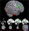
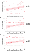
Results: The DBM analysis revealed decreased values (i.e., tissue atrophy) in the bilateral frontal regions in the HIV+ group, including bilateral superior frontal gyrus, left middle frontal gyrus, and their neighboring white matter tract, superior corona radiata. The functional connectivity between the right superior frontal gyrus and the right inferior temporal region was enhanced in the HIV+ group, the connectivity strength of which was significantly correlated with the global deficit scores (r = 0.214, P = 0.034), and deficits in learning (r = 0.246, P = 0.014) and recall (r = 0.218, P = 0.031). Increased DBM indexes (i.e., tissue enlargement) of the right cerebellum were also observed in the HIV+ group.
Conclusion: The current study revealed both gray and white matter volume changes in frontal regions and cerebellum in HIV+ individuals using DBM, complementing previous voxel-based morphological studies. Structural alterations were not limited to the local regions but were accompanied by disrupted functional connectivity between them and other relevant regions. Disruptions in neural networks were associated with cognitive performance, which may be related to HIV-associated neurocognitive disorders.
Keywords: HIV; MRI; cognitive assessment; deformation-based morphometry; functional connectivity.
Copyright © 2022 Chen, Cai, Song, Zhan, Wei, Shi, Wang and Shi.
Conflict of interest statement
The authors declare that the research was conducted in the absence of any commercial or financial relationships that could be construed as a potential conflict of interest.
Figures


References
Grants and funding
LinkOut - more resources
Full Text Sources

