

Different Cells of the Human Body: Categories and Morphological Characters
- PMID: 35832316
- PMCID: PMC9272693
- DOI: 10.4103/jmau.jmau_74_20
Different Cells of the Human Body: Categories and Morphological Characters
Abstract
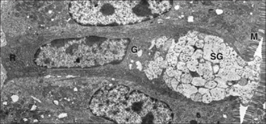
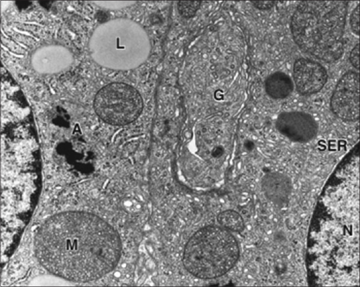
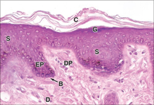
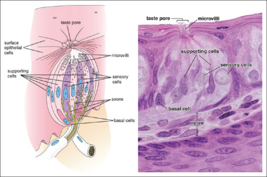
The cell is the basic structural unit of all living organisms. Most of the cells forming the human body share the basic components, but there are many categories that have specific light and electron microscopic characteristics. This review shed a light on these cell categories and their morphologies. Stem cell category is the cells responsible for the regeneration of damaged or lost cells, whereas protein-secreting cells are those responsible for the production and secretion of proteins. Protein-secreting cells have specific characters such as basophilic cytoplasm and vesicular nucleus by light microscope; these are confirmed by an electron microscope that shows rough endoplasmic reticulum, Golgi apparatus, secretory granules, and mitochondria. Steroid secreting, ion transporting, and contracting categories have specific morphology. Phagocytic cells such as macrophages and neutrophils are characterized by the presence of many lysosomes and phagosomes. Supporting cells are very important category as they not only support but also usually have another function such as myelin formation in Schwann, blood-brain barrier in astrocytes, or modification of response as in Pacinian corpuscle. Many cells showed interrelated characters between different categories, for example, phagocytic cells are able to contract to perform their function in fighting microorganism. Although we have trillions of cells, yet they only fall in some categories. Each cell category has specific morphological characters confirmed by ultrastructural characters. They all adapted to perform the desired functions.
Keywords: Categories; cells; human body; morphology.
Copyright: © 2021 Journal of Microscopy and Ultrastructure.
Conflict of interest statement
There are no conflicts of interest.
Figures
References
-
- Bianconi E, Piovesan A, Facchin F, Beraudi A, Casadei R, Frabetti F, et al. An estimation of the number of cells in the human body. Ann Hum Biol. 2013;40:463–71. - PubMed
-
- Mescher AL Junqueira LC. Junqueira’s Basic Histology: Text and Atlas. 2018
-
- Alberts, Bruce, Alexander D, Johnson, Julian Lewis, David Morgan, Martin Raff, Keith Roberts, Peter Walter. Molecular Biology of the Cell. Sixth edition. New York, NY: W. W. Norton & Company; 2014.
-
- Gartner LP. 7th ed. Philadelphia: Wolters Kluwer; 2018. Color Atlas and Text of Histology.
-
- Ross MH, Pawlina W. 7th ed. China: Wolters Kluwer Health; 2016. Histology: A Text and Atlas : With Correlated Cell and Molecular Biology.
Publication types
LinkOut - more resources
Full Text Sources