. 2022 Jun 27:10:867127.
doi: 10.3389/fcell.2022.867127. eCollection 2022.

Parental High-Fat High-Sugar Diet Intake Programming Inflammatory and Oxidative Parameters of Reproductive Health in Male Offspring

Affiliations

Parental High-Fat High-Sugar Diet Intake Programming Inflammatory and Oxidative Parameters of Reproductive Health in Male Offspring

Marcela Nascimento Sertorio et al. Front Cell Dev Biol. .

Abstract

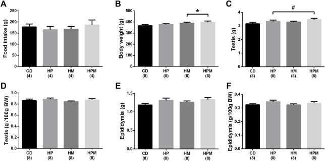
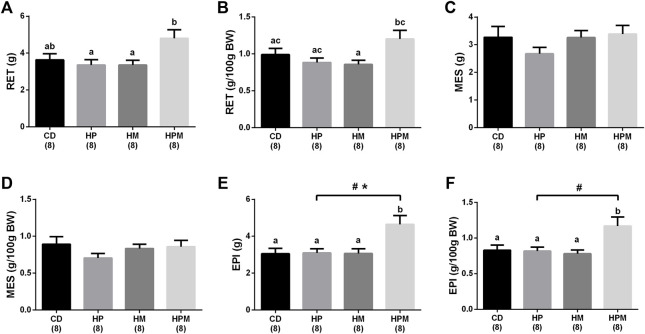
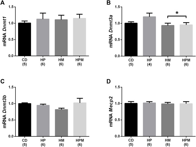
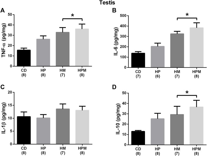
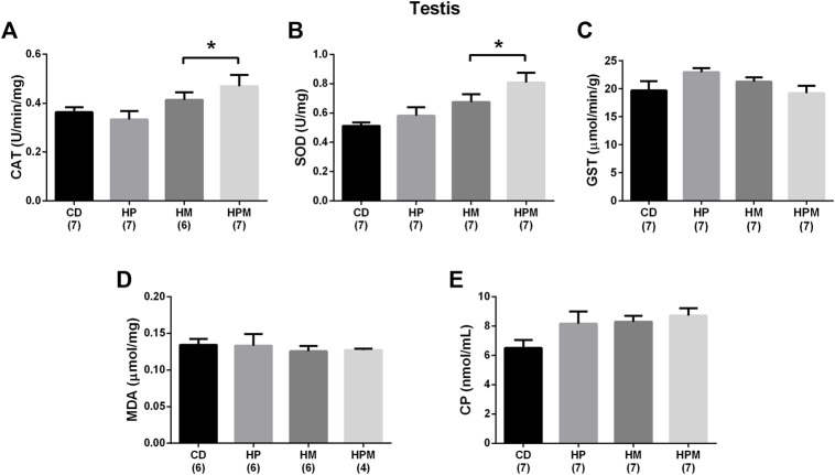
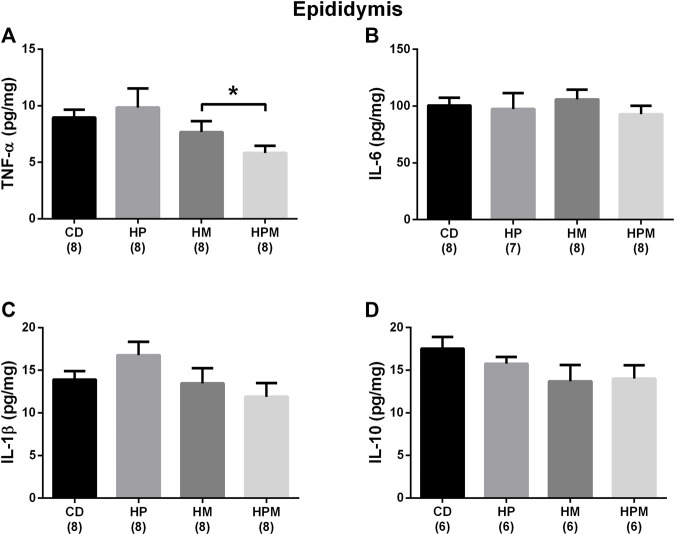
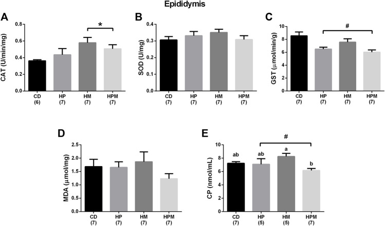
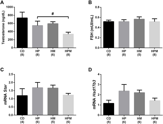
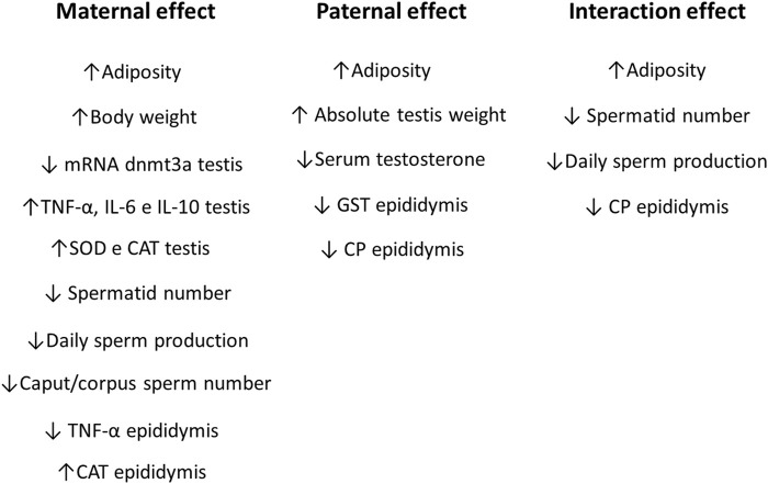
Parental nutrition can impact the health of future generations, programming the offspring for the development of diseases. The developing germ cells of the offspring could be damaged by the maternal or the paternal environment. The germ cells in development and their function could be affected by nutritional adversity and therefore, harm the health of subsequent generations. The paternal or maternal intake of high-fat diets has been shown to affect the reproductive health of male offspring, leading to imbalance in hypothalamic-pituitary-gonadal axis, testicular oxidative stress, low testosterone production, and changes in sperm count, viability, motility, and morphology. There is a need for studies that address the combined effects of diets with a high-fat and high-sugar (H) content by both progenitors on male reproduction. In this context, our study evaluated epigenetic parameters and the inflammatory response that could be associated to oxidative stress in testis and epididymis of adult offspring. 90 days-old male rats were divided according to the combination of the parental diet: CD (control paternal and maternal diet), HP (H paternal diet and control maternal diet), HM (H maternal diet and control paternal diet) and HPM (H paternal and maternal diet).We evaluated serum levels of testosterone and FSH; testicular gene expression of steroidogenic enzymes Star and Hsd17b3 and epigenetic markers Dnmt1, Dnmt3a, Dnmt3b, and Mecp2; testicular and epididymal levels of TNF-α, IL-6, IL-10, and IL-1β; testicular and epididymal activity of SOD, CAT, and GST; the oxidative markers MDA and CP; the daily sperm production, sperm transit time, and sperm morphology. Testicular epigenetic parameter, inflammatory response, oxidative balance, and daily sperm production of the offspring were affected by the maternal diet; paternal diet influenced serum testosterone levels, and lower daily sperm production was exacerbated by the interaction effect of both parental intake of high-fat high-sugar diet in the testis. There was isolated maternal and paternal effect in the antioxidant enzyme activity in the cauda epididymis, and an interaction effect of both parents in protein oxidative marker. Maternal effect could also be observed in cytokine production of cauda epididymis, and no morphological effects were observed in the sperm. The potential programming effects of isolated or combined intake of a high-fat high-sugar diet by the progenitors could be observed at a molecular level in the reproductive health of male offspring in early adulthood.

Keywords: epididimys; epigenetics; fetal programming; high-fat diet; inflammation; oxidative stress; sperm production; testis.


Conflict of interest statement

The authors declare that the research was conducted in the absence of any commercial or financial relationships that could be construed as a potential conflict of interest.

Figures

FIGURE 1
FIGURE 1
Biometric data and food intake of male rat offspring from progenitors fed on high-fat high-sugar diet. Mean ± SEM; symbols indicate significant difference caused by parental diet (p < 0.05): #statistically significant by the effect of paternal diet; *statistically significant by the effect of maternal diet. H: high-fat and high-sugar diet; CD (control paternal and maternal diet), HP (H paternal diet and control maternal diet), HM (H maternal diet and control paternal diet) and HPM (H paternal and maternal diet); BW: body weight. (A): food intake; (B): body weight (g); (C): testis (g); (D): testis (g/100 g BW); (E): epididymis (g); epididymis (g/100 g BW); (n = 8).
FIGURE 2
FIGURE 2
Adipose tissue data of male rat offspring from progenitors fed on high-fat high-sugar diet. Mean ± SEM; symbols indicate significant difference caused by the effect of parental diet (p < 0.05): #statistically significant by paternal diet; *statistically significant by the effect of maternal diet. a,b,c Different letters represent significant difference between groups (p < 0.05). H: high-fat and high-sugar diet; CD (control paternal and maternal diet), HP (H paternal diet and control maternal diet), HM (H maternal diet and control paternal diet) and HPM (H paternal and maternal diet); BW: body weight; (A): retroperitoneal adipose tissue (RET) (g); (B): RET (g/100 g BW); (C): mesenteric adipose tissue (MES) (g); (D): MES (g/100 g BW); (E): epididymal adipose tissue (EPI) (g); (F): EPI (g/100 g BW); (n = 8).
FIGURE 3
FIGURE 3
Testicular gene expression of epigenetic markers in male rat offspring from progenitors fed on high-fat high-sugar diet. Mean ± SEM; symbols indicate significant difference caused by parental diet (p < 0.05): *statistically significant by the effect of maternal diet. H: high-fat and high-sugar diet; CD (control paternal and maternal diet), HP (H paternal diet and control maternal diet), HM (H maternal diet and control paternal diet) and HPM (H paternal and maternal diet). (A): DNA methyltransferase 1 (Dnmt1) (n = 5-6); (B): DNA methyltransferase 3a (Dnmt3a) (n = 4-6); (C): DNA methyltransferase 3b (Dnmt3b) (n = 5-6); (D): methyl CpG binding protein 2 (Mecp2) (n = 5-6).
FIGURE 4
FIGURE 4
Testicular cytokine levels in male rat offspring from progenitors fed on high-fat high-sugar diet. Mean ± SEM; symbols indicate significant difference caused by parental diet (p < 0.05): *statistically significant by the effect of maternal diet. H: high-fat and high-sugar diet; CD (control paternal and maternal diet), HP (H paternal diet and control maternal diet), HM (H maternal diet and control paternal diet) and HPM (H paternal and maternal diet). (A): tumor necrosis factor α (TNF-α) (pg/mg) (n = 7-8); (B): interleukin 6 (IL-6) (pg/mg) (n = 6-8); (C): interleukin 1β (IL-1β) (pg/mg) (n = 7-8); (D): interleukin 10 (IL-10) (pg/mg) (n = 7-8).
FIGURE 5
FIGURE 5
Testicular antioxidant enzyme activity and oxidative stress markers in male rat offspring from progenitors fed on high-fat high-sugar diet. Mean ± SEM; symbols indicate significant difference caused by parental diet (p < 0.05): *statistically significant by the effect of maternal diet. H: high-fat and high-sugar diet; CD (control paternal and maternal diet), HP (H paternal diet and control maternal diet), HM (H maternal diet and control paternal diet) and HPM (H paternal and maternal diet). (A) catalase (CAT) (U/min/mg) (n = 6-7); (B): superoxide dismutase (SOD) (U/mg) (n = 7); (C): glutathione S-transferase (GST) (μmol/min/g) (n = 7); (D): malondialdehyde (MDA) (μmol/mg) (n = 4-6); (E): CP (carbonyl protein) (nmol/mL) (n = 7).
FIGURE 6
FIGURE 6
Epididymal cytokine levels in male rat offspring from progenitors fed on high-fat high-sugar diet. Mean ± SEM; symbols indicate significant difference caused by parental diet (p < 0.05): *statistically significant by the effect of maternal diet. H: high-fat and high-sugar diet; CD (control paternal and maternal diet), HP (H paternal diet and control maternal diet), HM (H maternal diet and control paternal diet) and HPM (H paternal and maternal diet). A: tumor necrosis factor α (TNF-α) (pg/mg) (n = 8); B: interleukin 6 (IL-6) (pg/mg) (n = 7-8); C: interleukin 1β (IL-1β) (pg/mg) (n = 8); D: interleukin 10 (IL-10) (pg/mg) (n = 6).
FIGURE 7
FIGURE 7
Epidydimal antioxidant enzyme activity and oxidative stress markers in male rat offspring from progenitors fed on high-fat high-sugar diet. Mean ± SEM; symbols indicate significant difference caused by parental diet (p < 0.05): #statistically significant by the effect of paternal diet; *statistically significant by the effect of maternal diet. a,b Different letters represent significant difference between groups (p < 0.05). H: high-fat and high-sugar diet; CD (control paternal and maternal diet), HP (H paternal diet and control maternal diet), HM (H maternal diet and control paternal diet) and HPM (H paternal and maternal diet). (A): catalase (CAT) (U/min/mg) (n = 6-7); (B): superoxide dismutase (SOD) (U/mg) (n = 7); (C): glutathione S-transferase (GST) (μmol/min/g) (n = 7); (D): malondialdehyde (MDA) (μmol/mg) (n = 7); (E): carbonyl protein (CP) (nmol/mL) (n = 5-7).
FIGURE 8
FIGURE 8
Serum levels of sex hormones and gene expression of testicular steroidogenic enzymes in male rat offspring from progenitors fed on high-fat high-sugar diet. Mean ± SEM; symbols indicate significant difference caused by parental diet (p < 0.05): #statistically significant by the effect of paternal diet. H: high-fat and high-sugar diet; CD (control paternal and maternal diet), HP (H paternal diet and control maternal diet), HM (H maternal diet and control paternal diet) and HPM (H paternal and maternal diet). (A): serum testosterone: n = 6–8; (B): serum follicle stimulating hormone (FSH) (n = 5–8); (C): steroidogenic acute regulatory protein (Star) (n = 4–6); (D): 17β-hydroxysteroid dehydrogenase 3(Hsd17b3) (n = 5–6).
FIGURE 9
FIGURE 9
Maternal, paternal, and interaction effects in male rat offspring from progenitors fed on high-fat and high-sugar diet.

References

    1. Allis C. D., Jenuwein T. (2016). The Molecular Hallmarks of Epigenetic Control. Nat. Rev. Genet. 17 (8), 487–500. 10.1038/nrg.2016.59 - DOI - PubMed
    1. Azenabor A., Ekun A. O., Akinloye O. (2015). Impact of Inflammation on Male Reproductive Tract. J. Reprod. Infertil. 16 (3), 123–129. - PMC - PubMed
    1. Barker D. J. P. (1998). In Utero programming of Chronic Disease. Clin. Sci. 95 (2), 115. 10.1042/cs19980019 - DOI - PubMed
    1. Bautista C. J., Rodríguez-González G. L., Morales A., Lomas-Soria C., Cruz-Pérez F., Reyes-Castro L. A., et al. (2017). Maternal Obesity in the Rat Impairs Male Offspring Aging of the Testicular Antioxidant Defence System. Reprod. Fertil. Dev. 29 (10), 1950–1957. 10.1071/rd16277 - DOI - PubMed
    1. Bradford M. M. (1976). A Rapid and Sensitive Method for the Quantitation of Microgram Quantities of Protein Utilizing the Principle of Protein-Dye Binding. Anal. Biochem. 72 (1–2), 248–254. 10.1006/abio.1976.9999 - DOI - PubMed