. 2022 Jul;35(3):275-279.
doi: 10.1293/tox.2022-0008. Epub 2022 May 7.

A simple specimen preparation method for histopathological evaluation of vestibular organs

Affiliations

A simple specimen preparation method for histopathological evaluation of vestibular organs

Shinichi Onishi et al. J Toxicol Pathol. 2022 Jul.

Abstract

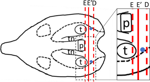
Vestibular organs consist of the maculae staticae, which are located in both the utricle and saccule, as well as the semicircular ducts and their ampullas. There have been no reports on specimen preparation methods for vestibular organs, including maculae staticae or semicircular ducts. In this study, we investigated highly reproducible methods of preparing vestibular organ specimens for histopathological examinations. We established a method that allows researchers to observe the utricle and saccule, including otoliths, the ampulla of a semicircular duct, and parts of semicircular ducts. This highly reproducible method is useful for histopathological analysis of mice with symptoms of abnormal equilibrium caused by medical toxicity and genetic modification.

Keywords: maculae staticae; mice; semicircular ducts; specimen preparation; vestibular organs.


Figures

Fig. 1.
Fig. 1.
Schema of dissecting direction (red line) of methods A–E (left figure). Structure of labyrinth (right). Labels: Pituitary gland (p), Tympanic cavity (t), Trigeminal nerve (tn). Maculae staticae (blue dots).
Fig. 2.
Fig. 2.
(A–E) Schema of methods A–E, respectively. (A–D) Dissecting (dotted line) and sectioning directions (arrow) (left). Dissected surface (right). (E) Dissecting (dotted line) and sectioning directions (arrow) (left). Dissected surface (right). Labels: Pituitary gland (p), Tympanic cavity (t), Trigeminal nerve (tn), Orbit (o). Bar=5 mm.
Fig. 3.
Fig. 3.
Specimens in low magnification (left) and the areas highlighted by squares in high magnification (right). (A) Saccule and parts of semicircular ducts stained with hematoxylin and eosin (HE). (B) Saccule, parts of semicircular ducts, and cochlea stained with HE. (C) Utricle, saccule, and parts of semicircular ducts stained with HE. (D) Utricle, saccule, the ampulla of a semicircular duct (arrowhead), and parts of semicircular ducts stained with HE. (D-300 μm) All compartments of vestibular organs disappeared after further 300 μm sectioning; stained with HE. Labels: Tympanic cavity (t), Utricle (u), Saccule (s), Semicircular ducts (d), Cochlea (c).
Fig. 4.
Fig. 4.
Schema of dissecting direction of methods E and E′. The dissection site for method E′ was 1 mm posterior to that of method E. Dissection direction (red line). Maculae staticae (blue dots).
Fig. 5.
Fig. 5.
Histology at 160 μm sectioned from the dissected surface. (A) Dissecting direction (dotted line) and sectioning direction (arrow) of method Eʹ. (B) The specimen in loupe view stained with HE. (C) Overview of the vestibular organ; stained with HE. (D) The saccule, including sensory epithelium, otolithic membrane, and otoliths stained with HE. The utricle, including sensory epithelium, otolithic membrane, and otoliths stained with HE. (F) The ampulla of a semicircular duct, including sensory epithelium stained with HE. (G) Parts of semicircular ducts stained with HE. Labels: Utricle (u), Saccule (s), Ampulla of semicircular duct (a), Semicircular ducts (d), Sensory epithelium (se), Otoliths (o), Otolithic membrane (om).

References

    1. Ekdale EG. Form and function of the mammalian inner ear. J Anat. 228: 324–337. 2016. - PMC - PubMed
    1. Paken J, Govender CD, Pillay M, and Sewram V. Cisplatin-associated ototoxicity: a review for the health professional. J Toxicol. 2016: 1809394. 2016. - PMC - PubMed
    1. Ding D, Liu H, Qi W, Jiang H, Li Y, Wu X, Sun H, Gross K, and Salvi R. Ototoxic effects and mechanisms of loop diuretics. J Otol. 11: 145–156. 2016. - PMC - PubMed
    1. Schirmer M, Kaiser A, Lessenich A, Lindemann S, Fedrowitz M, Gernert M, and Löscher W. Auditory and vestibular defects and behavioral alterations after neonatal administration of streptomycin to Lewis rats: Similarities and differences to the circling (ci2/ci2) Lewis rat mutant. Brain Res. 1155: 179–195. 2007. - PubMed
    1. Harada Y, and Sugimoto Y. Metabolic disorder of otoconia after streptomycin intoxication. Acta Otolaryngol. 84: 65–71. 1977. - PubMed

LinkOut - more resources