

Do All Notochordal Lesions Require Proton Beam Radiotherapy? A Proposed Reclassification of Ecchordosis Physaliphora as Benign Notochord Cell Tumor
- PMID: 35832978
- PMCID: PMC9272248
- DOI: 10.1055/s-0040-1722717
Do All Notochordal Lesions Require Proton Beam Radiotherapy? A Proposed Reclassification of Ecchordosis Physaliphora as Benign Notochord Cell Tumor
Abstract
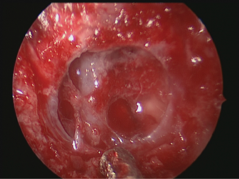
Objectives Ecchordosis physaliphora (EP) is a benign notochord lesion of the clivus arising from the same cell line as chordoma, its malignant counterpart. Although usually asymptomatic, it can cause spontaneous cerebrospinal fluid (CSF) rhinorrhea. Benign notochordal cell tumor (BNCT) is considered another indolent, benign variant of chordoma. Although aggressive forms of chordoma require maximal safe resection followed by proton beam radiotherapy, BNCT and EP can be managed with close imaging surveillance without resection or radiotherapy. However, while BNCT and EP can be distinguished from more aggressive forms of chordoma, differentiating the two is challenging as they are radiologically and histopathologically identical. This case series aims to characterize the clinicopathological features of EP and to propose classifying EP and BNCT together for the purposes of clinical management. Design Case series. Setting Tertiary referral center, United Kingdom. Participants Patients with suspected EP from 2015 to 2019. Main Outcome Measures Diagnosis of EP. Results Seven patients with radiological suspicion of EP were identified. Five presented with CSF rhinorrhea and two were asymptomatic. Magnetic resonance imaging features consistently showed T1-hypointense, T2-hyperintense nonenhancing lesions. Diagnosis was made on biopsy for patients requiring repair and radiologically where no surgery was indicated. The histological features of EP included physaliphorous cells of notochordal origin (positive epithelial membrane antigen, S100, CD10, and/or MNF116) without mitotic activity. Conclusion EP is indistinguishable from BNCT. Both demonstrate markers of notochord cell lines without malignant features. Their management is also identical. We therefore propose grouping EP with BNCT. Close imaging surveillance is required for both as progression to chordoma remains an unquantified risk.
Keywords: BNCTs; EP; benign notochordal cell tumors; chordoma; ecchordosis physaliphora; proton beam radiotherapy.
Thieme. All rights reserved.
Conflict of interest statement
Conflict of Interest None declared.
Figures


References
-
- Nishigaya K, Kaneko M, Ohashi Y, Nukui H. Intradural retroclival chordoma without bone involvement: no tumor regrowth 5 years after operation. Case report. J Neurosurg. 1998;88(04):764–768. - PubMed
-
- Filis A, Kalakoti P, Nanda A. Symptomatic ecchordosis physaliphora mimicking as an intracranial arachnoid cyst. J Clin Neurosci. 2016;28:171–174. - PubMed
