. 2022 Jul 8:10:e13721.
doi: 10.7717/peerj.13721. eCollection 2022.

Optimizing human coronavirus OC43 growth and titration

Affiliations

Optimizing human coronavirus OC43 growth and titration

Christopher Savoie et al. PeerJ. .

Abstract

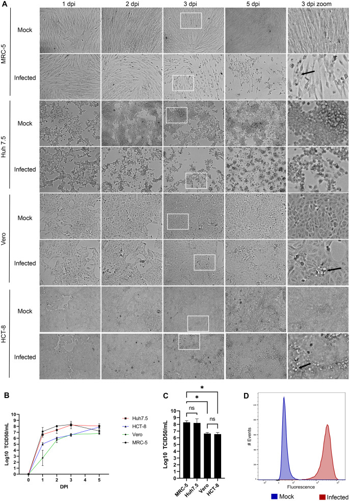
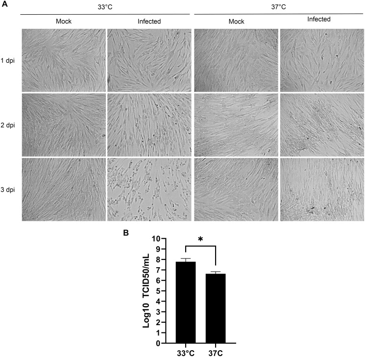
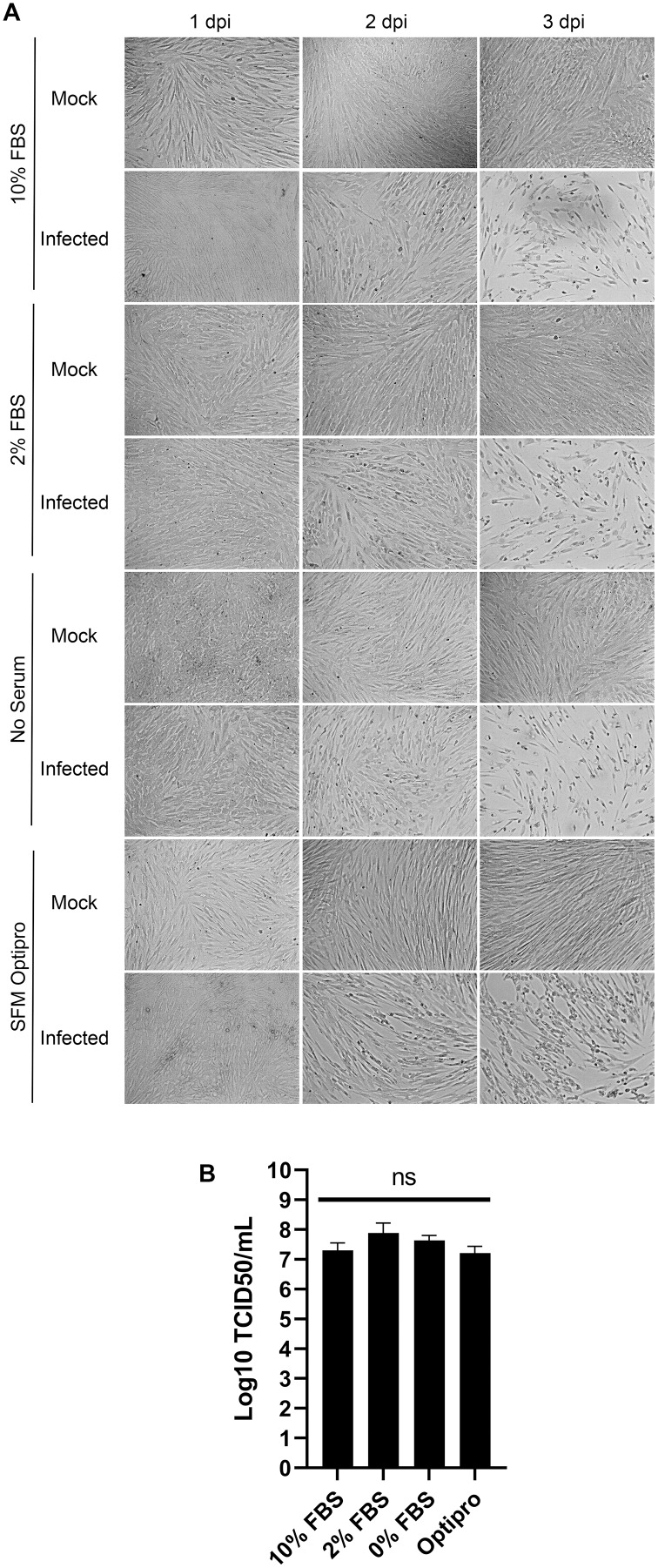
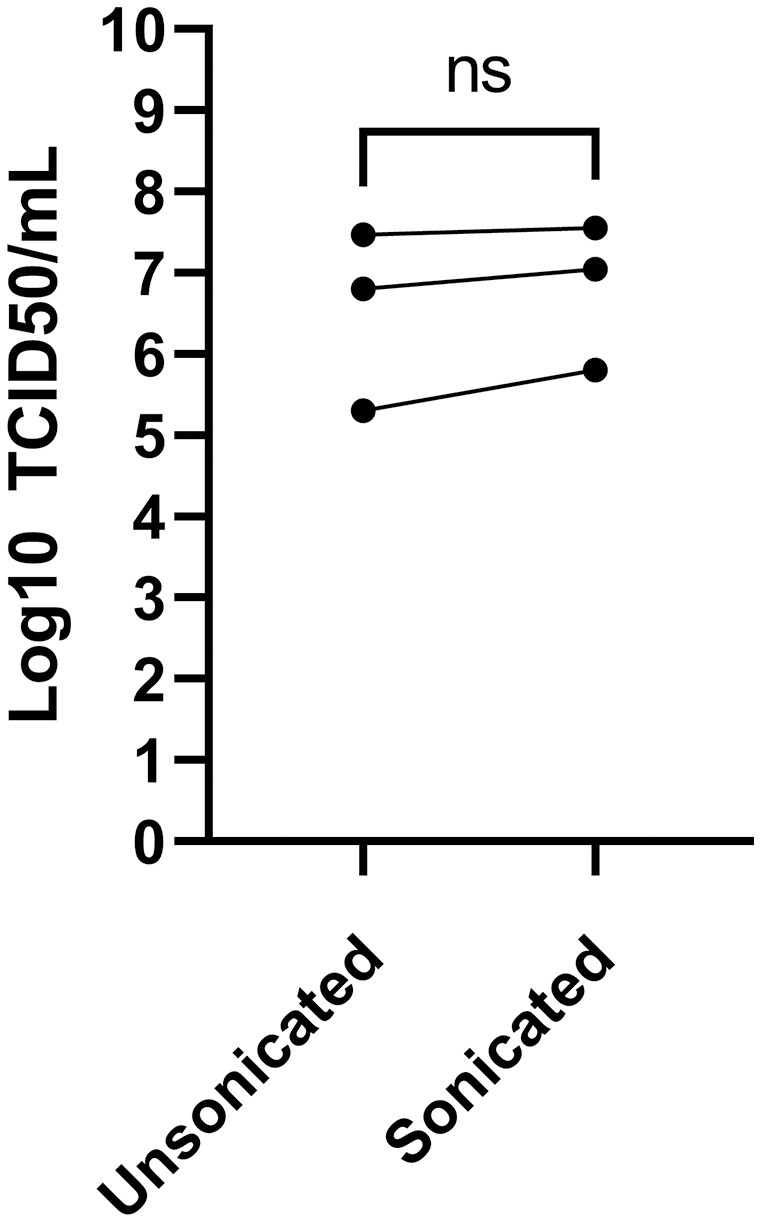
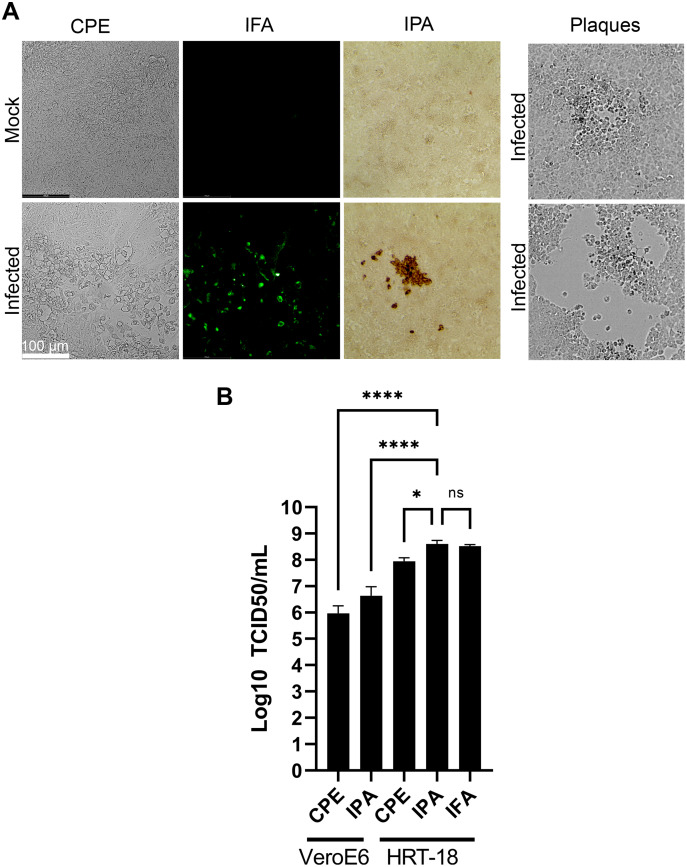
Coronaviruses have been at the forefront of the news for the last 2 years. Unfortunately, SARS-CoV-2, the etiologic agent for the COVID-19 pandemic, must be manipulated in biosecurity level 3 settings, which significantly limits research. Meanwhile, several less pathogenic human coronaviruses (HCoV) exist and can be studied in much more common biosafety level 2 laboratories. Among them, HCoV-OC43 is a good surrogate candidate for SARS-CoV-2 since both are phylogenetically related human Betacoronaviruses. However, one issue has been the lack of standardized means among laboratories to propagate and titer this less virulent coronavirus. The present study probes the optimal parameters to propagate HCoV-OC43. First, testing of five different cell lines (MRC-5, Huh7.5, Vero, HCT-8, HRT-18) indicated that the physiologically relevant MRC-5 human lung cell line produced among the highest viral titers. HRT-18 may however be an interesting alternative as they are quick growing cells that also led to higher viral titers and a better tropism for various HCoV-OC43 variants. We also probed the impact of serum and temperature during viral expansion and confirmed that the normal temperature of the upper respiratory track (33 °C) improves viral yields over the typical 37 °C used to grow many other viruses. Meanwhile, we did not notice any evidence that serum concentrations significantly affected the virus but interestingly noted that the virus grew quite efficiently in a serum-free media formulation. Meanwhile sonication of viral stocks somewhat improved viral titers. Four titration methods (plaque assays, TCID50-CPE, TCID50-IFA and TCID50-IPA) were also probed using two cell lines (VeroE6 and HRT-18). In our hands, plaque assays proved unreliable and quantification of the virus by scoring CPE positive wells was significantly less sensitive than antibody-based assays (IFA and IPA). While the latter methods were equally sensitive, we favor the TCID50-IPA method since simpler, faster and cheaper than the IFA protocol. Moreover, the HRT-18 cells appeared more sensitive to quantify the virus. Perhaps most importantly, these optimized protocols routinely led to high titer viral stocks in the order of 108 TCID50/ml magnitude, which should fulfill the requirements of most experimental settings.

Keywords: COVID; HCoV-OC43; OC43; Propagation; SARS-CoV-2; Titration; Viral stocks.


Conflict of interest statement

The authors declare that they have no competing interests.

Figures

Figure 1
Figure 1. HCoV-OC43 grows best on the MRC-5 and Huh7.5 cell lines.
For all panels, MRC-5, Huh7.5, Vero and HCT-8 cells were grown to 80% confluence on six-well plates then mock treated or infected with at an MOI of 0.7. The cells were then incubated for up to 5 days at 33 °C in DMEM containing 2% serum. (A) CPE was monitored by bright field microscopy at the indicated days post-infection (dpi). The right panels show a zoom view of CPE observed at 3 dpi. (B) To quantify the kinetics of propagation of HCoV-OC43, the extracellular virus produced by infected MRC-5 cells was quantified on HRT-18 cells using the TCID50 immunoperoxidase assay (TCID50-IPA) of Talbot and colleagues (see Materials and Methods). (C) MRC-5 cells infected for 3 dpi were fixed, permeabilized and HCoV-OC43 positive cells scored using viral specific antibodies (see Materials and Methods). (D) To compare viral yield among the cell lines, the extracellular virus harvested at 3 dpi was titered as above. Error bars represent SEM (n = 3). The titers were calculated with the Spearman and Karber method. Statistical analyses were done by one-way ANOVA with Dunnett multiple comparisons (*p < 0.05).
Figure 2
Figure 2. Optimal propagation of HCoV-OC43 at 33 °C.
MRC-5 cells were grown to 80% confluence on 6-well plates before being mock treated or infected at an MOI of 0.7 and grown for 3 days at either 33 °C or 37 °C in DMEM supplemented with 2% serum. (A) Monitoring of the cells by bright field microscopy over time. Note that the infection leads to more CPE at 33 °C compared to 37 °C. (B) Extracellular virus harvested at 3 dpi were titered by the TCID50-IPA method as in Fig. 1. Error bars represent SEM (n = 3). Statistical analyses were done with a Student t-test (*p < 0.05).
Figure 3
Figure 3. HCoV-OC43 can be grown in serum free media.
MRC-5 cells were grown to 80% confluence on six-well plates then mock treated or infected at an MOI of 0.7 in serum free media. After a 1h adsorption period, cells were incubated for up to 3 days at 33 °C in the presence of EMEM containing different concentrations of serum (0%, 2% or 10%) or Optipro, a serum-free media (SFM-Optipro). (A) Bright field monitoring over the course of the infection indicated the lack of noticeable effect of the serum. (B) Extracellular virus harvested on at 3 dpi were titered by the TCID50-IPA method. Error bars represent SEM. Statistical analyses were done by one-way ANOVA with Dunnett multiple comparisons (n = 3).
Figure 4
Figure 4. Effect of sonication of viral titers.
The media was harvested at 3 dpi from MRC-5 infected cells and were split into two and one of the samples sonicated while the other was kept on ice (see Materials and Methods). Viral titers were then determined by the TCID50-IPA method. Three independent experiments are shown individually in the figure (the line indicates the paired samples). Statistical analyses were done by a paired Student t-test.
Figure 5
Figure 5. Optimal TCID50 method for the titration of HCoV-OC43.
A common viral stock produced on MRC-5 cells was used to infect VeroE6 or HRT-18. (A) Typical images of the infected cells by bright field (CPE, IPA) or fluorescence microscopy (IFA). (B) Quantification of the viral yields by TCID50-CPE, TCID50-IPA and TCID50-IFA. Error bars represent SEM (n = 3). Statistical analyses were done by one-way ANOVA with Dunnett multiple comparisons (*p < 0.05; ****p < 0.0001).
Figure 6
Figure 6. The VR-1558 HCoV-OC43 variant has an increased tropism.
MRC-5 and HRT-18 cells were grown to 80% confluence on 6-well plates then infected at an MOI of 0.7 with three HCoV-OC43 variants (VR-759, the HCoV-OC43 rOC/US183-2 double mutant (VR-759 dm) and VR-1558). Three days later, the media was harvested and titered by the TCID50-IPA method on HRT-18 cells. Error bars represent SEM (n = 3). Statistical analyses were done by one-way ANOVA with Dunnett multiple comparisons (*p < 0.05, ***p < 0.001).

Similar articles

Cited by

References

    1. Baer A, Kehn-Hall K. Viral concentration determination through plaque assays: using traditional and novel overlay systems. Journal of Visualized Experiments. 2014;4(93):e52065. doi: 10.3791/52065. - DOI - PMC - PubMed
    1. Bracci N, Pan HC, Lehman C, Kehn-Hall K, Lin SC. Improved plaque assay for human coronaviruses 229E and OC43. PeerJ. 2020;8(93):e10639. doi: 10.7717/peerj.10639. - DOI - PMC - PubMed
    1. Brison E, Jacomy H, Desforges M, Talbot PJ. Novel treatment with neuroprotective and antiviral properties against a neuroinvasive human respiratory virus. Journal of Virology. 2014;88:1548–1563. doi: 10.1128/JVI.02972-13. - DOI - PMC - PubMed
    1. Butler N, Pewe L, Trandem K, Perlman S. Murine encephalitis caused by HCoV-OC43, a human coronavirus with broad species specificity, is partly immune-mediated. Virology. 2006;347(2):410–421. doi: 10.1016/j.virol.2005.11.044. - DOI - PMC - PubMed
    1. Chu H, Chan JF-W, Yuen TT-T, Shuai H, Yuan S, Wang Y, Hu B, Yip CC-Y, Tsang JO-L, Huang X, Chai Y, Yang D, Hou Y, Chik KK-H, Zhang X, Fung AY-F, Tsoi H-W, Cai J-P, Chan W-M, Ip JD, Chu AW-H, Zhou J, Lung DC, Kok K-H, To KK-W, Tsang OT-Y, Chan K-H, Yuen K-Y. Comparative tropism, replication kinetics, and cell damage profiling of SARS-CoV-2 and SARS-CoV with implications for clinical manifestations, transmissibility, and laboratory studies of COVID-19: an observational study. The Lancet Microbe. 2020;1(1):e14–e23. doi: 10.1016/S2666-5247(20)30004-5. - DOI - PMC - PubMed

Publication types

Grants and funding