

Primary muscular hydadit cyst: Case report
- PMID: 35833094
- PMCID: PMC9271982
- DOI: 10.1016/j.radcr.2022.06.021
Primary muscular hydadit cyst: Case report
Abstract
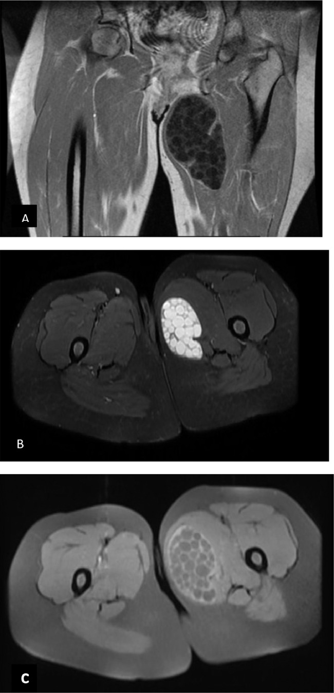
Primary hydatid disease of the skeletal muscle without systemic involvement is rare. Since the infection appears on clinical examination to be a soft-tissue tumor. It is important to have a preoperative radiological evaluation, particularly with magnetic resonance imaging (MRI) to avoid biopsy or improper cyst management during surgery. We present a unique case of a primary hydatid cyst manifesting as an expanding soft-tissue mass in a 31-year-old woman's left thigh with magnetic resonance imaging revealed a very suggestive aspect of hydatid cyst in the adductor muscles and shows the interaction between cysts and nearby structures. The cyst was surgically removed, and macroscopic and microscopic histopathological examinations confirmed the diagnosis of muscular hydatidosis.
Keywords: Intramuscular hydatid cyst; Primary hydatidosis; imaging.
© 2022 The Authors. Published by Elsevier Inc. on behalf of University of Washington.
Figures


References
-
- Pedrosa I., Saiz A., Arrazola J., Ferreiros J., Pedrosa C.S. Hydatid disease: radiologic and pathologic features and complications. Radiographics. 2000;3:795–817. 2000. - PubMed
-
- Comert R.B., Aydingoz U., Ucaner A., &Arikan M. Water-lily sign on MR imaging of primary intramuscular hydatidosis of sartorius muscle. Skeletal Radiol. 2003;32(7):420–423. - PubMed
-
- Guthrie JA, Lawton JO, Chalmers AG. Case report: the MR appearances of primary intramuscular hydatiddisease. Clin Radiol. 1996;51:377–379. - PubMed
-
- Tatari H., Baran Ö., Sanlıdağ T., Göre O., Ak D., Manisalı M., et al. Primary intramuscular hydatidosis of supraspinatus muscle. Arch Orthopaed Trauma Surg. 2001;121(1-2):93–94. - PubMed
-
- Bayram M, Sirikci A. Hydatic cyst locatedintermuscular area of the forearm: MR imaging findings. Eur J Radiol. 2000;36:130–132. - PubMed
Publication types
LinkOut - more resources
Full Text Sources
