

Thiol Modifications in the Extracellular Space-Key Proteins in Inflammation and Viral Infection
- PMID: 35833136
- PMCID: PMC9271835
- DOI: 10.3389/fimmu.2022.932525
Thiol Modifications in the Extracellular Space-Key Proteins in Inflammation and Viral Infection
Abstract
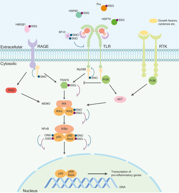
Posttranslational modifications (PTMs) allow to control molecular and cellular functions in response to specific signals and changes in the microenvironment of cells. They regulate structure, localization, stability, and function of proteins in a spatial and temporal manner. Among them, specific thiol modifications of cysteine (Cys) residues facilitate rapid signal transduction. In fact, Cys is unique because it contains the highly reactive thiol group that can undergo different reversible and irreversible modifications. Upon inflammation and changes in the cellular microenvironment, many extracellular soluble and membrane proteins undergo thiol modifications, particularly dithiol-disulfide exchange, S-glutathionylation, and S-nitrosylation. Among others, these thiol switches are essential for inflammatory signaling, regulation of gene expression, cytokine release, immunoglobulin function and isoform variation, and antigen presentation. Interestingly, also the redox state of bacterial and viral proteins depends on host cell-mediated redox reactions that are critical for invasion and infection. Here, we highlight mechanistic thiol switches in inflammatory pathways and infections including cholera, diphtheria, hepatitis, human immunodeficiency virus (HIV), influenza, and coronavirus disease 2019 (COVID-19).
Keywords: S-glutathionylation; S-nitrosylation; disulfide bond; extracellular; infection; inflammation; redox signaling; thiol switch.
Copyright © 2022 Brücksken, Loreto Palacio and Hanschmann.
Conflict of interest statement
The authors declare that the research was conducted in the absence of any commercial or financial relationships that could be construed as a potential conflict of interest.
Figures
Similar articles
-
Thiol-based posttranslational modifications in parasites.Antioxid Redox Signal. 2012 Aug 15;17(4):657-73. doi: 10.1089/ars.2011.4266. Epub 2012 Jan 16. Antioxid Redox Signal. 2012. PMID: 22085115 Review.
-
Thiol switches in membrane proteins - Extracellular redox regulation in cell biology.Biol Chem. 2020 Oct 28;402(3):253-269. doi: 10.1515/hsz-2020-0266. Print 2021 Feb 23. Biol Chem. 2020. PMID: 33108336 Review.
-
Sulfhydryl-mediated redox signaling in inflammation: role in neurodegenerative diseases.Arch Toxicol. 2015 Sep;89(9):1439-67. doi: 10.1007/s00204-015-1496-7. Epub 2015 Apr 1. Arch Toxicol. 2015. PMID: 25827102 Review.
-
Oxidation and S-nitrosylation of cysteines in human cytosolic and mitochondrial glutaredoxins: effects on structure and activity.J Biol Chem. 2007 May 11;282(19):14428-36. doi: 10.1074/jbc.M700927200. Epub 2007 Mar 13. J Biol Chem. 2007. PMID: 17355958
-
Cysteine thiol sulfinic acid in plant stress signaling.Plant Cell Environ. 2024 Aug;47(8):2766-2779. doi: 10.1111/pce.14827. Epub 2024 Jan 22. Plant Cell Environ. 2024. PMID: 38251793 Review.
Cited by
-
N-Acetyl Cysteine Restores the Diminished Activity of the Antioxidant Enzymatic System Caused by SARS-CoV-2 Infection: Preliminary Findings.Pharmaceuticals (Basel). 2023 Apr 14;16(4):591. doi: 10.3390/ph16040591. Pharmaceuticals (Basel). 2023. PMID: 37111348 Free PMC article.
-
Cysteine redoxome landscape in mouse brown adipose tissue under acute cold exposure.iScience. 2025 Feb 17;28(3):112051. doi: 10.1016/j.isci.2025.112051. eCollection 2025 Mar 21. iScience. 2025. PMID: 40104075 Free PMC article.
References
Publication types
MeSH terms
Substances
LinkOut - more resources
Full Text Sources
Medical
Miscellaneous