Review
. 2022 Jun 27:13:893365.
doi: 10.3389/fimmu.2022.893365. eCollection 2022.

N-Glycosylation and Inflammation; the Not-So-Sweet Relation

Affiliations
Review

N-Glycosylation and Inflammation; the Not-So-Sweet Relation

Barbara Radovani et al. Front Immunol. .

Abstract

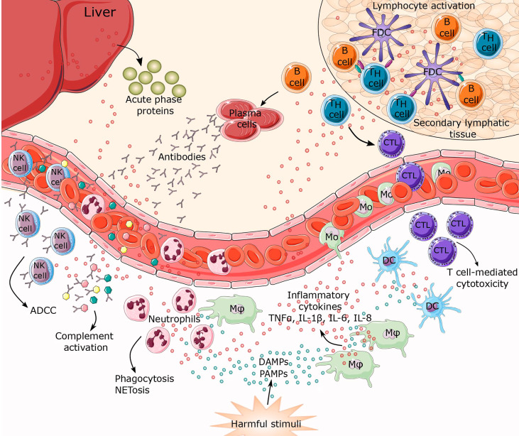
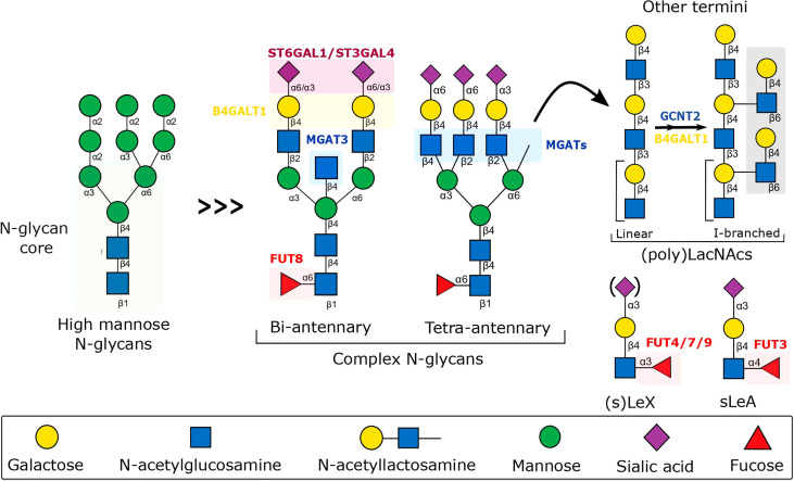
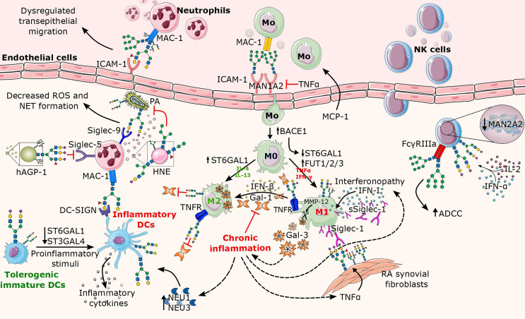
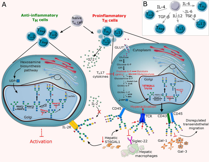
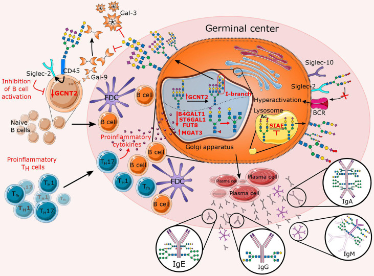
Chronic inflammation is the main feature of many long-term inflammatory diseases such as autoimmune diseases, metabolic disorders, and cancer. There is a growing number of studies in which alterations of N-glycosylation have been observed in many pathophysiological conditions, yet studies of the underlying mechanisms that precede N-glycome changes are still sparse. Proinflammatory cytokines have been shown to alter the substrate synthesis pathways as well as the expression of glycosyltransferases required for the biosynthesis of N-glycans. The resulting N-glycosylation changes can further contribute to disease pathogenesis through modulation of various aspects of immune cell processes, including those relevant to pathogen recognition and fine-tuning the inflammatory response. This review summarizes our current knowledge of inflammation-induced N-glycosylation changes, with a particular focus on specific subsets of immune cells of innate and adaptive immunity and how these changes affect their effector functions, cell interactions, and signal transduction.

Keywords: N-glycans; N-glycosylation; cytokines; immunity; immunoglobulins; inflammation; leukocytes.


Conflict of interest statement

Author IG was employed by Genos Glycoscience. The remaining author declares that the research was conducted in the absence of any commercial or financial relationships that could be construed as a potential conflict of interest.

Figures

Figure 1
Figure 1
Inflammatory response to harmful stimuli. When tissue or cellular damage occurs, danger-associated molecular patterns (DAMPS), pathogen associated molecular patterns (PAMPs) and myriad inflammatory cytokines (TNFα, IL-1β, IL-6, IL-8) are released. These biomolecules can initiate activation of inflammatory pathways resulting in leukocyte recruitment of innate and adaptive immunity, thus establishing a highly coordinated network of many cell types. Activated macrophages, together with damaged endothelial cells, release factors that attract neutrophils and monocytes to the site of inflammation. This represents the first line of defense characterized mostly by phagocytosis and NETosis. Macrophages, together with mature dendritic cells (DCs), are specialized in exposing antigens to lymphocytes (T and B cells), thereby activating antigen-specific adaptive immunity. Lymphocyte differentiation leads to T cell-mediated cytotoxicity, antibody secretion, and antibody dependent cell cytotoxicity (ADCC). Simultaneously, cytokines trigger synthesis and secretion of acute phase proteins from the liver. CTL, cytotoxic T lymphocytes; FDC, follicular dendritic cells; Mφ, macrophage; Mo, monocyte; NK cell, natural killer cell.
Figure 2
Figure 2
Schematic representation of the biosynthesis of N-glycans involved in the fine-tuning of the immune response to inflammation. The schematic includes the major N-glycan structures found on the surface of endothelium, immune cells, and secreted molecules, along with the relevant glycosyltransferases, whose expression has been shown to be modulated by inflammatory cytokines, dramatically affecting glycan-dependent interactions important for leukocyte immune regulation. B4GALT1, Beta-1,4-Galactosyltransferase 1; FUT, Fucosyltransferase; GCNT2, Glucosaminyl (N-acetyl) Transferase 2; MGAT, N-acetylglucosaminyltransferase; ST6GAL4, Beta-Galactoside Alpha-2,3-Sialyltransferase 4; ST6GAL1, Beta-Galactoside Alpha-2,6-Sialyltransferase 1.
Figure 3
Figure 3
Overview of altered N-glycosylation pathways in innate immune cells during chronic inflammation. The major factors contributing to the alterations in N-glycosylation are proinflammatory cytokines (e.g., TNFα, IL-2, IFN-α, IFN-γ) that are released in excess during inflammation. Here, the affected structural elements of N-glycans on the surface of innate leukocytes (neutrophils, macrophages, NK cells, and DCs) are shown along with their associated glycosyltransferases and glycosidases. In neutrophils, the increase of the Lex motif on the integrin MAC-1 leads to dysregulated neutrophil migration, whereas the binding of Lex decorated MAC-1 to DC-SIGN further triggers the activation of DCs. While neutrophilic granules (e.g., HNE) secreted by neutrophils carry truncated N-glycans, the presence of sialylated complex N-glycans and/or the sLex motif on Siglec counterbinding entities contributes to the inflammatory potential of neutrophils in a context-dependent manner. Proinflammatory cytokines enhance transport of monocytes and direct their differentiation into proinflammatory M1 macrophages, while contributing to the absence of sialylated N-glycans, cleavage of Gal-3, and increase in Siglec-1 expression. While surface-bound Siglec-1 is involved in the autoimmune response in rheumatoid arthritis (RA), soluble Siglec-1 is a marker in interferonopathy. In addition, the Gal-1/IFN-β feedback loop involved in termination of inflammation appears to be dysregulated in chronic inflammation. Similarly to macrophages, mature DCs also lack terminal sialic acids, plausibly due to inflammation-mediated decrease in sialyltransferase and/or increase in neuraminidase activity. As for NK cells, the presence of oligomannose N-glycans on FcγRIIIa significantly increases ADCC, whereas cytokine-induced increase in sialylation abrogates Siglec-9-dependent NK cell inhibition by cis-binding. BACE1, Beta-Site APP-Cleaving Enzyme 1; Gal, galectin; hAGP-1; hepatic α1-acid glycoprotein; HNE, human neutrophilic elastase; IFN, interferon; IL, interleukin; ICAM-1, intercellular adhesion molecule 1; MAC-1, macrophage-1 antigen; Man, Mannosidase; MMP-12, matrix metalloproteinase 12; MCP-1, monocyte chemoattractant protein-1; NEU, neuraminidase; NAGP-1, neutrophil α1-acid glycoprotein; PA, Pseudomonas aeruginosa.
Figure 4
Figure 4
Overview of altered N-glycosylation pathways regarding T cells during chronic inflammation. (A) Differentiation of lymphocytes and thus their surface N-glycome is under the direct influence of cytokines and stimulation by antigen presenting cells (APCs). Cytokines control differentiation in favor of proinflammatory T cells (Th1, Th17, Tfh), thereby altering their N-glycome by dysregulating the expression of glycosyltransferases such as MGAT5, ST6GAL1 and FUT8 and abrogating substrate availability for the hexosamine biosynthesis pathway (HBP). The resulting N-glycan changes significantly reduce the binding affinity of inhibitory galectins and Siglecs. (B) Schematic representation of the relevant cytokines responsible for the T cell differentiation. GLUT, glucose transporter; TCR, T cell receptor; Tfh, T follicular helper cell; Th, T helper cell; Treg, T regulatory cell.
Figure 5
Figure 5
Overview of altered N-glycosylation pathways regarding B cells during chronic inflammation. In the presence of proinflammatory stimuli, inflammatory T cells significantly affect B cell proliferation and their N-glycan profile by deregulating a specific subset of glycosyltransferases (B4GALT1, ST6GAL1, FUT8, MGAT3, and GCNT2). The latter is reflected in an increase in features such as bisecting GlcNAc, agalactosylation, afucosylation, and the presence of I-branches that have been shown to inhibit Gal-3 and Gal-9 binding. In addition to the affected Golgi enzymes, lysosomal sialic acid acetyl esterase (SIAE) is also downregulated so that it is unable to deacetylate sialic acids, which is necessary for immunomodulation of B cell receptor (BCR) signaling. This figure also summarizes the Fc N-glycome of secreted immunoglobulins, which reflects inflammation-related changes that may further contribute to disease progression.

Similar articles

Cited by

References

    1. Medzhitov R. Origin and Physiological Roles of Inflammation. Nature (2008) 454:428–35. doi: 10.1038/nature07201 - DOI - PubMed
    1. Feghali CA, Wright TM. Cytokines in Acute and Chronic Inflammation. Front Biosci (1997) 2:d12–26. doi: 10.2741/a171 - DOI - PubMed
    1. Gudelj I, Lauc G, Pezer M. Immunoglobulin G Glycosylation in Aging and Diseases. Cell Immunol (2018) 333:65–79. doi: 10.1016/j.cellimm.2018.07.009 - DOI - PubMed
    1. Groux-Degroote S, Cavdarli S, Uchimura K, Allain F, Delannoy P. Glycosylation Changes in Inflammatory Diseases. Adv Protein Chem Struct Biol (2020) 119:111–56. doi: 10.1016/bs.apcsb.2019.08.008 - DOI - PubMed
    1. Schjoldager KT, Narimatsu Y, Joshi HJ, Clausen H. Global View of Human Protein Glycosylation Pathways and Functions. Nat Rev Mol Cell Biol (2020) 21:729–49. doi: 10.1038/s41580-020-00294-x - DOI - PubMed

Publication types

LinkOut - more resources