. 2023 Feb;38(1):58-70.
doi: 10.1111/omi.12378. Epub 2022 Jul 25.

Increased sensitivity of Aggregatibacter actinomycetemcomitans to human serum is mediated by induction of a bacteriophage

Affiliations

Increased sensitivity of Aggregatibacter actinomycetemcomitans to human serum is mediated by induction of a bacteriophage

Gaoyan G Tang-Siegel et al. Mol Oral Microbiol. 2023 Feb.

Abstract

Aggregatibacter actinomycetemcomitans, a Gram-negative oral pathobiont causing aggressive periodontitis and systemic infections, demonstrates serum resistance. We have identified a dsDNA-tailed bacteriophage, S1249, which was found to convert from this microorganism inducible by human serum into a lytic state to kill the bacterium. This phage demonstrated active transcripts when exposed to human serum: 20% of genes were upregulated more than 10-fold, and 45% of them were upregulated 5-10-fold when the bacterium was grown in the presence of human serum compared to without the presence of human serum. Transcriptional activation when grown in equine serum was less pronounced. This phage demonstrated a tail with inner rigid tubes and an outer contractile sheath, features of Myoviridae spp. Further characterization revealed that the lysogenized integration of the phage in the chromosome of A. actinomycetemcomitans occurred between the genes encoding cold-shock DNA-binding domain-containing protein (csp) and glutamyl-tRNA synthetase (gltX). Both phage DNA integrated lysogeny and nonintegrated pseudolysogeny were identified in the infected bacterium. A newly generated, lysogenized strain using this phage displayed similar attributes, including 63% growth inhibition compared to its isogenic phage-free strain when in the presence of human serum. Our data suggest that bacteriophage S1249 can be induced in the presence of human serum and enters the lytic cycle, which reduces the viability of infected bacteria in vivo.

Keywords: Myoviridae; bacteriophages; gram-negative periodontal pathobionts; human serum; lytic cycle; pseudolysogeny.


Conflict of interest statement

The authors declare no conflict of interest.

Figures
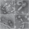
FIGURE 1
FIGURE 1
Aggregatibacter phage S1249 displays a contractile tail under transmission electron microscopy (TEM). Morphologies of phages at different assembly stages are presented. (a) Virus heads, (b) heads with inner rigid tubes of tails, (c and d) virions. This phage virion is composed of an oblate icosahedral head (∼60–70 nm in diameter) and a tail (∼100 nm in length) with tail fibers. The tail structure is composed of an inner rigid tube (∼15 nm in diameter, shown by the black arrow) and an outer contractile sheath (∼30 nm in diameter, shown by the write arrow). Scale bar: 100 nm.
FIGURE 2
FIGURE 2
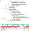
BLAST pairwise alignment using the phage S1249 sequence (GQ866233) against the database https://blast.ncbi.nlm.nih.gov/Blast.cgi. (a) Blast tree generated using neighbor‐joining method. Phage S1249 matches the DNA sequences of Myoviridae spp. (b) Distribution of the top 90 blast hits on 10 subjects of phages/prophage sequences identified from A. actinomycetemcomitans. Phage AaΦ23 demonstrates 75% query coverage, and two A. actinomycetemcomitans strains, 624 and NUM4039, show 76% and 65% coverage, respectively. The difference is mainly located in the region encoding the regulation of DNA replication, modification, and recombination.
FIGURE 3
FIGURE 3
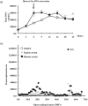
DNA replication of strain D11S‐1 and RNA‐seq analysis of phage S1249 under three growth conditions. (a) Total DNA replication and cell harvest for RNA extraction. Total DNA amounts were doubled in the presence of sera between 6 and 9 h. Cells were harvested at the 6‐h time point for RNA isolation. (b) RNA‐seq analysis. A total of 66 ORFs representing the whole phage genome are labeled with numbers on the X‐axis. Each dot represents an average transcription level of one gene based on duplicate independent experiments. Extensive transcriptional activation was observed, including 20% of the genes being upregulated greater than 10‐fold and 45% being upregulated between 5‐ and 10‐fold in the presence of human serum but not equine serum. One protein encoded by D11S_2259 was actively transcribed, especially in the presence of human serum (shown by hollow arrow).
FIGURE 4
FIGURE 4
Phage S1249 virions released in the culture medium containing human serum form aggregates. (a) Phage DNA was quantified by PCR from the filtered, bacterial culture spent medium of D11S‐1. Bacterial spent media were collected after 0‐, 3‐, 6‐, 9‐ and 24‐h bacterial growth, and a fivefold increase in phage DNA was detected after 6‐h growth in the presence of human serum (**p < 0.01). (b) Phage virions (indicated by arrows) released from the bacterium formed aggregates in human serum. Scale bar: 100 nm.
FIGURE 5
FIGURE 5
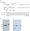
Intracellular location of phage DNA. (a) attB and attP sites: The attP site was identified from the phage located between ORF1 and ORF66 (the int gene), and the attB site was identified from A. actinomycetemcomitans between the csp and gltX genes. (b) Integration in strain D11S‐1. Lanes 1 and 2 were amplified using primers csp_F and gltX_R (1.5 kbp), and lanes 3 and 4 were amplified using primers int_F and gltX_R (2.2 kbp). The DNA of the phage‐free strain ATCC29523 was used as a control and amplified only with primers specific for the bacterium shown in lane 1 but not with primers int_F and gltX_R in lane 3. The DNA of D11S‐1 was displayed both without and with integrated phage DNA, as demonstrated by the 1.5‐ and 2.2‐kbp amplicons in lanes 2 and 4. (c) Circular dsDNA of phage S1249. To determine if phage DNA is circular when not integrated, PCR was performed using DNA isolated from bacteria, the isolated phage, and primers targeting the attP site, the first ORF1 and the last ORF66. Lanes 1 and 2: strain D11S‐1; lane 3: strain ATCC29523; lanes 4 and 5: virion S1249. PCR using primers attP_F and ORF1_R yielded a 450 bp product in both D11S‐1 (lane 1) and S1249 (lane 4), and PCR using int_F2 and ORF1_R yielded a 1.4 kbp amplicon in both D11S‐1 (lane 4) and S1249 (lane 5), indicating that a circular phage dsDNA is present in both infected strain D11S‐1 and the mature virus.
FIGURE 6
FIGURE 6
Genotype of phage S1249 in a generated infection model strain IDH84/S1249. Multiplex colony PCR was performed using four primers (csp_F, gltX_R, int_F, and int_R) targeting both bacterial and phage DNA at the same time. The 2.2 kb, 1.5 kb, and 1 kb bands represent phage DNA integrated, nonintegrated and infected by the phage. Cells from the same colony displayed both with and without phage DNA integrated into the bacterial chromosome in D11S‐1. The same genotype was observed in a newly generated, infected strain IDH84/S1249. IDH84: the parent strain without phage infection; IDH84/S1249_P1: single colony from the first passage after infection; IDH84/S1249_P2: single colony from the passaged strain of IDH84/S1249_P1. D7S‐1 and SCC1398 are two phage‐free strains demonstrating single bands only amplified by primers csp_F and gltX_R.
FIGURE 7
FIGURE 7
The infection model strain demonstrated human serum sensitivity. IDH84: the parent strain without phage infection; IDH84/S1249: strain IDH84 infected by phage S1249. (a) Grown in TSBYE. IDH84/S1249 demonstrated 1.5‐fold increased colony‐forming units (CFUs) compared to IDH84 after 6 h of growth in TSBYE. (b) Grown in 75% human serum. IDH84/S1249 demonstrated 33% and 63% reductions, respectively, in recovered CFUs compared to IDH84 after 6‐ and 22‐h exposure to 75% human serum. (*p < 0.05; **p < 0.01).

Similar articles

Cited by

References

    1. Akrivopoulou, C. , Green, I. M. , Donos, N. , Nair, S. P. , & Ready, D. (2017). Aggregatibacter actinomycetemcomitans serotype prevalence and antibiotic resistance in a UK population with periodontitis. Journal of Global Antimicrobial Resistance, 10, 54–58. - PubMed
    1. Anton‐Vazquez, V. , Dworakowski, R. , Cannata, A. , Amin‐Youssef, G. , Gunning, M. , Papachristidis, A. , MacCarthy, P. , Baghai, M. , Deshpande, R. , Khan, H. , Byrne, J. , & Fife, A. (2022). 16S rDNA PCR for the aetiological diagnosis of culture‐negative infective endocarditis. Infection, 50(1), 243–249. 10.1007/s15010-021-01690-x - DOI - PubMed
    1. Ardila, C. M. , & Bedoya‐García, J. A. (2020). Antimicrobial resistance of Aggregatibacter actinomycetemcomitans, Porphyromonas gingivalis and Tannerella forsythia in periodontitis patients. Journal of Global Antimicrobial Resistance, 22, 215–218. - PubMed
    1. Asakawa, R. , Komatsuzawa, H. , Kawai, T. , Yamada, S. , Goncalves, R. B. , Izumi, S. , Fujiwara, T. , Nakano, Y. , Suzuki, N. , Uchida, Y. , Ouhara, K. , Shiba, H. , Taubman, M. A. , Kurihara, H. , & Sugai, M. (2003). Outer membrane protein 100, a versatile virulence factor of Actinobacillus actinomycetemcomitans . Molecular Microbiology, 50(4), 1125–1139. 10.1046/j.1365-2958.2003.03748.x - DOI - PubMed
    1. Cahill, J. , & Young, R. (2019). Phage lysis: Multiple genes for multiple barriers. Advances in Virus Research, 103, 33–70. - PMC - PubMed

Publication types