. 2022 Nov;31(11):1699-1711.
doi: 10.1111/exd.14644. Epub 2022 Jul 25.

IL-38 orchestrates proliferation and differentiation in human keratinocytes

Affiliations

IL-38 orchestrates proliferation and differentiation in human keratinocytes

Loïc Mermoud et al. Exp Dermatol. 2022 Nov.

Abstract

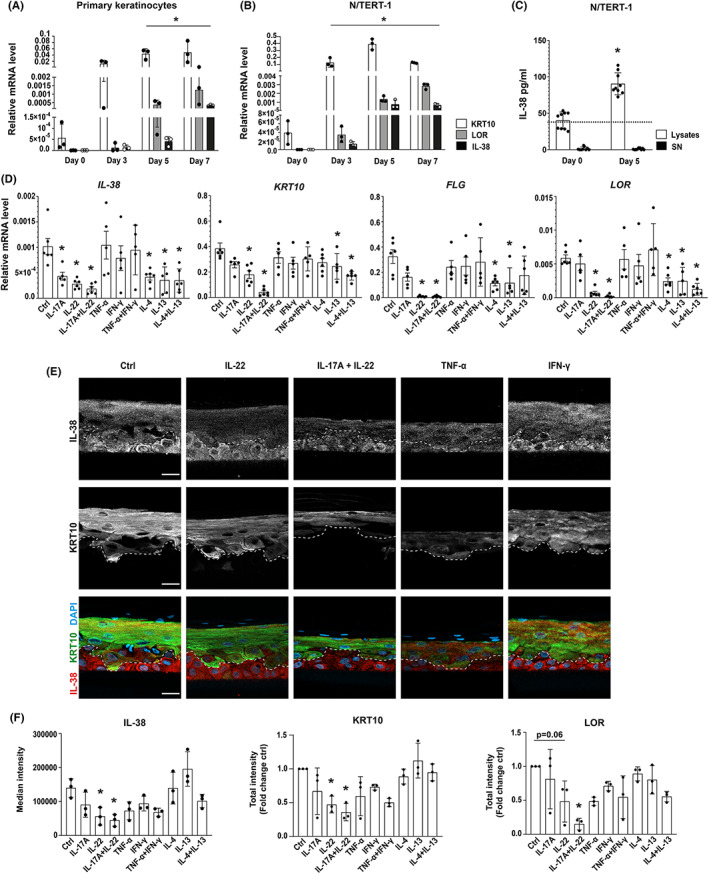
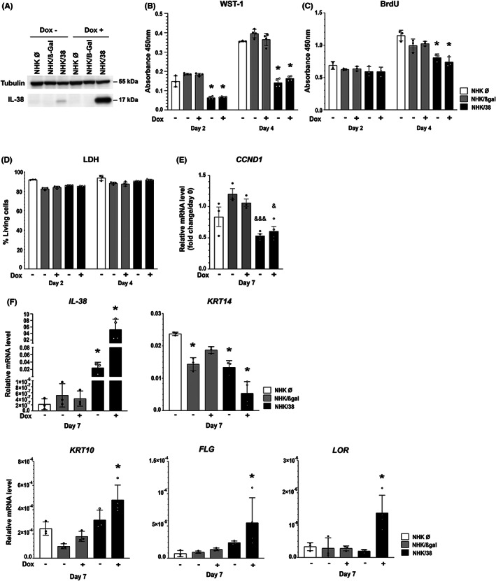
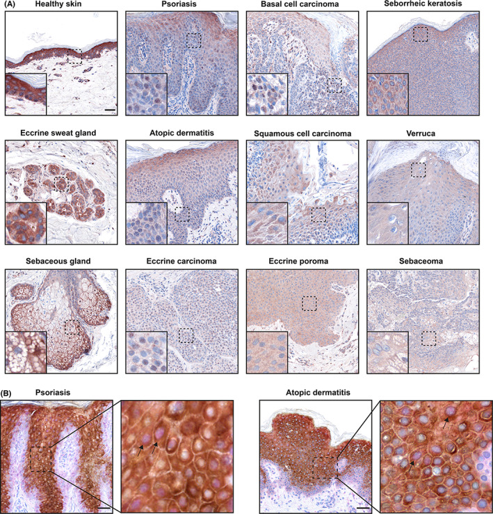
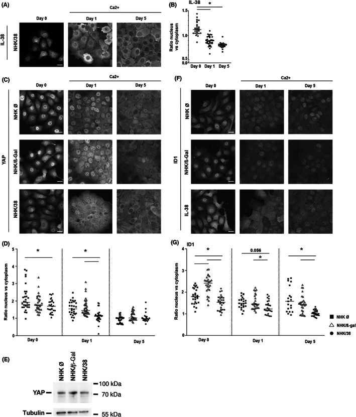
Interleukin (IL)-38 is a member of the IL-1 cytokine family with reported anti-inflammatory activity. The highest constitutive IL-38 expression is detected in the skin, where it is mainly produced by differentiating keratinocytes. However, little data are available regarding its biological functions. In this study, we investigated the role of IL-38 in skin physiology. We demonstrate here that dermal fibroblasts and epithelial cells of skin appendages, such as eccrine sweat glands and sebaceous glands, also express IL-38. Next, using two- and three-dimensional cell cultures, we show that endogenous expression of IL-38 correlates with keratinocyte differentiation and its ectopic overexpression inhibits keratinocyte proliferation and enhances differentiation. Accordingly, immunohistochemical analysis revealed downregulation of IL-38 in skin pathologies characterized by keratinocyte hyperproliferation, such as psoriasis and basal or squamous cell carcinoma. Finally, intracellular IL-38 can shuttle between the nucleus and the cytoplasm and its overexpression modulates the activity of the transcription regulators YAP and ID1. Our results indicate that IL-38 can act independently from immune system activation and suggest that it may affect the epidermis directly by decreasing proliferation and promoting differentiation of keratinocytes. These data suggest an important role of keratinocyte-derived IL-38 in skin homeostasis and pathologies characterized by epidermal alterations.

Keywords: IL-17A; IL-22; IL-38; differentiation; keratinocyte; nuclear localization; proliferation; psoriasis.


Conflict of interest statement

WHB received honoraria as advisor or invited speaker from Abbvie, Almirall, BMS, Celgene, Leo, Lilly, Novartis, UCB. The other authors have no conflict of interest related to the present manuscript to declare.

Figures

FIGURE 1
FIGURE 1
Regulation of IL‐38 expression in differentiating keratinocytes and in RHE stimulated with inflammatory cytokines. IL‐38, KRT10, and LOR mRNA levels, relative to 18S rRNA, assessed by RTqPCR in (A) primary keratinocytes and (B) the N/TERT‐1 cell line differentiated in monolayer cultures for 0, 3, 5 and 7 days. Data are means ± SEM (n = 3). (C) IL‐38 protein levels measured by ELISA in cell lysates and supernatants of N/TERT‐1 keratinocytes differentiated for 0 and 5 days. The dotted line represents the lower detection limit of the ELISA. Data are means ± SEM (n = 9). (D) IL‐38, KRT10, FLG and LOR mRNA levels, relative to the mean of 18S rRNA and GAPDH mRNA levels, assessed by RTqPCR in RHE stimulated with indicated cytokines for 24 h. Data are means ± SEM (n = 5–6). (E) Immunofluorescent staining for IL‐38 (upper panels, white; lower panels, red) and KRT10 (middle panels, white; lower panels, green) in RHE cultured in the presence of indicated cytokines for 72 h. Nuclei were counterstained with DAPI. The broken line represents the boundary between basal and suprabasal layers, based on KRT10 staining in suprabasal cells. Representative pictures chosen from two experiments out of three are shown. Scale bar, 50 μm. (F) Quantification of IL‐38, KRT10 and LOR immunofluorescent staining, shown as median intensity (IL‐38) or as total intensity in a 1000 μm2 area (KRT10 and LOR). Data are means ± SEM (n = 3). Data are (A, B, D, F) biological replicates or (C) technical triplicates of biological triplicates. *p < 0.05 as compared to (A, B, C) day 0 or (D, F) control. p values were calculated using a paired t‐test (C) or two‐way ANOVA with Dunnet's correction (a, B, D and F). KRT10, keratin 10; LOR, loricrin, FLG, filaggrin.
FIGURE 2
FIGURE 2
Effects of IL‐38 overexpression on NHK cells. (A) Western blot analysis of IL‐38 protein expression in empty vector transfected NHK cells (NHK Ø), and in NHK/β‐Gal and NHK/38 cells without and with Dox induction for 24 h. Tubulin levels are shown as a loading control. (B) Metabolic activity, assessed by WST‐1 colorimetric assay, in NHK Ø, NHK/β‐Gal and NHK/38 cells cultured for 2 and 4 days after induction without or with Dox. Data are means ± SEM (n = 3–4). (C) Cell proliferation assessed by BrdU assay in NHK Ø, NHK/β‐Gal and NHK/38 cells cultured for 2 and 4 days after induction without or with Dox. Data are means ± SEM (n = 3). (D) Cell viability, assessed by measuring lactate dehydrogenase (LDH) activity in supernatants of NHK Ø, NHK/β‐Gal and NHK/38 cells incubated during 2, 4 or 6 days after induction without or with Dox. Results are shown as the percentage of living cells. Data are means ± SEM (n = 3–4). (E) Fold change in CCND1 mRNA levels, normalized to 18S rRNA, relative to day 0, assessed by RTqPCR in NHK Ø, NHK/β‐Gal and NHK/38 cells differentiated for 7 days after induction without or with Dox. Data are means ± SEM (n = 3–4). (F) IL‐38, KRT14, KRT10, FLG and LOR mRNA levels, relative to 18S rRNA, assessed by RTqPCR in NHK Ø, NHK/β‐Gal and NHK/38 cells differentiated for 7 days after induction without or with Dox. Data are means ± SEM (n = 3–5). Data are (B‐D) technical triplicates or technical duplicates of biological duplicates, or (E, F) biological replicates. *p < 0.05 as compared to NHK Ø cells, & p < 0.05, &&& p < 0.001 as compared to NHK/β‐Gal cells, p values were calculated using a two‐way ANOVA with Tukey's multiple comparison.
FIGURE 3
FIGURE 3
IL‐38 expression in human skin pathologies. (A) Immunohistochemical analysis of IL‐38 expression in human skin. (B) Double immunohistochemical staining of IL‐38 (pink) and KRT10 (brown) in psoriasis and atopic dermatitis. The insets demonstrate higher magnification of the highlighted areas. The arrows point to examples of IL‐38 nuclear localization. Scale bar, 50 μm. One representative donor of one to three is shown.
FIGURE 4
FIGURE 4
Localization of IL‐38, YAP and ID1 in NHK cells. (A) IL‐38 protein localization assessed by immunofluorescence in Dox‐treated NHK/38 cells cultured without (=day 0) or with 2 mM Ca2+ for 1 or 5 days. Scale bar 20 μm (B) IL‐38 protein localization quantified as the ratio of nuclear/cytoplasmic staining intensity on days 0, 1 and 5. Data are individual values and means (n ≥ 21). (C) YAP protein localization assessed by immunofluorescence in Dox‐treated NHK Ø, NHK/β‐Gal and NHK/38 cells cultured without (=day 0) or with 2 mM Ca2+ for 1 or 5 days. (D) YAP protein localization quantified as the ratio of nuclear/cytoplasmic staining intensity on days 0, 1 and 5. Data are individual values and means (n ≥ 21). (E) YAP expression assessed by Western blotting in Dox‐treated NHK Ø, NHK/β‐Gal and NHK/38 cells cultured without Ca2+ (=day 0). Tubulin levels are shown as loading control. (F) ID1 protein localization assessed by immunofluorescence in Dox‐treated NHK Ø, NHK/β‐Gal and NHK/38 cells cultured without (=day 0) or with 2 mM Ca2+ for 1 or 5 days. (G) ID1 protein localization quantified as the ratio of nuclear/cytoplasmic staining on days 0, 1 and 5. Data are individual values and means (n ≥ 19). Data are values for individual cells (n ≥ 19) from biological triplicates. (B, D and G) *p < 0.05. p values were calculated using a two‐way ANOVA with Tukey's multiple comparison.

Similar articles

Cited by

References

    1. Lin H, Ho AS, Haley‐Vicente D, et al. Cloning and characterization of IL‐1HY2, a novel interleukin‐1 family member. J Biol Chem. 2001;276:20597‐20602. - PubMed
    1. Bensen JT, Dawson PA, Mychaleckyj JC, Bowden DW. Identification of a novel human cytokine gene in the interleukin gene cluster on chromosome 2q12‐14. J Interf Cytok Res. 2001;21:899‐904. - PubMed
    1. van de Veerdonk FL, Stoeckman AK, Wu G, et al. IL‐38 binds to the IL‐36 receptor and has biological effects on immune cells similar to IL‐36 receptor antagonist. Proc Natl Acad Sci USA. 2012;109:3001‐3005. - PMC - PubMed
    1. Mora J, Schlemmer A, Wittig I, et al. Interleukin‐38 is released from apoptotic cells to limit inflammatory macrophage responses. J Mol Cell Biol. 2016;8:426‐438. - PubMed
    1. Han Y, Mora J, Huard A, et al. IL‐38 ameliorates skin inflammation and limits IL‐17 production from γδ T cells. Cell Rep. 2019;27:835‐846. - PubMed

Publication types

Associated data