

Effects of Scaffold Shape on Bone Regeneration: Tiny Shape Differences Affect the Entire System
- PMID: 35833725
- PMCID: PMC9413413
- DOI: 10.1021/acsnano.2c03776
Effects of Scaffold Shape on Bone Regeneration: Tiny Shape Differences Affect the Entire System
Abstract
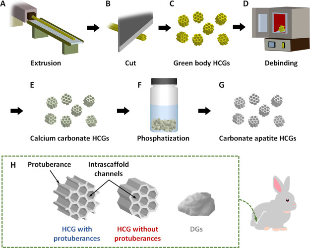
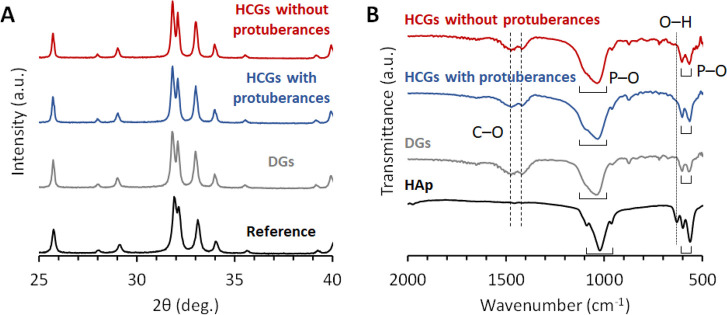
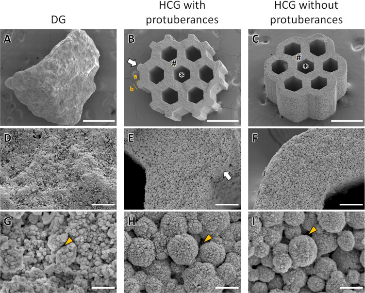
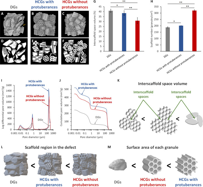
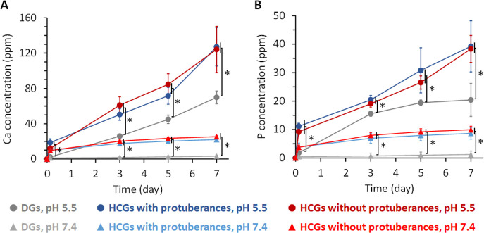
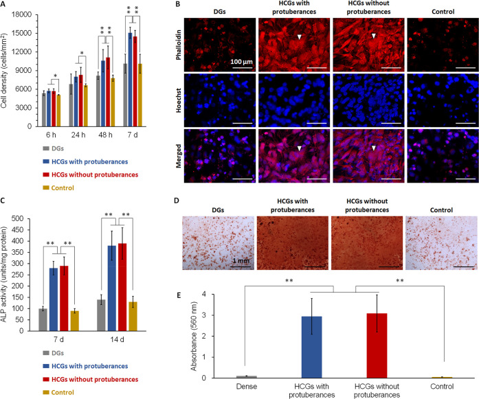
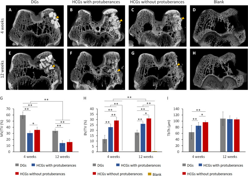
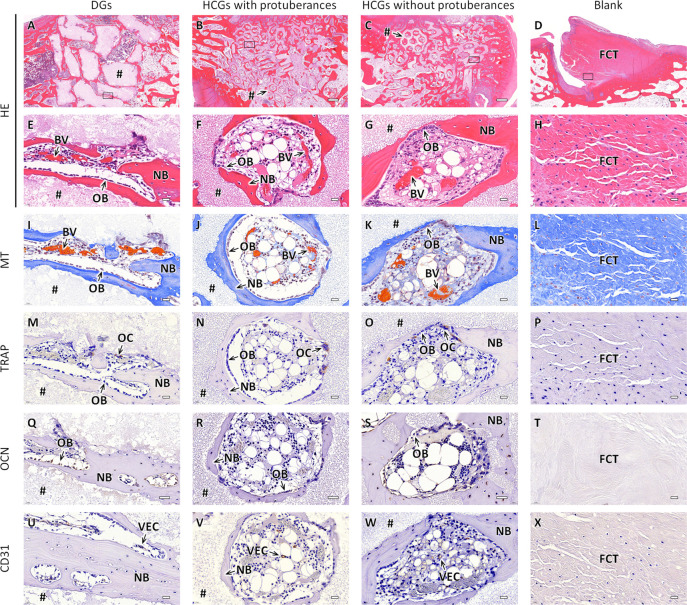
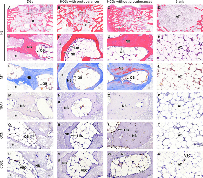
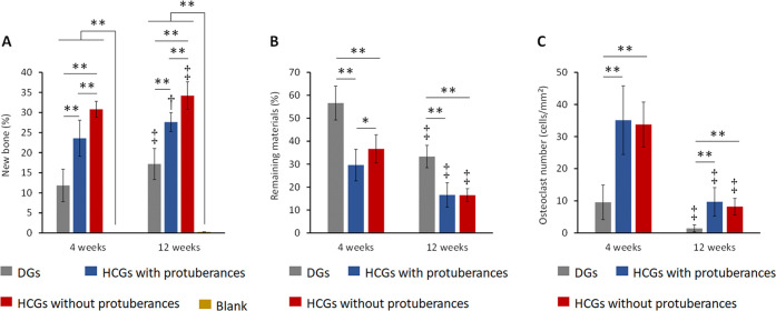
Although studies on scaffolds for tissue generation have mainly focused on the chemical composition and pore structure, the effects of scaffold shape have been overlooked. Scaffold shape determines the scaffold surface area (SA) at the single-scaffold level (i.e., microscopic effects), although it also affects the amount of interscaffold space in the tissue defect at the whole-system level (i.e., macroscopic effects). To clarify these microscopic and macroscopic effects, this study reports the osteogenesis abilities of three types of carbonate apatite granular scaffolds with different shapes, namely, irregularly shaped dense granules (DGs) and two types of honeycomb granules (HCGs) with seven hexagonal channels (∼255 μm in length between opposite sides). The HCGs possessed either 12 protuberances (∼75 μm in length) or no protuberances. Protuberances increased the SA of each granule by 3.24 mm2 while also widening interscaffold spaces and increasing the space percentage in the defect by ∼7.6%. Interscaffold spaces were lower in DGs than HCGs. On DGs, new bone formed only on the surface, whereas on HCGs, bone simultaneously formed on the surface and in intrascaffold channels. Interestingly, HCGs without protuberances formed approximately 30% more new bone than those with protuberances. Thus, even tiny protuberances on the scaffold surface can affect the percentage of interscaffold space, thereby exerting dominant effects on osteogenesis. Our findings demonstrate that bone regeneration can be improved by considering macroscopic shape effects beyond the microscopic effects of the scaffold.
Keywords: bone; granule; honeycomb; regenerative medicine; scaffold; tissue engineering.
Conflict of interest statement
The authors declare no competing financial interest.
Figures
Similar articles
-
Effects of macropore size in carbonate apatite honeycomb scaffolds on bone regeneration.Mater Sci Eng C Mater Biol Appl. 2020 Jun;111:110848. doi: 10.1016/j.msec.2020.110848. Epub 2020 Mar 13. Mater Sci Eng C Mater Biol Appl. 2020. PMID: 32279778
-
Bone formation on the apatite-coated zirconia porous scaffolds within a rabbit calvarial defect.J Biomater Appl. 2008 May;22(6):485-504. doi: 10.1177/0885328207078075. Epub 2007 May 10. J Biomater Appl. 2008. PMID: 17494967
-
A Novel 3D-bioprinted Porous Nano Attapulgite Scaffolds with Good Performance for Bone Regeneration.Int J Nanomedicine. 2020 Sep 22;15:6945-6960. doi: 10.2147/IJN.S254094. eCollection 2020. Int J Nanomedicine. 2020. PMID: 33061361 Free PMC article.
-
Porosity of 3D biomaterial scaffolds and osteogenesis.Biomaterials. 2005 Sep;26(27):5474-91. doi: 10.1016/j.biomaterials.2005.02.002. Biomaterials. 2005. PMID: 15860204 Review.
-
Gradient scaffolds for osteochondral tissue engineering and regeneration.J Mater Chem B. 2020 Sep 23;8(36):8149-8170. doi: 10.1039/d0tb00688b. J Mater Chem B. 2020. PMID: 32776030 Review.
Cited by
-
Lamellar Septa-like Structured Carbonate Apatite Scaffolds with Layer-by-Layer Fracture Behavior for Bone Regeneration.Biomimetics (Basel). 2024 Feb 14;9(2):112. doi: 10.3390/biomimetics9020112. Biomimetics (Basel). 2024. PMID: 38392158 Free PMC article.
-
Hollow Hydroxyapatite Microspheres Loaded with rhCXCL13 to Recruit BMSC for Osteogenesis and Synergetic Angiogenesis to Promote Bone Regeneration in Bone Defects.Int J Nanomedicine. 2023 Jun 29;18:3509-3534. doi: 10.2147/IJN.S408905. eCollection 2023. Int J Nanomedicine. 2023. PMID: 37404852 Free PMC article.
-
Carbonate Apatite Honeycomb Scaffold-Based Drug Delivery System for Repairing Osteoporotic Bone Defects.ACS Appl Mater Interfaces. 2024 Sep 4;16(35):45956-45968. doi: 10.1021/acsami.4c08047. Epub 2024 Aug 25. ACS Appl Mater Interfaces. 2024. PMID: 39182190 Free PMC article.
-
Reconstruction of Load-Bearing Segmental Bone Defects Using Carbonate Apatite Honeycomb Blocks.ACS Mater Au. 2023 Apr 26;3(4):321-336. doi: 10.1021/acsmaterialsau.3c00008. eCollection 2023 Jul 12. ACS Mater Au. 2023. PMID: 38090126 Free PMC article.
-
Silver phosphate-modified carbonate apatite honeycomb scaffolds for anti-infective and pigmentation-free bone tissue engineering.Mater Today Bio. 2024 Jul 18;27:101161. doi: 10.1016/j.mtbio.2024.101161. eCollection 2024 Aug. Mater Today Bio. 2024. PMID: 39155941 Free PMC article.
References
-
- Aida J.; Nakade M.; Hanibuchi T.; Hirai H.; Osaka K.; Kondo K.. Impact of Oral Health Status on Healthy Life Expectancy in Community-dwelling Population: The AGES Project Cohort Study. In Interface Oral Health Science 2009; Sasano T., Suzuki O., Eds.; Springer: Berlin, 2010; pp 326–328.
Publication types
MeSH terms
LinkOut - more resources
Full Text Sources
