

Autoimmunity and SARS-CoV-2 infection: Unraveling the link in neurological disorders
- PMID: 35833748
- PMCID: PMC9350097
- DOI: 10.1002/eji.202149475
Autoimmunity and SARS-CoV-2 infection: Unraveling the link in neurological disorders
Abstract
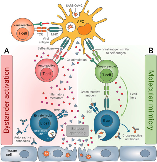
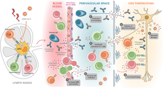
According to the World Health Organization, severe acute respiratory syndrome coronavirus 2 (SARS-CoV-2) has already infected more than 400 million people and caused over 5 million deaths globally. The infection is associated with a wide spectrum of clinical manifestations, ranging from no signs of illness to severe pathological complications that go beyond the typical respiratory symptoms. On this note, new-onset neurological and neuropsychiatric syndromes have been increasingly reported in a large fraction of COVID-19 patients, thus potentially representing a significant public health threat. Although the underlying pathophysiological mechanisms remain elusive, a growing body of evidence suggests that SARS-CoV-2 infection may trigger an autoimmune response, which could potentially contribute to the establishment and/or exacerbation of neurological disorders in COVID-19 patients. Shedding light on this aspect is urgently needed for the development of effective therapeutic intervention. This review highlights the current knowledge of the immune responses occurring in Neuro-COVID patients and discusses potential immune-mediated mechanisms by which SARS-CoV-2 infection may trigger neurological complications.
Keywords: SARS-CoV-2; autoimmunity, COVID-19, neuro-COVID, neuroimmunology.
© 2022 The Authors. European Journal of Immunology published by Wiley-VCH GmbH.
Conflict of interest statement
The author declares no commercial or financial conflicts of interest.
Figures
Similar articles
-
A Disease Hidden in Plain Sight: Pathways and Mechanisms of Neurological Complications of Post-acute Sequelae of COVID-19 (NC-PASC).Mol Neurobiol. 2025 Feb;62(2):2530-2547. doi: 10.1007/s12035-024-04421-z. Epub 2024 Aug 12. Mol Neurobiol. 2025. PMID: 39133434 Review.
-
Neurological Complications of COVID-19: Unraveling the Pathophysiological Underpinnings and Therapeutic Implications.Viruses. 2024 Jul 24;16(8):1183. doi: 10.3390/v16081183. Viruses. 2024. PMID: 39205157 Free PMC article. Review.
-
SARS-CoV-2 infection as a trigger of autoimmune response.Clin Transl Sci. 2021 May;14(3):898-907. doi: 10.1111/cts.12953. Epub 2021 Jan 21. Clin Transl Sci. 2021. PMID: 33306235 Free PMC article.
-
Myoclonus status revealing COVID 19 infection.Seizure. 2023 Jan;104:12-14. doi: 10.1016/j.seizure.2022.11.010. Epub 2022 Nov 22. Seizure. 2023. PMID: 36446232 Free PMC article.
-
Emerging COVID-19 Neurological Manifestations: Present Outlook and Potential Neurological Challenges in COVID-19 Pandemic.Mol Neurobiol. 2021 Sep;58(9):4694-4715. doi: 10.1007/s12035-021-02450-6. Epub 2021 Jun 24. Mol Neurobiol. 2021. PMID: 34169443 Free PMC article. Review.
Cited by
-
From infection to autoimmunity: can COVID-19 spark new auto-immune conditions?Respir Med Case Rep. 2025 Apr 26;55:102216. doi: 10.1016/j.rmcr.2025.102216. eCollection 2025. Respir Med Case Rep. 2025. PMID: 40415761 Free PMC article.
-
Mendelian randomization supports causality between COVID-19 and glaucoma.Medicine (Baltimore). 2024 Jun 14;103(24):e38455. doi: 10.1097/MD.0000000000038455. Medicine (Baltimore). 2024. PMID: 38875430 Free PMC article.
-
Severe Stiff-Person Syndrome After COVID: The First Video-Documented COVID Exacerbation and Viral Implications.Neurol Neuroimmunol Neuroinflamm. 2024 Mar;11(2):e200192. doi: 10.1212/NXI.0000000000200192. Epub 2023 Dec 26. Neurol Neuroimmunol Neuroinflamm. 2024. PMID: 38147623 Free PMC article.
-
Autoreactive T cells target peripheral nerves in Guillain-Barré syndrome.Nature. 2024 Feb;626(7997):160-168. doi: 10.1038/s41586-023-06916-6. Epub 2024 Jan 17. Nature. 2024. PMID: 38233524 Free PMC article.
-
Editorial: Knocking on neuroimmunology's doors: an entrechat concerning the immune system balance and its cell metabolism orchestration.Front Immunol. 2023 Jun 26;14:1236217. doi: 10.3389/fimmu.2023.1236217. eCollection 2023. Front Immunol. 2023. PMID: 37435063 Free PMC article. No abstract available.
References
-
- Spudich, S. and Nath, A. , Nervous system consequences of COVID‐19. Science 2022. 375: 267–269. - PubMed
-
- Chou, S. H. , Beghi, E. , Helbok, R. , Moro, E. , Sampson, J. , Altamirano, V. , Mainali, S. et al., Global incidence of neurological manifestations among patients hospitalized with COVID‐19‐A report for the GCS‐NeuroCOVID consortium and the ENERGY Consortium. JAMA Netw. Open 2021. 4: e2112131. - PMC - PubMed
-
- Ross Russell, A. L. , Hardwick, M. , Jeyanantham, A. , White, L. M. , Deb, S. , Burnside, G. , Joy, H. M. et al., Spectrum, risk factors and outcomes of neurological and psychiatric complications of COVID‐19: a UK‐wide cross‐sectional surveillance study. Brain Commun. 2021. 3: fcab168. - PMC - PubMed
Publication types
MeSH terms
LinkOut - more resources
Full Text Sources
Medical
Miscellaneous
